Key Points
Question
When performing sclerotherapy, are there differences between 0.2% polidocanol diluted in 70% hypertonic glucose (HG) and 75% HG alone to treat reticular veins in the lower limbs?
Findings
We found that the combination of 0.2% polidocanol diluted in 70% glucose was superior to 75% HG alone in sclerotherapy of reticular veins, with no statistical difference for complications.
Meaning
The combination 0.2% polidocanol diluted in 70% HG is a good treatment option for reticular veins in lower limbs.
Abstract
Importance
Reticular veins are subdermal veins located in the lower limbs and are mainly associated with aesthetic complaints. Although sclerotherapy is the treatment of choice for reticular veins in the lower limbs, no consensus has been reached regarding to the optimal sclerosant.
Objective
To compare the efficacy and safety of 2 sclerosants used to treat reticular veins: 0.2% polidocanol diluted in 70% hypertonic glucose (HG) (group 1) vs 75% HG alone (group 2).
Design, Setting, and Participants
Prospective, randomized, triple-blind, controlled, parallel-group clinical trial with patients randomly assigned in a 1:1 ratio between the 2 treatment groups from March through December 2014, with 2 months’ follow-up. The study was conducted in a single academic medical center. Eligible participants were all women, aged 18 to 69 years, who had at least 1 reticular vein with a minimum length of 10 cm in 1 of their lower limbs.
Interventions
The patients underwent sclerotherapy in a single intervention with either 0.2% polidocanol plus 70% HG or 75% HG alone to eliminate reticular veins.
Main Outcomes and Measures
The primary efficacy end point was the disappearance of the reticular veins within 60 days after treatment with sclerotherapy. The reticular veins were measured on images obtained before treatment and after treatment using ImageJ software. Safety outcomes were analyzed immediately after treatment and 7 days and 60 days after treatment and included serious adverse events (eg, deep vein thrombosis and systemic complications) and minor adverse events (eg, pigmentation, edema, telangiectatic matting, and hematomas).
Results
Ninety-three women completed the study, median (interquartile range) age 43.0 (24.0-61.0) years for group 1 and 41.0 (27.0-62.0) years for group 2. Sclerotherapy with 0.2% polidocanol plus 70% HG was significantly more effective than with 75% HG alone in eliminating reticular veins from the treated area (95.17% vs 85.40%; P < .001). No serious adverse events occurred in either group. Pigmentation was the most common minor adverse event, with a 3.53% treated-vein pigmentation length for group 1 and 7.09% for group 2, with no significant difference between the groups (P = .09).
Conclusions and Relevance
Sclerotherapy with 0.2% polidocanol diluted in 70% HG was superior to 75% HG alone in sclerosing reticular veins, with no statistical difference for complications. Pigmentation occurred in both groups, with no statistical difference between them. No serious adverse events occurred in either group.
Trial Registration
clinicaltrials.gov Identifier: NCT02054325
This randomized clinical trial compares sclerotherapy of reticular veins of the lower limb with 2% polidocanol diluted in 70% hypertonic glucose vs hypertonic glucose alone.
Introduction
Reticular veins are flat and bluish subdermal veins less than 4 mm in diameter located in the lower limbs. They have been designated as class 1 (C1) by the American Venous Forum in the CEAP (clinical, etiologic, anatomic, and pathophysiologic) classification system. The prevalence of reticular veins is extremely high, reaching up to 60% in some populations, being more common in women, and increasing with age. Their etiology is attributed to failure of microvenous valves and/or transmission of reflux from incompetent superficial venous systems.
The primary goal of treating reticular veins is to improve aesthetics, which is a frequent cause of embarrassment for patients, but treatment can also prevent disease progression, improve local venous hemodynamics, relieve local symptoms, and improve quality of life. Several techniques have been used to treat small veins and reticular veins of the lower limbs, including stab phlebectomy, laser treatment, and chemical sclerotherapy, which is the least invasive.
In sclerotherapy, the choice of sclerosing agent and mode of administration depends on factors related to the potency of the sclerosant and type of vein to be treated. The currently available sclerosants are categorized according to their mechanism of action into hyperosmolar agents (eg, hypertonic glucose [HG] and hypertonic saline [HS]), detergent agents (eg, polidocanol and sodium tetradecyl sulfate [STS]), and chemical irritants (eg, chromated glycerin).
The ideal sclerosant, which induces venous fibrosis without any adverse effects, has yet to be developed. A number of sclerotherapy-related minor adverse events have been reported, including increased pigmentation, scars, cough, superficial thrombophlebitis, telangiectatic matting, allergies, lipothymia, and scotomas. Serious adverse events, such as chest pain, transient neurological abnormalities, anaphylaxis, accidental arterial puncture, tissue necrosis, deep vein thrombosis (DVT), and pulmonary embolism, are rare.
Sclerotherapy has evolved and improved over several years. However, there is still a wide variety of treatments for reticular veins, and no consensus has yet been reached. To find more precise answers, the present trial was designed to compare the efficacy and safety of the 2 most commonly used sclerosants in Brazil, 0.2% polidocanol diluted in 70% HG vs 75% HG alone, to treat reticular veins.
Methods
Participants
The study population consisted of a calculated sample of adult women consecutively recruited among patients seeking treatment at the specialized outpatient clinic of our institution. Eligible participants were all women aged 18 to 69 years who had at least 1 reticular vein with a minimum length of 10 cm in 1 of their lower limbs with mild venous disease classified as CEAP C1, and all were available to attend the appointments. Exclusion criteria were venous disease with a CEAP clinical class other than C1, pregnancy or puerperium, allergy to polidocanol or glucose, restricted mobility, peripheral arterial disease, diabetes mellitus, uncontrolled systemic disorders, dermatitis at the treatment site, asthma, migraine, previous DVT, family history of DVT, acute thrombophlebitis, known thrombophilia or any hypercoagulable state, and use of anticoagulants. Patients who failed to attend the treatment session or the follow-up visits were also excluded.
Study Design
This was a single-center, prospective, randomized, triple-blind, controlled, parallel-group clinical trial in which participants were randomly assigned in a 1:1 ratio to undergo sclerotherapy in a single intervention with either 0.2% polidocanol plus 70% HG (group 1) or 75% HG alone (group 2). The 0.2% polidocanol plus 70% HG is a commercially available ready-to-use solution and has 2 mg/mL of polidocanol and 700 mg/mL of glucose (Health Tech Laboratory).
The study was conducted in accordance with the international ethical standards of the Declaration of Helsinki and approved by the Universidade Estadual Paulista “Júlio de Mesquita Filho” research ethics committee (CONEP). Written informed consent was obtained from all individual participants prior to their inclusion in the study, and they were allowed to withdraw from the trial at any time. The trial is registered at clinicaltrials.gov (NCT02054325). This study was approved by the Institutional Review Board (IRB) with registration number 4127.2012. The trial protocol is available in Supplement 1, and the CONSORT checklist and eAppendix are available in Supplement 2.
Sample Size
Based on an α = .05 and assuming a standard deviation (SD) in the primary end point score of 1.5, a sample size of 48 participants in each arm was required to detect a minimum difference in scores of 0.9 with more than 80% power. Only 1 lower limb was included per patient. Based on an expected loss-to-follow-up rate of 10% among participants and to promote the most statistically rigorous method, a sample size of at least 106 patients was recruited.
Randomization
Participants were randomly assigned using an open-source, web-based randomization software (Stat Trek, http://stattrek.com/statistics/random-number-generator.aspx) to 1 of 2 treatment groups: group 1 to receive 0.2% polidocanol diluted in 70% HG; group 2 to receive 75% HG. The computer-generated allocation sequence was kept by an independent nurse, who prepared opaque, sealed envelopes for each group. The nurse prepared the medications in a room separate from the treatment room.
Intervention
The treatment area on the participant’s lower limb was defined as a rectangle of approximately 600 cm2 (25 cm long × 15 cm wide) on the lateral aspect of the distal mid-thigh and the proximal and middle leg of 1 of the limbs. The lower limbs were photographed before and after treatment using a high-definition digital camera (D7000 Nikon Lens, AF-S Nikkor 18-105 mm, 1:3.5-5.6 G). The images were obtained under natural light with the patient standing on a platform at a distance of 1.0 m from the camera.
All sclerotherapy procedures were performed by the same physician. Both medications were in liquid form and identical in appearance within the syringes (colorless, odorless, and with similar viscosity). The maximum volume per puncture was 0.3 mL, and punctures were performed until whitening of the reticular veins occurred in the treatment area. After the procedure, elastic compression bandages (Atadress; Adamed) were applied directly over the treated area for 24 hours.
Patients were given the following instructions: (1) to remove the elastic bandages after 24 hours; (2) to rest with their leg in the Trendelenburg position if they observed edema at the treatment site; (3) to apply a small layer of cream containing 0.5% sodium heparin to treat possible bruising (twice a day for 2 weeks, 30g/patient); (4) not to expose the treated leg to sunlight during the study period; (5) to attend the scheduled return visits; and (6) to contact the research team at any time if they had any problems.
Monitoring
The appointments were scheduled as follows: (1) screening visit for patient selection; (2) visit for venous duplex scanning; (3) visit to collect clinical data, obtain pretreatment images, and perform the single-session treatment (day 0); (4) follow-up visit 7 days after treatment to collect clinical data and obtain the first posttreatment image (day 7); and (5) return visit 60 days after treatment for final clinical evaluation and final posttreatment image (day 60).
Immediately after the treatment session (day 0), all patients were asked to complete a questionnaire on their level of dissatisfaction with the disease (discomfort) and procedure-related pain. For pain assessment, patients were asked to rate pain intensity on a visual analogue scale ranging from 0 (no pain) to 10 (worst possible pain). All clinical data collected during the visits as well as data on the volume of medication used, number of punctures, and allergic reactions were recorded.
Outcome Measures
The primary efficacy end point was complete elimination of the reticular veins by 60 days after sclerotherapy treatment with the study medications. To assess this outcome, the reticular veins were measured on images obtained before treatment (day 0) and after treatment (day 60) using ImageJ software. The linear measurements of reticular veins before treatment and residual veins after treatment were captured in pixels, which were then converted to millimeters. Each image was analyzed by 2 independent observers who were blinded to the medication used.
Still blindly, the safety outcomes were analyzed at each posttreatment visit for the occurrence of serious adverse events (chest pain, transient neurological abnormalities, anaphylaxis, accidental arterial puncture, tissue necrosis, DVT, and pulmonary embolism), minor adverse events (scars, cough, superficial thrombophlebitis, telangiectatic matting, allergies, lipothymia, and scotomas), and particularly pigmentation running the course of the treated veins, which was analyzed based on direct measurements performed on posttreatment images using ImageJ software.
Other treatment-related data were also assessed, still blindly, as secondary outcomes: skin color, number of punctures, volume of medication used, occurrence of hematomas, residual veins in relation to pigmentation, comparison of current pain with pain at the time of previous treatments, treatment-related cough, migraine, edema, early (day 7) and late (day 60) phlebitis, telangiectatic matting, and lipothymia.
Statistical Analysis
Continuous variables were expressed as mean (SD) or median and interquartile range (IQR) values, and categorical variables were expressed as absolute and relative frequencies. The Student t test or the nonparametric Mann-Whitney test was used to compare continuous variables. The Fisher exact test or the χ2 test was used to compare categorical variables. Groups were compared using the Goodman test for multinomial populations. Intraclass correlation coefficients (ICCs) were calculated to assess interobserver reproducibility of measurements for efficacy outcomes and the main safety outcome (frequency of pigmentation, not related to the severity of the event). All statistical analyses were performed using STATA software, version 11.0 (StataCorp LLC), and the level of significance was set at less than 5% (P < .05). For clinical relevance, the number needed to treat was calculated based on results indicating a rate of 100% sclerosis.
Results
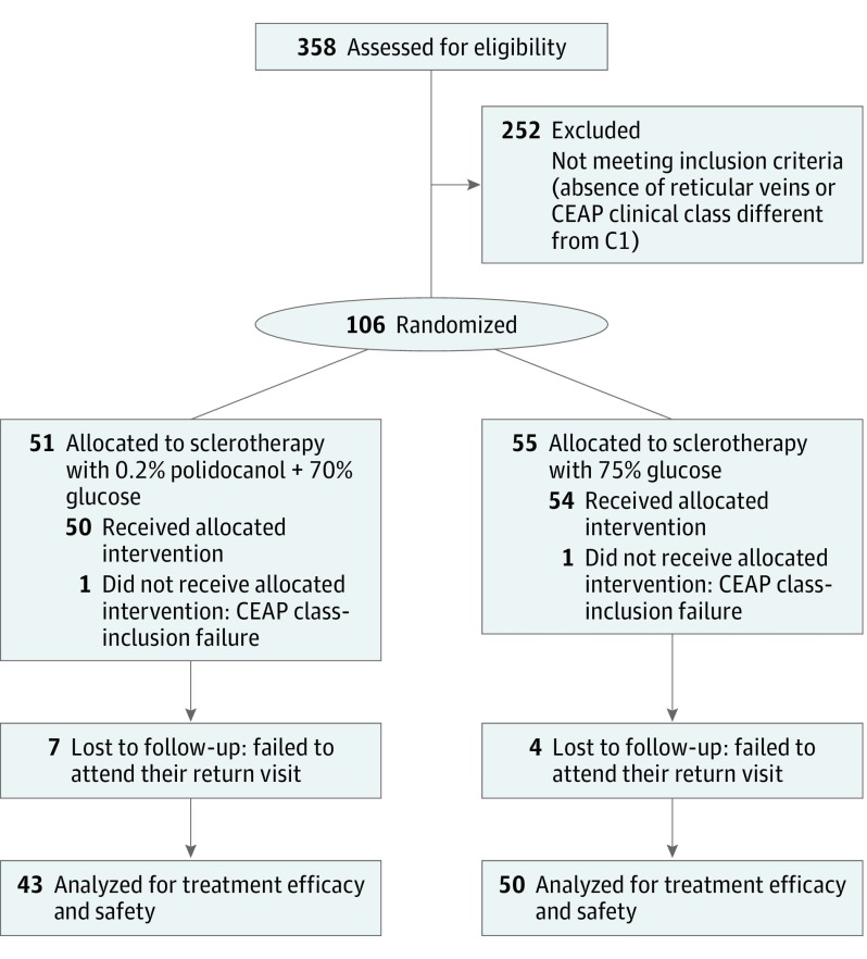
From March to December 2014, 358 patients were assessed for eligibility. Of 106 eligible patients, 51 were randomized to receive 0.2% polidocanol diluted in 70% HG (group 1), and 55 to receive 75% HG alone (group 2) for the treatment of reticular veins in the lower limb. Figure 1 shows the study flow diagram for this clinical trial, including the reasons for exclusions. The demographic characteristics of the study sample are reported in Table 1.
Figure 1. Study Flow Diagram.
CEAP indicates clinical, etiologic, anatomic, and pathophysiologic.
Table 1. Baseline Characteristics of Study Participantsa.
| Variables | Group 1 (n = 43) |
Group 2 (n = 50) |
P Value |
|---|---|---|---|
| Age, median (IQR), yb | 43.0 (24.0-61.0) | 41.0 (27.0-62.0) | .51 |
| Female sex, No. (%) | 43 (100) | 50 (100) | >.99 |
| Unilateral involvement, No. (%) | 43 (100) | 50 (100) | >.99 |
| BMI, mean (SD)c | 25.31 (3.48) | 26.05 (4.06) | .32 |
| Not physically inactive, No. (%)d | 21 (48.8) | 22 (44.0) | .32 |
| Smoking, No. (%)d | 4 (9.3) | 5 (10.0) | .46 |
| Hypertension, No. (%)d | 8 (18.6) | 7 (14.0) | .27 |
| Dyslipidemia, No. (%)d | 5 (11.6) | 5 (10.0) | .40 |
| Hypothyroidism, No. (%)d | 4 (9.3) | 7 (14.0) | .24 |
| Family history of varicose veins, No. (%)d | 34 (79.1) | 42 (84.0) | .27 |
| European ethnicity (skin type I, II, or III), No. (%)d,e | 37 (86.1) | 42 (84.0) | .39 |
| Mediterranean, Asian, Latin, East Indian, African, Native American, or Aboriginal ethnicity (skin type IV, V, or VI), No. (%)d,e | 6 (13.9) | 8 (16.0) | .39 |
| No. of pregnancies, median (IQR)c | 2.0 (0.0-5.0) | 1.0 (0.0-4.0) | .72 |
Abbreviations: BMI, body mass index (calculated as weight in kilograms divided by height in meters squared); IQR, interquartile range.
Group 1 treatment was 0.2% polidocanol plus 70% glucose; group 2 was 75% glucose alone.
Data did not have a normal distribution—nonparametric Mann-Whitney test.
Data had a normal distribution—Student t test for independent samples.
Goodman test for multinomial populations.
Skin color was classified according to the Fitzpatrick skin type scale.
A high interobserver agreement was obtained for all measurements of efficacy outcomes (ICCs of 0.95 for disappearance of the treated reticular veins, 0.95 for length of reticular veins, and 0.88 for length of residual reticular veins 60 days after treatment), and safety outcomes (ICCs of 0.82 and 0.83 for length in centimeters and percentage length of pigmentation, respectively). For subsequent measurements, the average of the interobserver results was used, and this was chosen to correct possible distortions of individual analysis. The average diameter of reticular veins was 3 mm.
Primary Efficacy End Point
Sclerotherapy with 0.2% polidocanol plus 70% HG (group 1) was significantly more effective than 75% HG alone (group 2) in eliminating reticular veins from the treatment area (95.17% vs 85.40%; P < .001). The mean length of residual veins was 3.07 cm in group 1 and 8.30 cm in group 2, a significant difference between the groups (P = .003) (Table 2). The calculated number needed to treat, considering a 100% sclerosis rate, was 4.
Table 2. Results for Efficacy and Safety End Points at 60 Days After Treatmenta.
| Characteristic | Group 1 (n = 43) |
Group 2 (n = 50) |
P Value |
|---|---|---|---|
| Efficacy | |||
| Residual reticular veins, median (IQR), cmb | 3.07 (0.0-33.32) | 8.30 (0.0-29.77) | .003 |
| Eliminated reticular veins, mean (SD), %c | 95.17 (9.09) | 85.4 (17.04) | <.001 |
| Patients with up to 60% sclerosis, No. (%)d | 0 | 4 (8) | .04 |
| Patients with 60%-80% sclerosis, No. (%)d | 4 (9) | 11 (22) | .08 |
| Patients with 80%-99% sclerosis, No. (%)d | 15 (35) | 21 (42) | .48 |
| Patients with 100% sclerosis, No. (%)d | 24 (56) | 14 (28) | .005 |
| Safety, median (IQR)b | |||
| Pigmentation, cm | 3.35 (0.00-20.52) | 3.67 (0.00-24.70) | .09 |
| Pigmentation, % | 3.53 (0.00-26.08) | 7.09 (0.00-50.82) | .06 |
Abbreviation: IQR, interquartile range.
Group 1 treatment was 0.2% polidocanol plus 70% glucose; group 2 was 75% glucose alone.
Data did not have a normal distribution—nonparametric Mann-Whitney test.
Data had a normal distribution—Student t test for independent samples.
Goodman test for multinomial populations.
Safety Outcomes
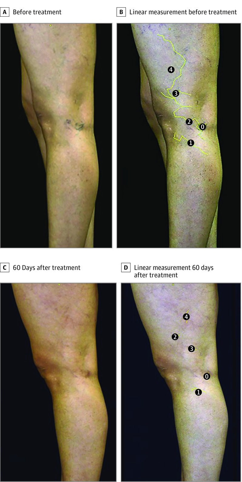
Pigmentation following sclerotherapy was frequently observed in the 2 groups (Figure 2), affecting 55.8% of patients in group 1 and 70.0% of patients in group 2, but with no significant difference (P = .08). In the qualitative analysis, the mean percentage length of pigmentation along the treated vein was 3.53% for group 1 and 7.09% for group 2, also not significantly different between the groups (P = .06) (Table 2). Secondary analysis showed a statistically positive correlation between the occurrence of pigmentation and the presence of residual veins in both centimeters and in percentage length for group 2 (ICC = −0.275; P = .04 and ICC = −0.281; P = .04, respectively) but not for group 1 (ICC = 0.046; P = .87 and ICC = 0.023; P = .77, respectively), indicating that pigmentation was more likely to occur when the veins were not completely sclerosed in group 2.
Figure 2. Images of a Patient Who Underwent Sclerotherapy for Reticular Veins Using 0.2% Polidocanol Diluted in 70% Hypertonic Glucose.
Linear measurements of reticular veins were conducted using Image J before treatment (A and B) and at 60 days after treatment (C and D). In the posttreatment images, pigmentation was also measured.
Secondary Outcomes
No serious adverse events occurred following sclerotherapy. One patient (group 1) presented with edema in the treated leg, but color duplex ultrasonographic imaging performed 5 days after treatment excluded DVT.
Data related to the treatment and complications of patients are reported in Table 3. There was no significant difference in minor or serious adverse events between the 2 treatment groups.
Table 3. Results for Patient Treatment and Monitoringa.
| Characteristic | Group 1 (n = 43) |
Group 2 (n = 50) |
P Value |
|---|---|---|---|
| Punctures, median (IQR) No.b | 14.0 (9.0-21.0) | 15.0 (4.0-19.0) | .27 |
| Volume of medication, mean (SD), mLc | 4.6 (0.6) | 4.6 (0.8) | >.99 |
| Intensity of treatment-related pain, median (IQR)b | 3.0 (0.0-8.0) | 4.0 (0.0-9.0) | .07 |
| Current pain > pain at the time of previous treatments, No. (%)d | 20 (48) | 25 (50) | .37 |
| Initial length of reticular veins, mean (SD), cmc | 66.20 (22.01) | 61.61 (21.06) | .31 |
| Medication-induced pain, No. (%)d | 35/41 (85) | 40/47 (85) | .49 |
| Edema in the foot (<4 d) | 4 (9) | 5 (10) | .45 |
| Edema in the calf (<4 d) | 1 (2) | 4 (8) | .10 |
| Edema at the treatment site (<4 d) | 8 (19) | 12 (24) | .26 |
| Phlebitis at 7 days, No. (%)d | 20 (47) | 23 (46) | .48 |
| Hematoma at 7 days, No. (%)d | 23 (54) | 33 (66) | .11 |
| Late phlebitis at 60 days, No. (%)d | 7 (16) | 9 (18) | .41 |
| Pigmented spots at 60 days, median (IQR) No.b | 1.0 (0.0-7.0) | 2.0 (0.0-16.0) | .28 |
| Absence of telangiectatic matting at 60 days, No. (%)d | 30 (70) | 30 (60) | .16 |
| Mild telangiectatic matting at 60 days, No. (%)d | 12 (28) | 16 (32) | .33 |
| Major telangiectatic matting at 60 days, No. (%)d | 1 (2) | 4 (8) | .10 |
Abbreviation: IQR, interquartile range.
Group 1 treatment was 0.2% polidocanol plus 70% glucose; group 2 was 75% glucose alone.
Data did not have a normal distribution—nonparametric Mann-Whitney test.
Data had a normal distribution—Student t test for independent samples.
Goodman test for multinomial populations.
Discussion
The sclerosing agents investigated in the present study, 0.2% polidocanol solution diluted in 70% HG and 75% HG alone, have been approved for use in sclerotherapy by the Brazilian National Health Surveillance Agency (ANVISA) and are the 2 most commonly used in clinical practice in Brazil. However, to our knowledge, no randomized clinical trials for sclerotherapy of reticular veins have compared these 2 treatments. We also know of no other study using method of objectively measuring the reticular veins with ImageJ software tools.
The initial hypothesis of this study was that 0.2% polidocanol solution diluted in 70% HG would be advantageous because of the low concentrations of polidocanol and maintenance of the high viscosity of HG, which may contribute to a prolonged contact time between the sclerosant and the vessel wall. Indeed, the glucose viscosity in this combination is greater than that of other sclerosants, and so as a vehicle for polidocanol, it could slow its flow in the vessel. Whether or not is this characteristic is able to improve results should be tested in another experimental set.
Because most patients seeking this type of treatment are women, exclusively women were chosen for inclusion in the present study, avoiding selection bias and making the sample more homogeneous. Several clinical trials have been conducted with a similar objective but using different medications, concentrations, and physical forms, and most of them have reported results concerning chromated glycerin, polidocanol and STS in liquid form, or polidocanol and STS foam.
Rabe et al (EASI study) demonstrated a high success rate in the treatment of reticular veins, in up to 3 sessions, using liquid 1% polidocanol or 1% STS, which showed significantly superior results compared with the control group (saline), but no difference between each other, based on subjective image analysis. However, polidocanol had fewer adverse effects and induced less pain. Other studies have demonstrated that liquid 1% polidocanol injections allow less painful administration during sclerotherapy sessions than STS and HS.
Peterson et al found similar results for 1% polidocanol and 23.4% HS in reticular vein treatment, but patients reported significantly greater pain intensity with HS than polidocanol. In the present study, using a more quantitative evaluation, we found that the solution with 0.2% polidocanol and 70% glucose had a superior technical success rate compared with 75% HG alone and a rate similar to that reported in the 2 earlier studies. The number needed to treat showed that about 1 in every 4 patients will benefit from treatment. This means that the clinical relevance for 0.2% polidocanol plus 70% glucose is good.
As for safety, Guex et al, in a large French study of patients treated with polidocanol, concluded that polidocanol was a safe sclerosing agent and that it should be preferably used in liquid form. Engelberger et al, however, reported 1 case of a cardiotoxic event and myocardial infarction following the use of polidocanol foam for the treatment of varicose veins, reinforcing the notion that it should be preferably used in liquid form and at low concentrations. In the present study, no serious adverse events were observed in any patient.
Pigmentation is a common minor adverse event following sclerotherapy, affecting 10% to 80% of patients undergoing sclerotherapy with different sclerosants and for different veins. Many authors have not considered this event important, mainly because it usually resolves spontaneously and tends to disappear within 1 year after treatment. In the present study, pigmentation was observed in both treatment groups, but a little less frequently in patients treated with 0.2% polidocanol plus 70% HG, with no significant difference between the groups (55.8% vs 70.0%; P = .08). ImageJ software also showed very small pigmented spots in the combination group and HG group, but considering only the measurements in percentage of pigmentation along the reticular vein, we found that this frequency was not high (3.53% and 7.09%, respectively).
Because increased pigmentation is probably considered the most undesirable minor complication by patients and physicians, it often serves as a justification to those who argue that reticular veins should be treated surgically. Multiple stab phlebectomy is believed to cause less pigmentation than sclerotherapy, but it is a surgical procedure that requires some type of anesthesia (local, locoregional, or general), is time-consuming, and is associated with risks inherent in surgical procedures, such as infections and loss of local sensitivity. For these reasons, sclerotherapy is considered the treatment of choice, provided the patient accepts some risk of temporary or even permanent skin pigmentation.
Limitations
Some limitations of the present study should be emphasized. First, while no serious adverse events occurred in either group, it must be remembered that this outcome is not a frequent event for sclerotherapy. Second, this study did not include a sham-procedure placebo group. Third, different concentrations of these drugs might produce different results. Fourth, the 3-week compression therapy was not used in this study.
Conclusions
In conclusion, sclerotherapy of reticular veins with 0.2% polidocanol diluted in 70% HG was significantly superior to sclerotherapy with 75% HG alone in this study population at 60 days after treatment. Both treatments were considered safe. There were no serious adverse events in either group. Pigmentation was the most common minor adverse event and occurred in both groups, with no statistical difference between them.
Trial Protocol
eTable. CONSORT checklist
eAppendix. Additional study information [in Portuguese]
References
- 1.Albanese AR, Albanese AM, Albanese EF. Lateral subdermic varicose vein system of the legs: its surgical treatment by the chiseling tube method. Vasc Surg. 1969;3(2):81-89. [DOI] [PubMed] [Google Scholar]
- 2.Somjen GM. Anatomy of the superficial venous system. Dermatol Surg. 1995;21(1):35-45. [DOI] [PubMed] [Google Scholar]
- 3.Eklöf B, Rutherford RB, Bergan JJ, et al. ; American Venous Forum International Ad Hoc Committee for Revision of the CEAP Classification . Revision of the CEAP classification for chronic venous disorders: consensus statement. J Vasc Surg. 2004;40(6):1248-1252. [DOI] [PubMed] [Google Scholar]
- 4.Robertson L, Evans C, Fowkes FGR. Epidemiology of chronic venous disease. Phlebology. 2008;23(3):103-111. [DOI] [PubMed] [Google Scholar]
- 5.Caggiati A, Phillips M, Lametschwandtner A, Allegra C. Valves in small veins and venules. Eur J Vasc Endovasc Surg. 2006;32(4):447-452. [DOI] [PubMed] [Google Scholar]
- 6.Wienert V, Simon H, Böhler U. Angioarchitecture of spider veins Scanning electron microscope study of corrosion spider veins. Phlebologie. 2006;35(1):24-29. [Google Scholar]
- 7.Parsi K. Interaction of detergent sclerosants with cell membranes. Phlebology. 2015;30(5):306-315. [DOI] [PubMed] [Google Scholar]
- 8.Rabe E, Breu FX, Cavezzi A, et al. ; Guideline Group . European guidelines for sclerotherapy in chronic venous disorders. Phlebology. 2014;29(6):338-354. [DOI] [PubMed] [Google Scholar]
- 9.Weiss MA, Hsu JT, Neuhaus I, Sadick NS, Duffy DM. Consensus for sclerotherapy. Dermatol Surg. 2014;40(12):1309-1318. [DOI] [PubMed] [Google Scholar]
- 10.Ramelet AA. Phlebectomy: technique, indications and complications. Int Angiol. 2002;21(2)(suppl 1):46-51. [PubMed] [Google Scholar]
- 11.Sadick NS. Long-term results with a multiple synchronized-pulse 1064 nm Nd:YAG laser for the treatment of leg venulectasias and reticular veins. Dermatol Surg. 2001;27(4):365-369. [DOI] [PubMed] [Google Scholar]
- 12.Rogachefsky AS, Silapunt S, Goldberg DJ. Nd:YAG laser (1064 nm) irradiation for lower extremity telangiectases and small reticular veins: efficacy as measured by vessel color and size. Dermatol Surg. 2002;28(3):220-223. [DOI] [PubMed] [Google Scholar]
- 13.Duffy DM. Sclerosants: a comparative review. Dermatol Surg. 2010;36(s2)(suppl 2):1010-1025. [DOI] [PubMed] [Google Scholar]
- 14.American Academy of Dermatology Guidelines of care for sclerotherapy treatment of varicose and telangiectatic leg veins. J Am Acad Dermatol. 1996;34(3):523-528. [DOI] [PubMed] [Google Scholar]
- 15.Goldman MP, Bennett RG. Treatment of telangiectasia: a review. J Am Acad Dermatol. 1987;17(2 Pt 1):167-182. [DOI] [PubMed] [Google Scholar]
- 16.Bourgeois A, Quillard J, Constantin JM, et al. 66% Glucose, a safe sclerosant: experimental study [in French]. J Mal Vasc. 1984;9(2):97-99. [PubMed] [Google Scholar]
- 17.Salomon JL, Bourgeois A, Le Baleur A, Pillot P, Gillot C, Frileux C. Product of choice for maintenance sclerosis: 66% glucose solution [in French]. Phlebologie. 1983;36(3):249-254. [PubMed] [Google Scholar]
- 18.Goldman MP, Sadick NS, Weiss RA. Cutaneous necrosis, telangiectatic matting, and hyperpigmentation following sclerotherapy: etiology, prevention, and treatment. Dermatol Surg. 1995;21(1):19-29. [DOI] [PubMed] [Google Scholar]
- 19.Ceulen RP, Sommer A, Vernooy K. Microembolism during foam sclerotherapy of varicose veins. N Engl J Med. 2008;358(14):1525-1526. [DOI] [PubMed] [Google Scholar]
- 20.Marrocco-Trischitta MM, Guerrini P, Abeni D, Stillo F. Reversible cardiac arrest after polidocanol sclerotherapy of peripheral venous malformation. Dermatol Surg. 2002;28(2):153-155. [DOI] [PubMed] [Google Scholar]
- 21.Schwartz L, Maxwell H. Sclerotherapy for lower limb telangiectasias. Cochrane Database Syst Rev. 2011;(12):CD008826. [DOI] [PMC free article] [PubMed] [Google Scholar]
- 22.Tisi PV, Beverley C, Rees A. Injection sclerotherapy for varicose veins. Cochrane Database Syst Rev. 2006;(4):CD001732. [DOI] [PubMed] [Google Scholar]
- 23.Gloviczki P, Gloviczki ML. Guidelines for the management of varicose veins. Phlebology. 2012;27(s1)(suppl 1):2-9. [DOI] [PubMed] [Google Scholar]
- 24.Benigni JP, Bihari I, Rabe E, et al. ; UIP–Union Internationale de Phlébologie . Venous symptoms in C0 and C1 patients: UIP consensus document. Int Angiol. 2013;32(3):261-265. [PubMed] [Google Scholar]
- 25.Peterson JD, Goldman MP, Weiss RA, et al. Treatment of reticular and telangiectatic leg veins: double-blind, prospective comparative trial of polidocanol and hypertonic saline. Dermatol Surg. 2012;38(8):1322-1330. [DOI] [PubMed] [Google Scholar]
- 26.Rabe E, Schliephake D, Otto J, Breu FX, Pannier F. Sclerotherapy of telangiectases and reticular veins: a double-blind, randomized, comparative clinical trial of polidocanol, sodium tetradecyl sulphate and isotonic saline (EASI study). Phlebology. 2010;25(3):124-131. [DOI] [PubMed] [Google Scholar]
- 27.Kern P, Ramelet AA, Wutschert R, Mazzolai L. A double-blind, randomized study comparing pure chromated glycerin with chromated glycerin with 1% lidocaine and epinephrine for sclerotherapy of telangiectasias and reticular veins. Dermatol Surg. 2011;37(11):1590-1594. [DOI] [PubMed] [Google Scholar]
- 28.Figueiredo M, Figueiredo MF. Survey on liquid sclerotherapy of lower limb varicose veins [in Portuguese]. J Vasc Bras. 2013;12(1):10-15. [Google Scholar]
- 29.Bertanha M, Sobreira ML, Pinheiro Lúcio Filho CE, et al. Polidocanol versus hypertonic glucose for sclerotherapy treatment of reticular veins of the lower limbs: study protocol for a randomized controlled trial. Trials. 2014;15(1):497. [DOI] [PMC free article] [PubMed] [Google Scholar]
- 30.Willenberg T, Smith PC, Shepherd A, Davies AH. Visual disturbance following sclerotherapy for varicose veins, reticular veins and telangiectasias: a systematic literature review. Phlebology. 2013;28(3):123-131. [DOI] [PubMed] [Google Scholar]
- 31.Goldman MP. Treatment of varicose and telangiectatic leg veins: double-blind prospective comparative trial between aethoxyskerol and sotradecol. Dermatol Surg. 2002;28(1):52-55. [DOI] [PubMed] [Google Scholar]
- 32.Kern P, Ramelet AA, Wutschert R, Bounameaux H, Hayoz D. Single-blind, randomized study comparing chromated glycerin, polidocanol solution, and polidocanol foam for treatment of telangiectatic leg veins. Dermatol Surg. 2004;30(3):367-372. [DOI] [PubMed] [Google Scholar]
- 33.Rao J, Wildemore JK, Goldman MP. Double-blind prospective comparative trial between foamed and liquid polidocanol and sodium tetradecyl sulfate in the treatment of varicose and telangiectatic leg veins. Dermatol Surg. 2005;31(6):631-635. [DOI] [PubMed] [Google Scholar]
- 34.McCoy S, Evans A, Spurrier N. Sclerotherapy for leg telangiectasia—a blinded comparative trial of polidocanol and hypertonic saline. Dermatol Surg. 1999;25(5):381-385. [DOI] [PubMed] [Google Scholar]
- 35.Guex JJ, Schliephake DE, Otto J, Mako S, Allaert FA. The French polidocanol study on long-term side effects: a survey covering 3,357 patient years. Dermatol Surg. 2010;36(s2)(suppl 2):993-1003. [DOI] [PubMed] [Google Scholar]
- 36.Engelberger RP, Ney B, Clair M, et al. Myocardial infarction after ultrasound-guided foam sclerotherapy for varicose veins—a case report and review of the literature of a rare but serious adverse event. Vasa. 2016;45(3):255-258. [DOI] [PubMed] [Google Scholar]
- 37.Munavalli GS, Weiss RA. Complications of sclerotherapy In: Gloster HM, ed. Complications in Cutaneous Surgery. New York, NY: Springer; 2008:213-223. [Google Scholar]
- 38.Goldman MP, Kaplan RP, Duffy DM. Postsclerotherapy hyperpigmentation: a histologic evaluation. J Dermatol Surg Oncol. 1987;13(5):547-550. [DOI] [PubMed] [Google Scholar]
- 39.Georgiev M. Postsclerotherapy hyperpigmentations: a one-year follow-up. J Dermatol Surg Oncol. 1990;16(7):608-610. [DOI] [PubMed] [Google Scholar]
- 40.Nootheti PK, Cadag KM, Magpantay A, Goldman MP. Efficacy of graduated compression stockings for an additional 3 weeks after sclerotherapy treatment of reticular and telangiectatic leg veins. Dermatol Surg. 2009;35(1):53-57. [DOI] [PubMed] [Google Scholar]
Associated Data
This section collects any data citations, data availability statements, or supplementary materials included in this article.
Supplementary Materials
Trial Protocol
eTable. CONSORT checklist
eAppendix. Additional study information [in Portuguese]


