A 38-year-old Brazilian man was referred to our hospital because of widespread cutaneous lesions of 10-months duration. Initial lesions emerged on the face and were taken as acne. Multiple lesions followed, and only the scalp was spared. He looked for medical advice when the feet lesions became painful and led to difficulty in walking. The patient lived alone under conditions of extreme poverty in a rural area 200 km far from the city of Rio de Janeiro. Because there was no sewage disposal system, he used to dig cesspits nearby, often using his own hands. He did not have direct contact with cats, but some cats did wander around his dwelling.
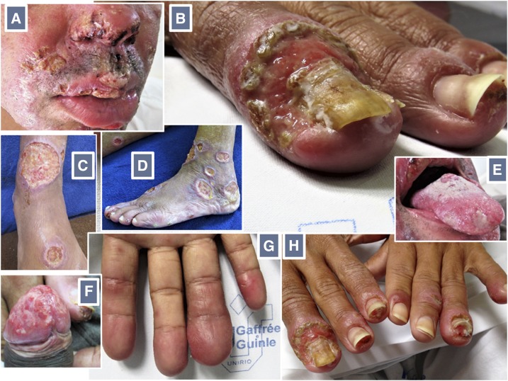
Clinical examination revealed a wasted, chronically ill patient with multiple tegumentary ulcerated lesions of variable sizes (Figure 1). Larger lesions drained a seropurulent discharge. Among the multiple lesions on the face, those on the lips and ala nasi were covered with a yellowish or hemorrhagic crust. There was balanitis, with a large painless ulcerated lesion covering the glans penis, and glossitis, with shallow ulcers on the dorsal aspect of the tongue. The helices and earlobes were reddened and infiltrated. Onychomycosis of multiple nails, paronychia, nail dystrophy, areas of onycholysis, hapalonychia, xantonychia, and onychauxis were recorded. Multiple digits were reddened, enlarged, and sausage-shaped, mainly over the distal phalanges.
Figure 1.
Multiple yellowish or hemorrhagic crust-covered lesions on the face, including the lips and ala nasi (A). Onychauxis (thickening and transverse ridging) of the right index fingernail, with a large surrounding area of paronychia (B). Multiple cutaneous ulcers of varying sizes, some surrounded by violaceous erythema and rolled borders (C, D). Shallow, fibrin-covered ulcers on the dorsum of the tongue (E). Ulcerative balanitis (F). Sausage-shaped fingers (G). Nail dystrophy and distal detachment from the nail bed (onycholysis) of several fingernails, with a bizarre aspect (H). This figure appears in color at www.ajtmh.org.
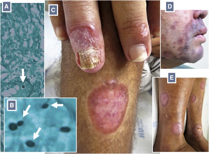
Laboratory evaluations were remarkable for a diagnosis of human immunodeficiency virus (HIV) infection, with a CD4 cell count of 249 mm−3. He tested negative for syphilis and viral hepatitis. There was no evidence of bone lytic lesions or systemic disease. Histopathological analyses of biopsied tissue revealed an architectural pattern of subcutaneous mycosis, but very few fungal elements were found, a pattern suggestive of sporotrichosis.1 These were spherical and cigar-shaped yeastlike structures, which is suggestive of Sporothrix spp. (Figure 2). Samples for fungal cultures (collected after initiation of antifungal therapy) were negative. A diagnosis of disseminated subcutaneous mycosis, most probably sporotrichosis, was made.
Figure 2.
Grocott’s methenamine silver stain unmasks few spherical (A) and cigar-shaped yeastlike structures with narrow-based budding (B) consistent with Sporothrix spp. (white arrows) on a skin biopsy section. Clinical resolution of the lesions after treatment (C–E). This figure appears in color at www.ajtmh.org.
Sporotrichosis is a neglected opportunistic infection in HIV-infected patients in southeast Brazil.2,3 It is transmitted mainly by traumatic inoculation of fungal elements.4 A daily regimen of amphotericin B deoxycholate was offered, starting with escalating doses, until a cumulative dose of 1 g was reached. Highly active antiretroviral therapy (HAART) was also started. Paradoxical worsening of the lesions (immune reconstitution inflammatory syndrome) developed 4 weeks later and was treated with corticosteroids.5 Three months later he was discharged without active lesions and on oral itraconazole and HAART.
REFERENCES
- 1.Quintella LP, Passos SR, do Vale AC, Galhardo MC, Barros MB, Cuzzi T, Reis Rdos S, de Carvalho MH, Zappa MB, Schubach AO, 2011. Histopathology of cutaneous sporotrichosis in Rio de Janeiro: a series of 119 consecutive cases. J Cutan Pathol 38: 25–32. [DOI] [PubMed] [Google Scholar]
- 2.Freitas DF, Valle AC, da Silva MB, Campos DP, Lyra MR, de Souza RV, Veloso VG, Zancopé-Oliveira RM, Bastos FI, Galhardo MC, 2014. Sporotrichosis: an emerging neglected opportunistic infection in HIV-infected patients in Rio de Janeiro, Brazil. PLoS Negl Trop Dis 28: e3110. [DOI] [PMC free article] [PubMed] [Google Scholar]
- 3.Moreira JA, Freitas DF, Lamas CC, 2015. The impact of sporotrichosis in HIV-infected patients: a systematic review. Infection 43: 267–276. [DOI] [PubMed] [Google Scholar]
- 4.Barros MB, de Almeida Paes R, Schubach AO, 2011. Sporothrix schenckii and sporotrichosis. Clin Microbiol Rev 24: 633–654. [DOI] [PMC free article] [PubMed] [Google Scholar]
- 5.Lyra MR, Nascimento ML, Varon AG, Pimentel MI, Antonio Lde F, Saheki MN, Bedoya-Pacheco SJ, Valle AC, 2014. Immune reconstitution inflammatory syndrome in HIV and sporotrichosis coinfection: report of two cases and review of the literature. Rev Soc Bras Med Trop 47: 806–809. [DOI] [PubMed] [Google Scholar]


