See the article by Rahme et al. on pp. 332–342.
Mouse models are essential tools for the study of the mechanisms of tumor initiation and progression and the testing of new therapies against glioblastoma (GBM), the most common and most deadly primary malignant brain tumor. Currently used mouse models can be classified into 4 categories: xenograft models, chemically induced models, genetically engineered models, and viral vector-induced models. Xenograft models, derived from the implantation of established GBM cell lines or primary GBM cells and cancer stem cells in immune-deficient mice, are easy to generate and are representative of the complexity of human tumors when established from primary cells but lack an immune system and a same-species microenvironment. Chemically induced models are syngeneic and immune-competent but are nonhuman and have undefined and uncontrolled genetic bases. Genetically engineered models are genetically precise and reproducible spontaneous models with an intact immune system but are time and effort consuming and generate nonhuman tumors with few genetic alterations that do not always mimic human alterations. Viral vector-induced models are a variation of genetically engineered models that use viral-based transfections to genetically modify regional somatic cells to generate mouse tumors. Compared with conventional genetically engineered models, vector-induced models are easier and faster to generate. Several mouse models of GBM have been developed, but due to the complexity and heterogeneity of the disease, no one model predominates and there remains a need for new models that reproduce specific aspects and/or subgroups of GBM.
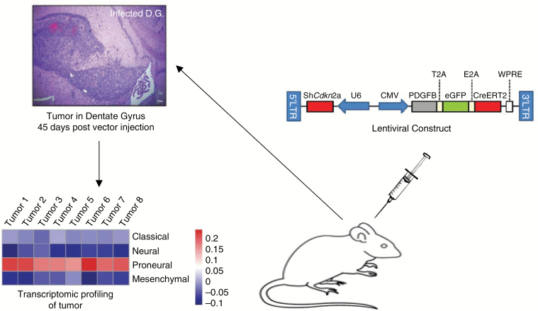
An article by Rahme et al1 in this issue uses a retroviral-based approach to efficiently generate a new practical and versatile mouse model of proneural GBM. An integrated genomic analysis of GBM data from The Cancer Genome Atlas classified tumors into 4 subtypes: classical, proneural, mesenchymal, and neural.2 Vector-based mouse models of mesenchymal GBM have been previously generated and published,3,4 but no such models are known for the proneural subtype, characterized by isocitrate dehydrogenase mutations, gain-of-function mutations of platelet derived growth factor (PDGF) receptor alpha, and loss-of-function mutations of cyclin-dependent kinase inhibitor (CDKN)2A/B and tumor suppressor protein 53.2 Rahme et al1 generated and validated the first virus-based model of proneural GBM. To develop a virus that would generate proneural-like GBM tumors, they constructed a lentivirus encoding PDGF subunit B and a short hairpin RNA against CDKN2A. To enable the study of additional genes of interest in mice with floxed alleles of these genes, they further engineered the vector to coexpress CreERT2 together with PDGFB. To be able to track infected cells, they also constructed a version of the above vector that encoded enhanced green fluorescent protein (eGFP) together with PDGFB and CreERT2. When the vector was injected into the dentate gyrus (a region known to harbor neural stem and progenitor cells) of immune-competent mice, it generated tumors with all the hallmarks of GBM (hypercellularity, pseudopalisading necrosis, invasion of surrounding brain, and hemorrhage). Hypercellularity in the dentate gyrus was observed one week after virus injection, tumor penetrance was very high (88.5%), and experimental mice had a significantly shorter median survival than controls. The tumors expressed molecular markers of stemness, proliferation, and angiogenesis consistent with those found in human GBM. Transcriptomic profiling of the tumors showed a strong correlation with the proneural subtype and no significant correlation with any of the other subtypes. Gene set enrichment analysis (GSEA) showed an enrichment of genes associated with the Wnt pathway that is known to be activated in proneural GBM. The authors also verified the capability of the vector to induce eGFP expression in transduced tumor cells as well as the activity of CreERT2 in a tdTomato floxed mouse model. Altogether, the authors convincingly describe the generation of a single lentiviral vector-mediated, efficient, representative, versatile, and immune-competent new mouse model of proneural GBM (Fig. 1).
Fig. 1.
Schematic of model generation and validation
The above-described mouse model possesses several advantageous characteristics that should facilitate its use by researchers studying the proneural subtype of GBM and those developing and testing new therapies against these tumors. First and foremost, the model is very efficient and easy to generate. Injections of a single virus into available immune-competent mice will generate tumors relatively quickly (median of 77 days) and with high penetrance (88.5%). The transduced tumor cells can be visualized with eGFP fluorescence, allowing the tracking of tumor initiation, progression, and invasion as well as responsiveness to therapy. Intriguingly, unlike other PDGF-driven glioma mouse models,5,6 eGFP-positive transduced tumor cells made up the vast majority of tumor cells, and no significant numbers of non-transduced cells were recruited to the tumor and transformed. The authors did not experimentally pursue the reasons for such a difference but speculated that it might be associated with different cells of origin in the different models. A second important advantage of the model is its very faithful representation of the human proneural subtype. Both transcriptomic and GSEA analyses unequivocally classify the virus-generated tumors as proneural. This model will thus complement previously generated models of mesenchymal GBM, which suggests that the generation of a classical subtype mouse model based on the same principles used by Rahme et al is feasible and desirable for the study of GBM subtypes. In this context, considering the demonstrated existence of all GBM subtypes in different regions and different cells of any given tumor7 and the lack of any significant clinical impact of molecular subtyping to date, the importance of studies focused on investigating molecular subtypes remains to be determined. A third advantageous and practical aspect of the new mouse model is its usefulness to study specific genes of interest when used in transgenic mice with floxed alleles of the gene in question. This capability substantially expands the use of the model and the breadth of questions it can be used to answer. The limitations of the model are similar to those of any other transgenic model, as discussed above.
In summary, Rahme et al succeeded in generating a new, efficient, representative, and versatile mouse model of proneural GBM that should be of interest to many researchers in the neuro-oncology field.
References
- 1. Rahme GJ, Luikart BW, Cheng C et al. . A recombinant lentiviral PDGF-driven mouse model of proneural glioblastoma. Neuro Oncol. 2017. doi: 10.1093/neuonc/nox129. [DOI] [PMC free article] [PubMed] [Google Scholar]
- 2. Verhaak RG, Hoadley KA, Purdom E et al. . Integrated genomic analysis identifies clinically relevant subtypes of glioblastoma characterized by abnormalities in PDGFRA, IDH1, EGFR, and NF1. Cancer Cell. 2010;17(1):98–110. [DOI] [PMC free article] [PubMed] [Google Scholar]
- 3. Marumoto T, Tashiro A, Friedmann-Morvinski D et al. . Development of a novel mouse glioma model using lentiviral vectors. Nat Med. 2009;15(1):110–116. [DOI] [PMC free article] [PubMed] [Google Scholar]
- 4. Niola F, Zhao X, Singh D et al. . Mesenchymal high-grade glioma is maintained by the ID-RAP1 axis. J Clin Invest. 2013;123(1):405–417. [DOI] [PMC free article] [PubMed] [Google Scholar]
- 5. Fomchenko EI, Dougherty JD, Helmy KY et al. . Recruited cells can become transformed and overtake PDGF-induced murine gliomas in vivo during tumor progression. PLoS One. 2011;6(7):e20605. [DOI] [PMC free article] [PubMed] [Google Scholar]
- 6. Assanah M, Lochhead R, Ogden A et al. . Glial progenitors in adult white matter are driven to form malignant gliomas by platelet-derived growth factor-expressing retroviruses. J Neurosci. 2006;26(25):6781–6790. [DOI] [PMC free article] [PubMed] [Google Scholar]
- 7. Patel AP, Tirosh I, Trombetta JJ et al. . Single-cell RNA-seq highlights intratumoral heterogeneity in primary glioblastoma. Science. 2014;344(6190):1396–1401. [DOI] [PMC free article] [PubMed] [Google Scholar]


