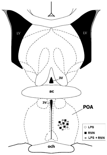
Fig. 4. Schematic representation of a coronal section through the POA 0.36 mm from bregma, illustrating the location of the tip of the microdialysis probes for RMN-treated (filled squares) and saline-treated control (open circles) animals.
ac indicates anterior commissure; LV, lateral ventricle; 3V, third ventricle; och, optic chiasm.

