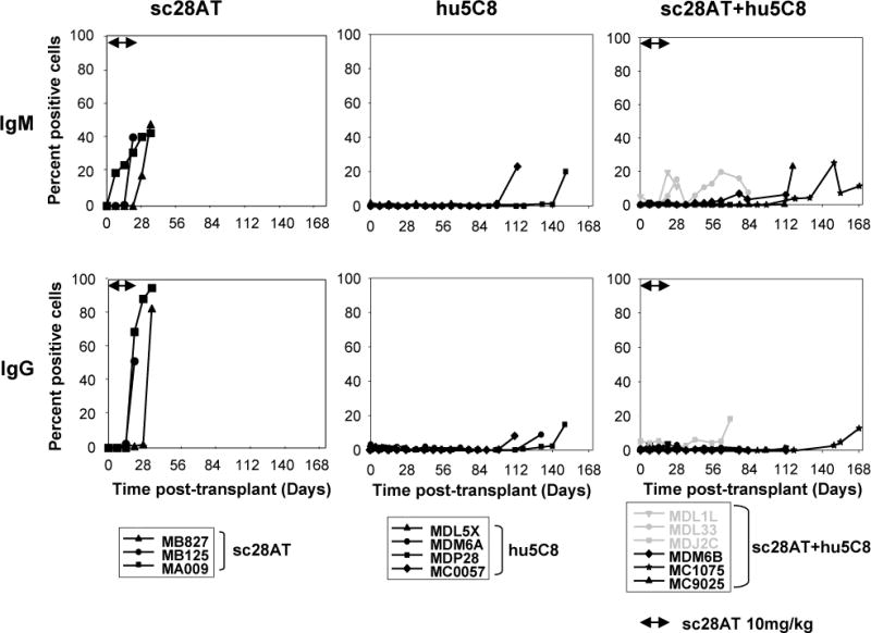
Figure 8. Donor-specific alloantibodies from the day of transplant to graft explant.

The presence of IgM (upper panel) and IgG (lower panel) anti-donor serum alloantibodies was measured by flow cytometry. In sc28AT+hu5C8 animals (right panels), weak IgM Abs were detected in recipients that suffered from malaria infection (MDL1L and MDL33, grey lines) and weak IgG Ab was detected in recipient (MDJ2C) on the day of euthanasia due to post biopsy intestinal complication (grey line). Significant (>20%) elaboration of donor-specific antibody was consistently prevented during treatment with either hu5C8 or sc28AT+hu5C8.
