Abstract
An 81-year-old woman was admitted with a nodular cutaneous melanoma of the abdominal wall involving the umbilicus. After performing wide excision with 2 cm margin of the melanoma, umbilical reconstruction and defect closure were planned. After careful consideration, we decided to use an island pedicle flap which allowed closure of the defect and reconstruction of the umbilicus.
Keywords: Island pedicle flap, reconstruction, umbilicus
CLINICAL CASE
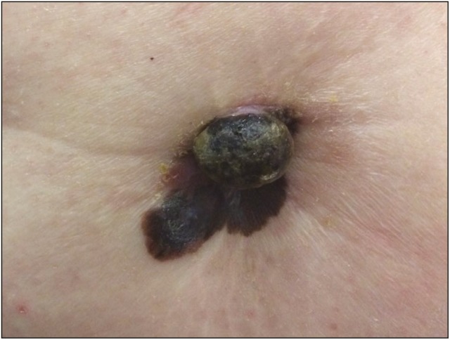
An 81-year-old woman was admitted with a nodular cutaneous melanoma of the abdominal wall involving the umbilicus. The primary tumor, 5.6 mm thick, was ulcerated [Figure 1]. Wide excision with 2 cm margin and sentinel lymph node biopsy were performed. The resultant defect measured 90 × 60 mm, resulting in total umbilical loss. How would you reconstruct this defect in order to repair the umbilicus?
Figure 1.
Nodular cutaneous melanoma, 5.6 mm thick, enclosing the totality of umbilicus
RESOLUTION
The umbilicus is the only naturally occurring scar on the body, having a significant aesthetic role.[1] Its absence or dysmorphia is deemed unattractive and may give rise to psychological distress, making it a common concern in surgical planning.[2] Great care must be taken with scar-line placement and tissue matching to give a cosmetically acceptable final result.
The main challenge of umbilicus reconstruction is to create a new and natural-looking structure at an adequate position with minimal scarring.[2] Although a wide range of configurations exist, the “ideal” umbilicus is apparently small, oval, and nonprotruberant.[3]
In our clinical case, the final defect was oval and occupied a significant portion of the abdominal wall and reconstruction of a total absent umbilicus was needed. Several neoumbilicoplasty techniques have been proposed, involving second-intention healing, skin grafts, cartilage, purse-string suture, and flaps.[2] Flaps can be either local and contiguous or islanded. The latter include the spiral rotation flap, the V-shaped flap, the tubularized flap, the umbilical C-V flap, the island flap, the reverse fan-shaped flap, the upper inverted omega-shaped flap, and the lower lazy M-shaped flap. There are also techniques using three triangular skin flaps, diamond-shaped skin flaps, superior-based single triangular flaps, transverse flaps, inferior-based vertical flaps, or the combination of skin flaps and cartilage grafts.[2] The use of local tissue for neoumbilicoplasty accords with the fundamental reconstructive principle of “like with like” tissue to give the most natural outcome.[3] Furthermore, flaps introduce well-vascularized tissue with wound-healing advantages and confine surgical morbidity to a single area.[3] Possible complications of these procedures include flap necrosis, infection, hematoma, suture dehiscence, hypertrophic scar, and umbilical flattening.[2,3,4]
After careful consideration, we decided to use an island pedicle flap that allowed closure of the defect and reconstruction of the umbilicus.
THE PROCEDURE
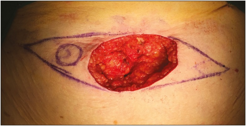
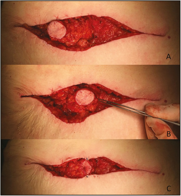
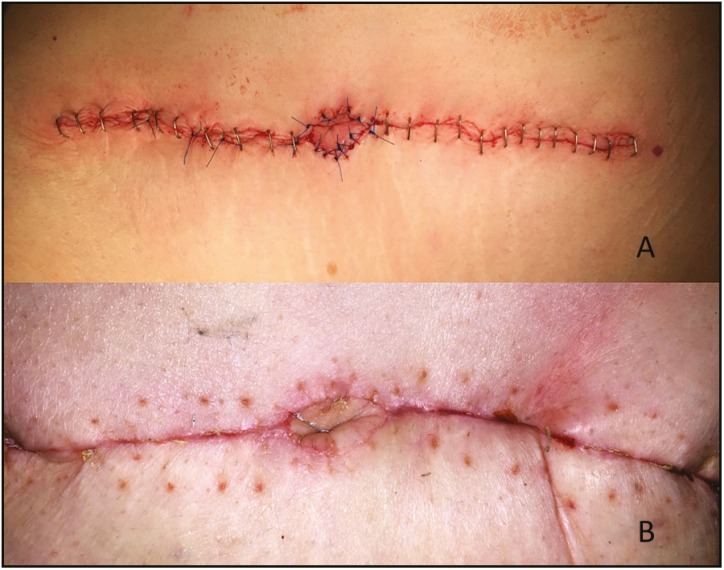
This procedure was performed with the patient under general anesthesia. After performing wide excision with 2 cm margin of the melanoma, umbilical reconstruction and defect closure were planned [Figure 2]. Donor skin of the lateral margin of the defect was outlined to match the size and shape of the intended umbilicus. An island flap was designed such that the subcutaneous pedicle was long enough to be transferred to the planned site, setting the pivot point on the left lateral side of the flap. The island flap was successfully transferred to the desired position and was fixed to the anterior sheath of the rectus abdominis muscle using a transfixive U stitch at the center of the flap with a 2/0 absorbable suture (polyglactin 910). With this stitch, the flap acquired the intended conic shape recreating the umblicus [Figure 3A–C]. The perimeter of the new umbilicus was sutured to the surrounding skin with 4/0 polypropylene. Meticulous placement of multiple subcutaneous sutures with 2.0 polyglactin 910 facilitated the approximation of the epidermis of the donor area without any tension. Epidermal approximation was obtained with surgical stainless steel staples [Figure 4A]. A pressure dressing was secured for 72 h and then routine wound care advice was given to the patient. To prevent surgical site infection, the patient was given cefazolin 1000 mg ev before surgery and cefuroxime 500 mg bid po for 5 days after. The patient returned 2 weeks later for suture removal [Figure 4B].
Figure 2.
Planning of reconstruction using an island pedicle flap using otherwise redundant skin of the lateral margin of the defect
Figure 3.
The island flap (A) was transferred to the desired position (B) and was fixed to the anterior sheath of the rectus abdominis muscle using a transfixive U stitch acquiring the intended conic shape (C).
Figure 4.
Final result immediately after surgery (A) and after removal of sutures 14 days after (B)
In summary, we present a repair option to reconstruct the umbilicus. The aim of this technique was to create a harmonious umbilicus of adequate size together with a natural shape and depth. It is a relatively simple procedure and uses otherwise redundant skin of the lateral margin of the defect. In addition, it offers a rapid recovery time for the patient. On the other hand, this umbilicoplasty technique needs to be combined with either a vertical or horizontal scar over the umbilical position.[4]
PRACTICE POINTS
- Preoperative positioning of the proposed umbilicus is essential for its proper reconstruction.
- An island flap facilitates reconstruction of the umbilicus at an ideal position.
- The island flap was designed such that the subcutaneous pedicle is long enough to be transferred to the desired position.
- An anchor suture on the bottom of the anterior sheath of the rectus abdominis muscle recreates the three-dimensional morphology of the umbilicus.
- This technique is easy and safe, and simultaneously recreates a natural-appearing umbilicus.
Financial support and sponsorship
Nil.
Conflicts of interest
None.
REFERENCES
- 1.Kakudo N, Kusumoto K, Fujimori S, Shimotsuma A, Ogawa Y. Reconstruction of a natural-appearing umbilicus using an island flap: Case report. J Plast Reconstr Aesthet Surg. 2006;59:999–1002. doi: 10.1016/j.bjps.2005.12.025. [DOI] [PubMed] [Google Scholar]
- 2.da Silva Júnior VV, de Sousa FR. Improvement on the neo-umbilicoplasty technique and review of the literature. Aesthetic Plast Surg. 2017 doi: 10.1007/s00266-017-0847-6. doi: 101007/s00266-017-0847-6 [Epub ahead of print] [DOI] [PubMed] [Google Scholar]
- 3.Southwell-Keely JP, Berry MG. Umbilical reconstruction: a review of techniques. J Plast Reconstr Aesthet Surg. 2011;64:803–8. doi: 10.1016/j.bjps.2010.11.014. [DOI] [PubMed] [Google Scholar]
- 4.Pfulg M, Van de Sijpe K, Blondeel P. A simple new technique for neo-umbilicoplasty. Br J Plast Surg. 2005;58:688–91. doi: 10.1016/j.bjps.2005.01.013. [DOI] [PubMed] [Google Scholar]


