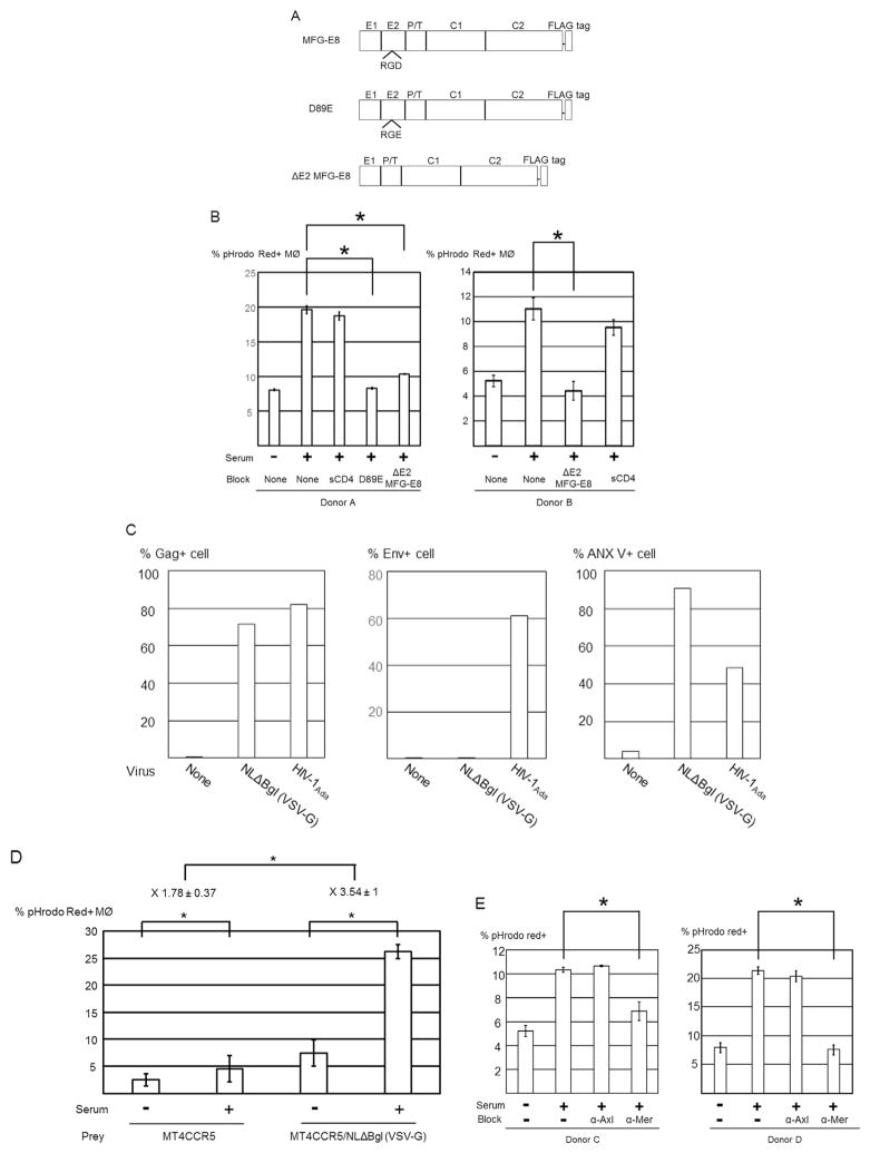
Fig. 3.
Human serum-induced phagocytosis of HIV-1-infected cells is dependent on PtdSer exposed on infected cells and Mer expressed on macrophages. (A) Schematic representation of recombinant MFG-E8 and two types of mutants of MFG-E8 (D89E and ΔE2 MFG-E8) that bind PtdSer but not integrins. E1 and E2, epidermal growth factor-like domains 1 and 2; P/T, proline/threonine-enriched motif; C1 and C2, discoidin domains 1 and 2. The C2 domain of MFG-E8 binds PtdSer, and the RGD motif in the E2 domain of MFG-E8 binds integrins. (B) pHrodo red-labeled MT4CCR5/NL4-3 cells were incubated with or without 20 μg/ml of D89E, ΔE2 MFG-E8 or sCD4, followed by co-culture with macrophages in the presence or absence of 3% human serum. Efferocytosis of MT4CCR5/NL4-3 cells was analyzed by flow cytometry. This experiment was repeated twice in triplicate as independent experiments, using cells from different donors for each independent experiment. The results shown are averages and standard deviations. Significance was calculated by comparing the values of the samples with 3% serum and no blocking reagents to the values of the samples with 3% serum with the blocking reagents indicated in the figures, using a one-sided Mann-Whitney U test. * p=0.05. (C) MT4CCR5 cells were infected with NLΔBgl (VSV-G) or HIV-1Ada at MOI 5. Three days post-infection, Gag and Env expression and PtdSer exposure were analyzed by flow cytometry. This experiment was repeated 5 times in singlicate as independent experiments. The representative results are shown. (D) pHrodo Red-labeled MT4CCR5/NLΔBgl (VSV-G) cells were co-cultured with equivalent numbers of macrophages in the absence or presence of 3% human serum for 90 min. The cells were then stained with APC-conjugated anti-CD14 antibody, and pHrodo Red signals in CD14-positive population were analyzed by flow cytometry. This experiment was repeated three times in triplicate, using cells from different donors, and the results shown are averages and standard deviations of the all experiments. (E) Macrophages were incubated with or without anti-Axl or anti-Mer antibodies (10 μg/ml), followed by co-culture with pHrodo Red-labeled MT4CCR5/NL4-3 cells in the presence or absence of 3% human serum. Efferocytosis of MT4CCR5/NL4-3 cells was analyzed by flow cytometry. This experiment was repeated once in singlicate and twice in triplicate as independent experiments, using cells from different donors for each independent experiment. The results shown are averages and standard deviations of the triplicate experiments. Significance was calculated by comparing the value of the samples with 3% serum and no blocking reagents to the value of the samples with 3% serum with the blocking reagents indicated in the figures, using a one-sided Mann-Whitney U test. * p=0.05.

