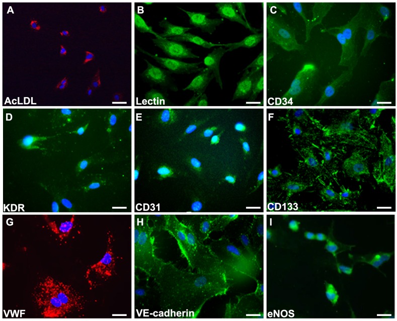
Figure 1. Characterization of late endothelial progenitor cells from peripheral blood.
Cells was characterized by immunofluorescence detection of (A) Dil-AcLDL (red), (B) Lectin, (C) CD34, (D) KDR, (E) CD31, (F) CD133, (G) VWF (red), (H) VE-cadherin, and (I) eNOS. Cells were counterstained with 4’,6-diamidino-2-phenylindole (DAPI) for nuclear (blue). Scale bar: 50 μm.

