Abstract
Purpose
To evaluate the adequacy of retreatment, including hypofractionated re-irradiation (HFReRT), after surgery for recurrent glioblastoma (GBM) and related prognosticators of outcomes.
Materials and Methods
From 2011 to 2014, 25 consecutive patients with recurrent (n=17) or secondary (n=7) disease underwent maximal surgery and subsequent HFReRT after meeting the following conditions: 1) confirmation of recurrent or secondary GBM after salvage surgery; 2) Karnofsky performance score (KPS) ≥60; and 3) interval of ≥12 months between initial radiotherapy and HFReRT. HFReRT was delivered using a simultaneous integrated boost technique, with total dose of 45 Gy in 15 fractions to the gross tumor volume (GTV) and 37.5 Gy in 15 fractions to the clinical target volume.
Results
During a median follow-up of 13 months, the median progression-free and overall survival (OS) were 13 and 16 months, respectively. A better KPS (p=0.026), no involvement of the eloquent area at recurrence (p=0.030), and a smaller GTV (p=0.005) were associated with better OS. Additionally, OS differed significantly between risk groups stratified by the National Institutes of Health Recurrent GBM Scale (low-risk vs. high-risk, p=0.025). Radiologically suspected radiation necrosis (RN) was observed in 16 patients (64%) at a median of 9 months after HFReRT, and 8 patients developed grade 3 RN requiring hospitalization.
Conclusion
HFReRT after maximal surgery prolonged survival in selected patients with recurrent GBM, especially those with small-sized recurrences in non-eloquent areas and good performance.
Keywords: Recurrent glioblastoma, retreatment, re-irradiation, surgery, survival
INTRODUCTION
Long-term local control of glioblastoma (GBM) is rarely achieved with standard treatments, which include maximal surgical resection followed by radiotherapy (RT) with concomitant and adjuvant temozolomide. Such cases nearly always experience a relapse within 5 years.1,2,3,4,5 Although a standard of care has not been established for recurrent GBM, various re-treatment approaches, including re-operation, re-irradiation, chemotherapy (CTx), and combined treatment, are used despite the increased risks of toxicity and uncertainty regarding survival advantages.6
Although re-operation is an effective option for patients with small tumor volumes, good Karnofsky performance scores (KPSs), no involvement at the eloquent site, and young ages, this treatment is indicated for only 10–30% of patients with recurrent GBM.7,8,9,10 Re-irradiation with a moderate dose (~40 Gy) and conventional fractionation has yielded acceptable complication rates; however, the efficacy of this treatment remains undetermined because of the relatively low radiation doses administered to GBMs. Recently, hypofractionated RT administered via intensity modulated RT (IMRT) and/or stereotactic RT has improved the outcomes of re-irradiation for recurrent GBM.11,12,13,14 However, almost all treated patients, particularly those treated with stereotactic radiosurgery (SRS), experienced recurrent GBMs with small volumes that could not be differentiated from treatment-related changes, pseudoprogression, or radiation necrosis (RN).15 Therefore, a recurrence should be pathologically confirmed before subjecting patients with recurrent GBM to salvage treatment.
In light of these earlier findings, our institution administers hypofractionated re-irradiation (HFReRT) to patients with recurrent GBM who had undergone maximal surgical resection. We hypothesized that for selected cases of recurrent GBM, this treatment combination likely comprises the most effective salvage treatment and provides the best chance of survival prolongation. In addition, we evaluated the adequacy of HFReRT after maximal surgical resection in patients with recurrent GBM.
MATERIALS AND METHODS
Study design
We prospectively collected and followed all cases of recurrent or secondary GBM treated with HFReRT after salvage surgery at our institutions between November 2011 and December 2014. All patients were presented to our institute's multidisciplinary neuro-oncology tumor conference, which included neurosurgeon, radiation oncologist, neuro-oncologist, and neuro-radiologist. Tumor recurrences were identified by the appearance of new contrast-enhanced lesions on T1-weighted magnetic resonance imaging (MRI) scans or increase in the volumes of enhanced lesions that had been recorded as treatment-related changes after initial treatment. Eventually, a diagnosis of recurrent GBM was established via histologic analysis after salvage surgery, and HFReRT was offered to selected patients who met the following inclusion criteria, based on our previous work: 1) pathologically confirmed recurrent GBM treated with salvage surgery; 2) age of 18–70 years; 3) KPS ≥60 at the time of referral for HFReRT; and 4) an interval of ≥12 months between initial RT and HFReRT.16 This study was approved by our Institutional Review Board (4-2017-0878).
Salvage re-treatment
Patients with recurrent or secondary GBM underwent maximal surgical resection of the enhancing lesions identified on MRI scans. HFReRT was offered to patients within 4 weeks after salvage surgery. Each patient underwent a computed tomography simulation in the supine position while immobilized with a customized thermoplastic mask device. MRI scans at recurrence and after salvage surgery for the recurrent tumor were transferred to the planning system; after image fusion, the gross tumor volume (GTV) was delineated on T1-weighted MRI scans, based on the operative cavity and residual contrast (gadolinium)-enhanced lesion plus a 0.5-cm margin. The clinical target volume (CTV) was defined as the GTV with a 1–2-cm expansion to account for peritumoral edema on T2 fluid-attenuated inversion recovery MRI sequences. HFReRT was delivered via helical tomotherapy with 6-MV photons (Tomo-Therapy, Madison, WI, USA) using a simultaneous integrated boost technique. Doses of 45 Gy in 15 fractions and 37.5 Gy in 15 fractions were delivered to the GTV and CTV, respectively.
Treatment outcomes and prognostic variables
All patients underwent MRI evaluations at 1 month after salvage treatment for recurrent GBM, and every 3 months thereafter. During follow-up, the treatment outcome and toxicity assessments included a thorough history taking, physical examination (particularly the KPS and neurological status), and analysis of all available imaging data, including contrast-enhanced brain MRI scans. Toxicity after salvage treatment was assessed according to the criteria of the Radiation Therapy Oncology Group. However, no specific tool was used to assess the patients' neurocognitive status or quality of life. The primary endpoints for analysis were overall survival (OS) and progression-free survival (PFS). OS was calculated from the date of surgery for recurrence to the date of death from any cause, and PFS was measured from the date of recurrence until the date of tumor progression or death. The following prognostic factors associated with OS were included: age, gender, KPS, initial histology, eloquent area involvement at recurrence, extent of surgery at recurrence, O6-methylguanine-DNA-methyltransferase status at recurrence, isocitrate dehydrogenase (IDH) mutation at recurrence, tumor volume at recurrence, GTV for HFReRT, and interval between RT courses. In addition, we divided patients into prognostic groups using the NIH recurrent GBM scale, which is a practical scoring system devised by the National Institutes of Health (NIH),9 and assessed OS by group. The secondary endpoints included any documented treatment-related toxicity observed during follow-up examinations and on MRI scans.
Statistical analysis
Survival rates were estimated using the Kaplan-Meier method. A univariate analysis of risk factors was performed by comparing survival rates using the log-rank test. A multivariate analysis was performed using a Cox regression model with a stepwise method to identify prognostic factors. A p value <0.05 was considered statistically significant. All analyses were performed using IBM SPSS, version 20.0 (IBM Corp., Armonk, NY, USA).
RESULTS
Patient and treatment characteristics
Our study population included 25 patients (11 men, 14 women) with a median age at recurrence of 45 years (range, 23–61 years) and median and minimum KPSs of 90 and 60, respectively. Among all patients, 8 patients had secondary GBM, who were initially diagnosed with WHO grade II/III glioma. The median recurrent tumor volume was 32 mL (range, 4–90 mL). All patients underwent surgery for pathologic diagnosis and maximal tumor debulking; 15 underwent gross total resection (GTR) and 10 underwent subtotal resection, defined as the removal of >90% of the contrast-enhanced tumor. All patients had received a median postoperative RT dose of 60 Gy (range, 54–70 Gy) at the time of the initial diagnosis, and the median time interval between the initial RT and re-irradiation courses was 30 months (range, 12–72 months; 29 months for primary recurrent GBM, 30 months for secondary GBM). At the time of HFReRT, the median GTV and CTV were 97 mL (range, 21–261 mL) and 270 mL (range, 101–1353 mL), respectively. Thirteen patients received sequential systemic therapy with temozolomide after HFReRT. Details of the baseline characteristics are described in Table 1.
Table 1. Patient and Treatment Characteristics.
| Characteristic | All patients (n = 25) | |
|---|---|---|
| n (%) | ||
| Age (yr) | Median (range) | 45 (23–61) |
| <45 | 10 (40) | |
| ≥45 | 15 (60) | |
| Sex | Male | 11 (44) |
| Female | 14 (56) | |
| Initial histology | WHO II glioma | 2 (8) |
| WHO III glioma | 6 (24) | |
| GBM | 17 (68) | |
| Initial RT dose (Gy) | Median (range) | 60 (54–70) |
| Initial CTx | No | 7 (28) |
| Yes | 18 (72) | |
| KPS at recurrence | Median (range) | 90 (60–100) |
| Eloquent area involvement at recurrence | No | 13 (52) |
| Yes | 12 (48) | |
| Extent of surgery at recurrence | Gross total | 15 (58) |
| Subtotal | 10 (42) | |
| MGMT status at recurrence | Unmethylated | 9 (36) |
| Methylated | 16 (64) | |
| IDH mutation at recurrence | Present | 8 (32) |
| Absent | 14 (56) | |
| Unknown | 3 (12) | |
| Tumor volume at recurrence (mL)* | Median (range) | 32 (4–90) |
| <50 | 15 (60) | |
| ≥50 | 10 (40) | |
| GTV of HFReRT (mL) | Median (range) | 97 (21–261) |
| <100 | 14 (56) | |
| ≥100 | 11 (44) | |
| CTV of HFReRT (mL) | Median (range) | 270 (101–1353) |
| CTx after HFReRT | No | 12 (48) |
| Yes, sequential | 13 (52) | |
| Cumulative dose of RT (Gy) | Median (range) | 105 (95.4–115) |
| <100 | 2 (8) | |
| ≥100 | 23 (92) | |
| Interval between RT (month) | Median (range) | 29 (12–72) |
| <30 | 13 (52) | |
| ≥30 | 12 (48) | |
WHO, World Health Organization; GBM, glioblastoma; RT, radiotherapy; KPS, Karnofsky performance score; MGMT, O6-methylguanine-DNA-methyltransferase; IDH, isocitrate dehydrogenase; GTV, gross tumor volume; CTV, clinical target volume; HFReRT, hypofractionated re-irradiation; CTx, chemotherapy.
*Classified using a tumor volume cut-off value of 50 mL at recurrence, according to the National Institutes of Health recurrent GBM scale.
Survival and prognostic variables
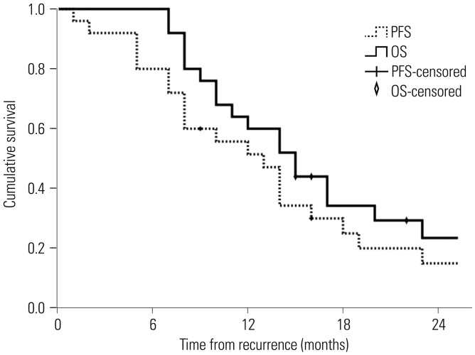
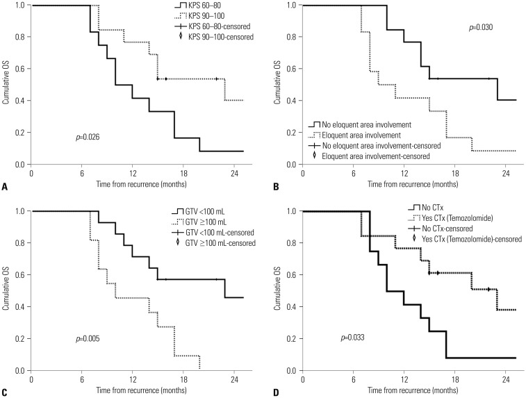
Patients were followed up for a median of 13 months (range, 4–41 months) after a recurrence. At the time of the last observation, 5 patients (20%) remained alive at 15, 16, 22, 28, and 29 months after recurrence; the other 20 (80%) had died. The median PFS duration and 1-year PFS rate were 13 months (range, 1–41 months) and 51.4%, respectively, and the median OS duration and 1-year OS rate were 16 months (range, 7–41 months) and 60%, respectively (Fig. 1). A univariate analysis of factors related to OS after a recurrence identified the KPS (p=0.026) (Fig. 2A), eloquent area involvement at recurrence (p=0.030) (Fig. 2B), GTV at HFReRT (p=0.005) (Fig. 2C), and administration of sequential CTx after HFReRT (p=0.033) (Fig. 2D) as significant factors. In contrast, age (p=0.385), initial histology (p=0.232), the extent of surgery at recurrence (p=0.391), IDH mutation status at recurrence (p=0.626), tumor volume at recurrence (p=0.075), and interval between RT (p=0.489) did not affect OS. All results from the univariate analysis are shown in Table 2. In a multivariate stepwise Cox regression model analysis, the GTV at HFReRT (≥100 mL vs. <100 mL: hazard ratio=3.717, 95% confidence interval: 1.356–10.187, p=0.011) was confirmed as an independent prognostic factor affecting OS (Table 2).
Fig. 1. Kaplan-Meier curves of PFS and OS. PFS, progression-free survival; OS, overall survival.
Fig. 2. OS according to the KPS (A), eloquent area involvement at recurrence (B), GTV (C), and CTx after hypofractionated re-irradiation (D). OS, overall survival; KPS, Karnofsky performance score; GTV, gross tumor volume; CTx, chemotherapy.
Table 2. Univariate and Stepwise Multivariate Cox Regression Analyses of Prognosticators of OS.
| Characteristics | Univariate analysis | Multivariate analysis† | ||||
|---|---|---|---|---|---|---|
| 1-year OS | p value | HR | 95% CI | p value | ||
| Age (yr) | <45 | 60.0 | 0.385 | NI | ||
| ≥45 | 60.0 | |||||
| Sex | Male | 63.6 | 0.565 | NI | ||
| Female | 57.1 | |||||
| KPS | 60–80 | 41.7 | 0.026* | Ref | 0.680 | |
| 90–100 | 76.9 | 0.759 | 0.204–2.815 | |||
| Initial histology | Non-GBM | 50.0 | 0.232 | NI | ||
| GBM | 64.7 | |||||
| Eloquent area involvement at recurrence | No | 76.9 | 0.030* | Ref | 0.584 | |
| Yes | 41.7 | 1.381 | 0.435–4.388 | |||
| Extent of surgery at recurrence | Gross total | 73.3 | 0.391 | NI | ||
| Subtotal | 40.0 | |||||
| MGMT status at recurrence | Unmethylated | 55.6 | 0.978 | NI | ||
| Methylated | 62.5 | |||||
| IDH mutation at recurrence | Present | 62.5 | 0.626 | NI | ||
| Absent | 57.1 | |||||
| Tumor volume at recurrence (mL) | <50 | 73.3 | 0.075 | NI | ||
| ≥50 | 40.0 | |||||
| GTV of HFReRT (mL) | <100 | 71.4 | 0.005* | Ref | 0.011 | |
| ≥100 | 45.5 | 3.717 | 1.356–10.187 | |||
| Interval between RT (month) | <30 | 61.5 | 0.489 | NI | ||
| ≥30 | 58.3 | |||||
| Sequential CTx after HFReRT | No | 41.7 | 0.033* | Ref | 0.231 | |
| Yes, Temozolomide | 76.9 | 0.528 | 0.185–1.503 | |||
OS, overall survival; HR, hazard ratio; CI, confidence interval; KPS, Karnofsky performance score; GBM, glioblastoma; MGMT, O6-methylguanine-DNA-methyltransferase; IDH, isocitrate dehydrogenase; GTV, gross tumor volume; HFReRT, hypofractionated re-irradiation; RT, radiotherapy; CTx, chemotherapy; Ref, reference; NI, not included.
*Included in the multivariate analysis, †Variables with a p value ≤0.05 were entered in the multivariate regression model in a stepwise manner and were removed at any point if the p value exceeded 0.05.
Survival according to prognostic groups as defined by the NIH recurrent GBM scale
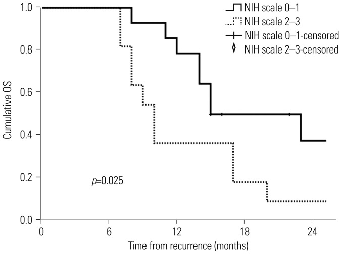
We classified patients into prognostic groups based on the NIH recurrent GBM scale, which included the involvement of eloquent regions, KPS ≤80, and recurrent tumor volume ≥50 mL.9 Because our cohort was small, we defined the following groups: NIH recurrent GBM scale of 0 or 1 (low-risk group, n=14) and NIH recurrent GBM scale of 2 or 3 (high-risk group, n=11). The NIH recurrent GBM scale was found to be a significant prognosticator of OS (low-risk group vs. high-risk group: 15 months vs. 10 months, p=0.025) (Fig. 3).
Fig. 3. OS according to prognostic groups, defined according to the NIH recurrent glioblastoma scale. OS, overall survival; NIH, National Institutes of Health.
Toxicity
During treatment, no severe acute toxicity events requiring the interruption of HFReRT were recorded. During the follow-up period, corticosteroids, antiepileptic drugs, and mannitol were administered in 22 (88%), 22 (88%), and 7 (28%) cases, respectively. At a median of 9 months after HFReRT, 16 patients (64%) harbored suspected areas of RN, based on gadolinium-enhanced T1-weighted MRI scans. However, these lesions could not be completely differentiated from tumor progression. Of these 16 patients, 8 were hospitalized for neurological deterioration with suspected grade 3 RN. Grade 3 RN developed in 18.8% (3/16) of cases involving an interval of >24 months between RT courses and in 55.6% (5/9) of cases with an interval of <24 months (p=0.062). No differences in the incidence of grade 3 RN according to GTV and CTV were observed.
DISCUSSION
According to several studies, a more aggressive local treatment comprising GTR and/or high-dose re-irradiation via advanced RT techniques may improve the survival of selected patients with recurrent GBM.6,7,13,17,18 However, the use of re-irradiation, which is associated with late side effects and tumoricidal effects, remains controversial. Accordingly, our institution has implemented very strict criteria regarding the administration of HFReRT to patients with recurrent GBM, as we expect that the benefits of salvage HFReRT in terms of increased tumor control would outweigh the costs of treatment-related toxicity.
Given the uncertainty of a differential diagnosis that includes both recurrent GBM and treatment-related lesions such as pseudoprogression or RN after initial treatment for malignant gliomas,19 we applied HFReRT only to patients with pathologically confirmed recurrent or secondary GBM. Previous re-irradiation series involving patients with non-pathologically confirmed recurrent GBM, especially those treated with SRS, might have included cases of treatment-related reactions rather than true GBM, which might have led to overestimations of the outcomes.6,13,15,20,21,22,23 Next, our study required the total or subtotal removal of all suspected recurrent GBMs with the aim of maximum debulking prior to HFReRT, which would thus improve the efficacy of this treatment given the radio-resistance of recurrent tumors and uncontrolled behaviors of bulky gross tumors in response to radiation.6,7,24,25 Notably, the extent of surgery did not affect survival in the current study, despite previous reports that the extent of resection is a well-known predictive factor for survival.24,25 This discrepancy may be attributable not only to the limited number of patients, but also to the fact that upfront HFReRT (i.e., within 4 weeks after salvage re-operation) may have temporarily controlled the small amount of residual tumor remaining after subtotal removal, which we defined as the resection of >90% of a tumor.25
The performance status has also been identified as a factor affecting the use of HFReRT in patients with recurrent GBM. Several studies have reported strong correlations between a good performance score and longer survival.16 Given the fundamental importance of this factor, particularly in a re-treatment setting, we set a poor KPS (<60) as an exclusion criterion to reduce the risks of physical and neurological deterioration after treatment. Notably, a higher performance status, no eloquent area involvement, and a smaller GTV were identified as positive prognostic factors related to OS in the selected patients who received HFReRT following surgical resection. These prognosticators were similar to those used in the abovementioned validated NIH scale to predict outcomes after the surgical resection of recurrent GBM.9 The difference in tumor volumes between the studies (GTV=100 mL vs. tumor volume at recurrence=50 mL) was a consequence of the time points used to determine the treatment modalities (postoperative status vs. preoperative status). Both studies also proposed that the subgroup of patients with a poor performance and large or diffuse recurrent tumors located in the eloquent brain area might benefit from best supportive care, rather than re-treatment.
Regarding dose fractionation in the context of re-irradiation, the prescribed doses in a recent review ranged from 20 Gy to 45.5 Gy, and the cumulative equivalent radiation doses (in 2-Gy fractions; EQD2) for both primary and second RT ranged from 81.6 Gy to 102.8 Gy.26 In most published studies, the doses prescribed for re-irradiation ranged from 30 Gy to 45 Gy, thus maintaining a cumulative EQD2 of approximately 100 Gy. More recently, however, advances in conformal radiation techniques, such as stereotactic RT and IMRT, have led to the delivery of higher therapeutic re-irradiation doses via hypofractionation within short treatment courses.26 In such situations, we prescribed 45 Gy in 15 fractions (EQD2, 56.25 Gy) to the operative cavity with/without gross residual tumors plus 0.5 cm. This was expected to reduce the treatment period along with the tumoricidal re-irradiation dose, as most patients with recurrent GBM have a short life expectancy, limited mobility, and considerable dependency.
Studies of surgery or conventional RT for recurrent GBM have reported mild or moderate survival benefits. Although most studies of surgery for recurrences were affected by a high probability of selection bias, the reported median intervals from salvage surgery to death remained <12 months,9,10 and the survival durations of patients treated with conventional re-irradiation for recurrent glioma were also <12 months.16,21 In the present study, HFReRT after maximal resection yielded a median survival duration of 16 months, although the follow-up period was short. We attribute these outcomes to a synergistic effect for the following reasons: 1) maximal surgical resection is the best approach for tumor eradication and 2) compared to conventional RT, higher RT dose with a precision technique was applied in our HFReRT strategy.
We additionally evaluated the safety of HFReRT after surgical resection for recurrent GBM. No acute toxicity events were recorded during HFReRT, and all patients completed treatment during a median period of 20 days. However, 16 (64%) patients developed late toxicities, including 8 cases each of grade 2 and grade 3 RN. This considerably high incidence of RN in our study, relative to other studies,26 could not completely be attributed to tumor progression. Re-irradiation is a well-known factor that contributes to the risk of RN in cases receiving a cumulative total irradiated dose of >100 Gy, a larger irradiated volume, and a higher fractional dose, as well as those involving a short interval between RT courses.27,28 Overall, our study cases exhibited the following risk factors: a median HFReRT GTV of 97 mL, median CTV of 270 mL, high median cumulative dose of 105 mL, and high fraction dose of 3 Gy. These results suggest that clinical feasibility of the current retreatment protocol should be investigated based on the clear evidence indicating late toxicity.
Despite large treated volumes, the current treatment scheme encouragingly prolonged survival in our cohort of selected patients with recurrent GBM, which was relatively homogenous regarding demographic and treatment characteristics. However, we did not address the meaningful benefit of retreatment in patients with recurrent GBM in terms of the negative effects of treatment-related toxicity on the quality of life or the caregivers' burdens during the end-of-life phase.29 Re-treatment for recurrent GBM should always be considered after thoroughly discussing the potential advantages and disadvantages with patients and their family.
We note that our current study was limited by a small cohort and the risk of selection bias. Furthermore, our study featured some flaws in patient composition. Our cohort comprised of patients with both secondary and recurrent GBM, which are different diseases; Secondary GBM might have better survival outcome. Additionally, our study lacked a comprehensive assessment of toxicity after re-treatment. The retrospective design precluded our ability to evaluate the quality of life, including systemic neurocognitive function, of patients in our cohort. Accordingly, our findings must be validated in well-designed studies with larger cohorts.
In conclusion, the combined use of HFReRT with maximal surgical resection has prolonged the survival of selected patients with recurrent GBM. This re-treatment strategy was especially beneficial for patients with smaller-sized recurrent GBMs, no eloquent area involvement, and good performance scores. However, studies with larger numbers of patients are needed to determine the efficacy of this treatment scheme in light of the significant risk of RN.
ACKNOWLEDGEMENTS
This study was supported by the Basic Science Research Program through the National Research Foundation of Korea (NRF) funded by the Ministry of Education (NRF-2014R1A1A2058058) and a faculty research grant of Yonsei University College of Medicine (6-2016-0070).
Footnotes
The authors have no financial conflicts of interest.
References
- 1.Stupp R, Mason WP, van den Bent MJ, Weller M, Fisher B, Taphoorn MJ, et al. European Organisation for Research and Treatment of Cancer Brain Tumor and Radiotherapy Groups; National Cancer Institute of Canada Clinical Trials Group. Radiotherapy plus concomitant and adjuvant temozolomide for glioblastoma. N Engl J Med. 2005;352:987–996. doi: 10.1056/NEJMoa043330. [DOI] [PubMed] [Google Scholar]
- 2.Milano MT, Okunieff P, Donatello RS, Mohile NA, Sul J, Walter KA, et al. Patterns and timing of recurrence after temozolomide-based chemoradiation for glioblastoma. Int J Radiat Oncol Biol Phys. 2010;78:1147–1155. doi: 10.1016/j.ijrobp.2009.09.018. [DOI] [PubMed] [Google Scholar]
- 3.Stupp R, Hegi ME, Mason WP, van den, Taphoorn MJ, Janzer RC, et al. European Organisation for Research and Treatment of Cancer Brain Tumour and Radiation Oncology Groups; National Cancer Institute of Canada Clinical Trials Group. Effects of radiotherapy with concomitant and adjuvant temozolomide versus radiotherapy alone on survival in glioblastoma in a randomised phase III study: 5-year analysis of the EORTC-NCIC trial. Lancet Oncol. 2009;10:459–466. doi: 10.1016/S1470-2045(09)70025-7. [DOI] [PubMed] [Google Scholar]
- 4.Brandes AA, Tosoni A, Franceschi E, Sotti G, Frezza G, Amistà P, et al. Recurrence pattern after temozolomide concomitant with and adjuvant to radiotherapy in newly diagnosed patients with glioblastoma: correlation with MGMT promoter methylation status. J Clin Oncol. 2009;27:1275–1279. doi: 10.1200/JCO.2008.19.4969. [DOI] [PubMed] [Google Scholar]
- 5.Sherriff J, Tamangani J, Senthil L, Cruickshank G, Spooner D, Jones B, et al. Patterns of relapse in glioblastoma multiforme following concomitant chemoradiotherapy with temozolomide. Br J Radiol. 2013;86:20120414. doi: 10.1259/bjr.20120414. [DOI] [PMC free article] [PubMed] [Google Scholar]
- 6.Taunk NK, Moraes FY, Escorcia FE, Mendez LC, Beal K, Marta GN. External beam re-irradiation, combination chemoradiotherapy, and particle therapy for the treatment of recurrent glioblastoma. Expert Rev Anticancer Ther. 2016;16:347–358. doi: 10.1586/14737140.2016.1143364. [DOI] [PMC free article] [PubMed] [Google Scholar]
- 7.Montemurro N, Perrini P, Blanco MO, Vannozzi R. Second surgery for recurrent glioblastoma: a concise overview of the current literature. Clin Neurol Neurosurg. 2016;142:60–64. doi: 10.1016/j.clineuro.2016.01.010. [DOI] [PubMed] [Google Scholar]
- 8.Barbagallo GM, Jenkinson MD, Brodbelt AR. ‘Recurrent’ glioblastoma multiforme, when should we reoperate? Br J Neurosurg. 2008;22:452–455. doi: 10.1080/02688690802182256. [DOI] [PubMed] [Google Scholar]
- 9.Park JK, Hodges T, Arko L, Shen M, Dello Iacono D, McNabb A, et al. Scale to predict survival after surgery for recurrent glioblastoma multiforme. J Clin Oncol. 2010;28:3838–3843. doi: 10.1200/JCO.2010.30.0582. [DOI] [PMC free article] [PubMed] [Google Scholar]
- 10.Park CK, Kim JH, Nam DH, Kim CY, Chung SB, Kim YH, et al. A practical scoring system to determine whether to proceed with surgical resection in recurrent glioblastoma. Neuro Oncol. 2013;15:1096–1101. doi: 10.1093/neuonc/not069. [DOI] [PMC free article] [PubMed] [Google Scholar]
- 11.Shi W, Palmer JD, Werner-Wasik M, Andrews DW, Evans JJ, Glass J, et al. Phase I trial of panobinostat and fractionated stereotactic re-irradiation therapy for recurrent high grade gliomas. J Neurooncol. 2016;127:535–539. doi: 10.1007/s11060-016-2059-3. [DOI] [PubMed] [Google Scholar]
- 12.Wuthrick EJ, Curran WJ, Jr, Camphausen K, Lin A, Glass J, Evans J, et al. A pilot study of hypofractionated stereotactic radiation therapy and sunitinib in previously irradiated patients with recurrent high-grade glioma. Int J Radiat Oncol Biol Phys. 2014;90:369–375. doi: 10.1016/j.ijrobp.2014.05.034. [DOI] [PMC free article] [PubMed] [Google Scholar]
- 13.Fogh SE, Andrews DW, Glass J, Curran W, Glass C, Champ C, et al. Hypofractionated stereotactic radiation therapy: an effective therapy for recurrent high-grade gliomas. J Clin Oncol. 2010;28:3048–3053. doi: 10.1200/JCO.2009.25.6941. [DOI] [PMC free article] [PubMed] [Google Scholar]
- 14.Nieder C, Astner ST, Mehta MP, Grosu AL, Molls M. Improvement, clinical course, and quality of life after palliative radiotherapy for recurrent glioblastoma. Am J Clin Oncol. 2008;31:300–305. doi: 10.1097/COC.0b013e31815e3fdc. [DOI] [PubMed] [Google Scholar]
- 15.Kong DS, Lee JI, Park K, Kim JH, Lim DH, Nam DH. Efficacy of stereotactic radiosurgery as a salvage treatment for recurrent malignant gliomas. Cancer. 2008;112:2046–2051. doi: 10.1002/cncr.23402. [DOI] [PubMed] [Google Scholar]
- 16.Lee J, Cho J, Chang JH, Suh CO. Re-irradiation for recurrent gliomas: treatment outcomes and prognostic factors. Yonsei Med J. 2016;57:824–830. doi: 10.3349/ymj.2016.57.4.824. [DOI] [PMC free article] [PubMed] [Google Scholar]
- 17.Vogelbaum MA. The benefit of surgical resection in recurrent glioblastoma. Neuro Oncol. 2016;18:462–463. doi: 10.1093/neuonc/now004. [DOI] [PMC free article] [PubMed] [Google Scholar]
- 18.Scorsetti M, Navarria P, Pessina F, Ascolese AM, D'Agostino G, Tomatis S, et al. Multimodality therapy approaches, local and systemic treatment, compared with chemotherapy alone in recurrent glioblastoma. BMC Cancer. 2015;15:486. doi: 10.1186/s12885-015-1488-2. [DOI] [PMC free article] [PubMed] [Google Scholar]
- 19.Na A, Haghigi N, Drummond KJ. Cerebral radiation necrosis. Asia Pac J Clin Oncol. 2014;10:11–21. doi: 10.1111/ajco.12124. [DOI] [PubMed] [Google Scholar]
- 20.Minniti G, Armosini V, Salvati M, Lanzetta G, Caporello P, Mei M, et al. Fractionated stereotactic reirradiation and concurrent temozolomide in patients with recurrent glioblastoma. J Neurooncol. 2011;103:683–691. doi: 10.1007/s11060-010-0446-8. [DOI] [PubMed] [Google Scholar]
- 21.Amelio D, Amichetti M. Radiation therapy for the treatment of recurrent glioblastoma: an overview. Cancers (Basel) 2012;4:257–280. doi: 10.3390/cancers4010257. [DOI] [PMC free article] [PubMed] [Google Scholar]
- 22.Combs SE, Thilmann C, Edler L, Debus J, Schulz-Ertner D. Efficacy of fractionated stereotactic reirradiation in recurrent gliomas: long-term results in 172 patients treated in a single institution. J Clin Oncol. 2005;23:8863–8869. doi: 10.1200/JCO.2005.03.4157. [DOI] [PubMed] [Google Scholar]
- 23.Grosu AL, Weber WA, Franz M, Stärk S, Piert M, Thamm R, et al. Reirradiation of recurrent high-grade gliomas using amino acid PET (SPECT)/CT/MRI image fusion to determine gross tumor volume for stereotactic fractionated radiotherapy. Int J Radiat Oncol Biol Phys. 2005;63:511–519. doi: 10.1016/j.ijrobp.2005.01.056. [DOI] [PubMed] [Google Scholar]
- 24.Suchorska B, Weller M, Tabatabai G, Senft C, Hau P, Sabel MC, et al. Complete resection of contrast-enhancing tumor volume is associated with improved survival in recurrent glioblastoma-results from the DIRECTOR trial. Neuro Oncol. 2016;18:549–556. doi: 10.1093/neuonc/nov326. [DOI] [PMC free article] [PubMed] [Google Scholar]
- 25.Grabowski MM, Recinos PF, Nowacki AS, Schroeder JL, Angelov L, Barnett GH, et al. Residual tumor volume versus extent of resection: predictors of survival after surgery for glioblastoma. J Neurosurg. 2014;121:1115–1123. doi: 10.3171/2014.7.JNS132449. [DOI] [PubMed] [Google Scholar]
- 26.Sminia P, Mayer R. External beam radiotherapy of recurrent glioma: radiation tolerance of the human brain. Cancers (Basel) 2012;4:379–399. doi: 10.3390/cancers4020379. [DOI] [PMC free article] [PubMed] [Google Scholar]
- 27.Lawrence YR, Li XA, el Naqa, I, Hahn CA, Marks LB, Merchant TE, et al. Radiation dose-volume effects in the brain. Int J Radiat Oncol Biol Phys. 2010;76(3 Suppl):S20–S27. doi: 10.1016/j.ijrobp.2009.02.091. [DOI] [PMC free article] [PubMed] [Google Scholar]
- 28.Mayer R, Sminia P. Reirradiation tolerance of the human brain. Int J Radiat Oncol Biol Phys. 2008;70:1350–1360. doi: 10.1016/j.ijrobp.2007.08.015. [DOI] [PubMed] [Google Scholar]
- 29.Flechl B, Ackerl M, Sax C, Oberndorfer S, Calabek B, Sizoo E, et al. The caregivers' perspective on the end-of-life phase of glioblastoma patients. J Neurooncol. 2013;112:403–411. doi: 10.1007/s11060-013-1069-7. [DOI] [PubMed] [Google Scholar]


