To the Editor
With interest we read the recent publication of Dude et al. (2017) on the evaluation of somatostatin receptor (SSTR) agonists and an antagonist for SSTR-mediated imaging of breast cancer using positron emission tomography. In this study the authors compared 2 SSTR agonists (DOTA-Tyr3-octreotide and DOTA-Tyr3-octreotate) and the SSTR antagonist (NODAGA-JR11) in in vitro binding and saturation studies and in in vivo imaging and biodistribution studies. To our surprise their results demonstrated both agonists to have a more favorable receptor binding affinity and a better tumor uptake in vivo, whereas the saturation assay resulted in more binding sites for 67/natGa-DOTA-Tyr3-octreotide on the used breast cancer cell line (ZR75–1) than natGa-NODAGA-JR11 and 67/natGa-DOTA-Tyr3-octreotate.
The reported results are in contrast with previously published studies comparing radiolabeled DOTA-Tyr3-octreotate and DOTA-JR11 in various tumor models (Dalm et al., 2016; Nicolas et al., 2017; Reubi et al., 2017; Wild et al., 2014), including our recent publication on the use of SSTR agonists and antagonists for targeting of breast cancer (Dalm et al., 2017). The main explanation given by the authors for the contradicting results is the use of an endogenously SSTR expressing breast cancer cell line, ZR75–1, in contrast to transfected cell lines, cell lines of other cancer types and non-cancerous cell lines used in earlier studies evaluating SSTR-targeting radiotracers.
Concerning the above mentioned explanation of the authors, we have the following remarks:
First, some of the non-cancerous cell lines and cell lines of other cancer types used in previous studies also have endogenous SSTR expression. One example is our previously published study in which we reported better therapeutic efficacy with 177Lu-DOTA-JR11 compared to 177Lu-DOTA-Tyr3-octreotate in a xenograft model generated with the human small cell lung cancer cell line, H69 (Dalm et al., 2016).
Furthermore, previously published studies comparing the use of radiolabeled JR11 and radiolabeled DOTA-Tyr3-octreotate or DOTA-Tyr3-octreotide were not only performed preclinically in tumor models, but also clinically in patients with neuroendocrine tumors. In the latter mentioned study published by Wild et al. (2014) 177Lu-DOTA-JR11 tumor uptake was superior to that of 177Lu-DOTA-Tyr3-octreotate. Although this study was not performed in breast cancer patients, it again demonstrates superiority of the SSTR antagonist vs the agonist in tumors that have endogenous SSTR expression.
Concerning breast cancer, Reubi et al. (2017) demonstrated that binding of 125I–DOTA-JR11 to human breast cancer tissue was much higher than that of 125I–DOTA-Tyr3-octreotide. We also demonstrated higher binding of 111In-DOTA-JR11 vs 111In-DOTA-Tyr3-octreotate to 40 human breast cancer tissue samples (Dalm et al., 2017). Furthermore, in the same study we also showed higher in vivo tumor uptake of 177Lu-DOTA-JR11 vs 177Lu-DOTA-Tyr3-octreotate in an estrogen receptor positive patient derived breast cancer mouse model with endogenous SSTR expression.
Differences between the study of Dude et al. (2017) and our previous study (Dalm et al., 2017) include the use of different radionuclides and application of DOTA-JR11 instead of NODAGA-JR11. The authors chose NODAGA-JR11 because DOTA-JR11 has a lower receptor affinity when labeled with 68Ga. Similar to 111In-DOTA-JR11, 177Lu-DOTA-JR11 and 177Lu-DOTA-Tyr3-octreotate, 68Ga-NODAGA-JR11, 68Ga-DOTA-Tyr3-octreotate and 68Ga-DOTA-Tyr3-octreotide have comparable receptor affinity (Fani et al., 2012; Reubi et al., 2000).
Aspects concerning the methodology that to our opinion might influence the results when comparing different radiotracers, include:
The use of different peptide amounts as also addressed by the authors in the discussion. The peptide amount of 68Ga-DOTA-Tyr3-octreotate used in the study was twice as high as the peptide amount of 68Ga-NODAGA-JR11. Although the authors mention that previous studies showed that within a range of 10–60 pmol tumor uptake of 111In-DOTA-Tyr3-octreotide is >80% of the maximum in rats (de Jong et al., 1999), this might be different in the model currently applied and this needs to be determined for the other radiotracers as well.
The use of different peptide amounts for imaging and biodistribution studies.
The use of an agonist (SRIF-28) in the competition assay to determine the binding affinity of the tracers. This would only be correct if the antagonist and the agonist have the same binding site, which is unclear.
Also, in the study by Dude et al. (2017) imaging and biodistribution studies were performed at early time points (55 min and 60 min p.i., respectively), presumably because of the short half-life of 68Ga. However, in another study by Nicolas et al. (2017) it was reported that optimal tumor uptake of 177Lu-DOTA-JR11 and 177Lu-DOTA-Tyr3-octreotate was reached at 4 h p.i. as determined by biodistribution studies. Although there might be a difference in optimal tumor uptake when the same tracer is labeled with different radionuclides, the time point at which the imaging and biodistribution studies were performed might have contributed to the contradictory findings reported by in the study by Dude et al. (2017).
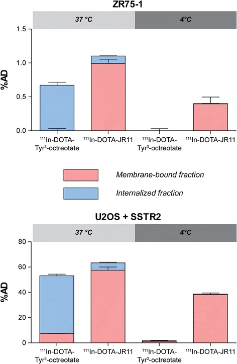
We recently compared binding of 111In-DOTA-JR11 and 111In-DOTA-Tyr3-octreotate to ZR75–1 and U2OS + SSTR2 (the latter is a human osteosarcoma cell line transfected with the SSTR2 receptor) in an internalization assay to investigate differences in SSTR agonist and antagonist binding to cell lines with endogenous and exogenous SSTR expression. The used method can be found in our previous paper (Dalm et al., 2016). Our results demonstrated that 111In-DOTA-JR11 is superior to 111In-DOTA-Tyr3-octreotate (even though the agonist is internalized) when applied for targeting of an endogenous SSTR-expressing cell line (ZR75–1) as well as for targeting the SSTR2 transfected cell line (Fig. 1).
Fig. 1.

Binding and internalization of 111In-DOTA-Tyr3-octreotate and 111In-DOTA-JR11 to the endougenously SSTR expressing cell line ZR75–1 and the SSTR2 transfected cell line U2OS + SSTR2. Cells were incubated for 1 h at 37 °C or 4 °C with 10−9 M of the agonist or the antagonist. The bars represent the percentage added dose (%AD) bound and/or internalized (in)to the cells. The %AD is corrected for unspecific binding determined by blocking studies in which 10−6 M of the unlabeled agonist or antagonist was added
Based on the above we conclude that endogenous SSTR expression of the model used in the study by Dude et al. (2017) does not explain the contradictory results obtained in this study. Multiple experiments in their study had a similar outcome and additional experiments are needed to determine what the reason is for these findings. However, in line with previous studies from our and other groups, the SSTR antagonist JR11 clearly shows superiority to the SSTR agonist octreotate for targeting breast cancer, also in the endogenously SSTR2 expressing breast cancer cell line ZR75–1.
Acknowledgements
The publication of this article was supported by funds of the European Association of Nuclear Medicine (EANM).
Funding
No funding was received for writing this letter or performing the experiments described in this letter. The publication costs were covered by the European Association of Nuclear Medicine.
Availability of data and materials
Data can be obtained from the authors.
Ethics approval and consent to participate
Not applicable.
Consent for publication
Not applicable.
Competing interests
The authors declare that they have no competing interests.
Publisher’s Note
Springer Nature remains neutral with regard to jurisdictional claims in published maps and institutional affiliations.
References
- Dalm SU, Nonnekens J, Doeswijk GN, de Blois E, van Gent DC, Konijnenberg MW, et al. Comparison of the therapeutic response to treatment with a 177Lu-labeled Somatostatin receptor agonist and antagonist in preclinical models. J Nucl Med. 2016;57(2):260–265. doi: 10.2967/jnumed.115.167007. [DOI] [PubMed] [Google Scholar]
- Dalm SU, Haeck J, Doeswijk GN, de Blois E, de Jong M, van Deurzen C. SSTR-mediated breast cancer imaging: is there a role for Radiolabeled SSTR antagonists? J Nucl Med. 2017; [epub ahead of print] [DOI] [PubMed]
- Dude I, Zhang Z, Rousseau J, Hundal-Jabal N, Colpo N, Merkens H, et al. Evaluation of agonist and antagonist radioligands for somatostatin receptor imaging of breast cancer using positron emission tomography. EJNMMI Radiopharmacy and Chemistry. 2017;2(1):4. doi: 10.1186/s41181-017-0023-y. [DOI] [PMC free article] [PubMed] [Google Scholar]
- Fani M, Braun F, Waser B, Beetschen K, Cescato R, Erchegyi J, et al. Unexpected sensitivity of sst2 antagonists to N-terminal radiometal modifications. J Nucl Med. 2012;53(9):1481–1489. doi: 10.2967/jnumed.112.102764. [DOI] [PubMed] [Google Scholar]
- de Jong M, Breeman WA, Bernard BF, van Gameren A, de Bruin E, Bakker WH, et al. Tumour uptake of the radiolabelled somatostatin analogue [DOTA0, TYR3]octreotide is dependent on the peptide amount. Eur J Nucl Med. 1999;26(7):693–698. doi: 10.1007/s002590050439. [DOI] [PubMed] [Google Scholar]
- Nicolas GP, Mansi R, McDougall L, Kaufmann J, Bouterfa H, Wild D, et al. Biodistribution, pharmacokinetics and dosimetry of 177Lu-, 90Y- and 111In-labeled somatostatin receptor antagonist OPS201 in comparison to the agonist 177Lu-DOTA-TATE: the mass effect. J Nucl Med. 2017;58(9):1436–41. doi: 10.2967/jnumed.117.191684. [DOI] [PubMed] [Google Scholar]
- Reubi JC, Schar JC, Waser B, Wenger S, Heppeler A, Schmitt JS, et al. Affinity profiles for human somatostatin receptor subtypes SST1-SST5 of somatostatin radiotracers selected for scintigraphic and radiotherapeutic use. Eur J Nucl Med. 2000;27(3):273–282. doi: 10.1007/s002590050034. [DOI] [PubMed] [Google Scholar]
- Reubi JC, Waser B, Macke H, Rivier J. Highly increased 125I-JR11 antagonist binding in vitro reveals novel indications for sst2 targeting in human cancers. J Nucl Med. 2017;58(2):300–306. doi: 10.2967/jnumed.116.177733. [DOI] [PubMed] [Google Scholar]
- Wild D, Fani M, Fischer R, Del Pozzo L, Kaul F, Krebs S, et al. Comparison of somatostatin receptor agonist and antagonist for peptide receptor radionuclide therapy: a pilot study. J Nucl Med. 2014;55(8):1248–1252. doi: 10.2967/jnumed.114.138834. [DOI] [PubMed] [Google Scholar]
Associated Data
This section collects any data citations, data availability statements, or supplementary materials included in this article.
Data Availability Statement
Data can be obtained from the authors.


