

Abstract
Objective
Previous mortality studies of U.S. Gulf War veterans through 2000 and 2004 have shown an increased risk of brain cancer mortality among some deployed individuals. When veterans possibly exposed to environmental contaminants associated with demolition of the Khamisiyah Ammunition Storage Facility at Khamisiyah, Iraq, have been compared to contemporaneously deployed unexposed veterans, the results have suggested increased risk for mortality from brain cancer among the exposed. Brain cancer mortality risk in this cohort has not been updated since 2004.
Methods
This study analyzes the risk for brain cancer mortality between 1991–2011 through two series of comparisons: U.S. Gulf War deployed and non-deployed veterans from the same era; and veterans possibly exposed to environmental contaminants at Khamisiyah compared to contemporaneously deployed but unexposed U.S. Gulf War veterans. Risk of brain cancer mortality was determined using logistic regression. Life test hazard models were created to plot comparisons of annual hazard rates. Joinpoint regression models were applied to assess trends in hazard rates for brain cancer mortality.
Results
U.S. Army veterans possibly exposed at Khamisiyah had similar rates of brain cancer mortality compared to those not possibly exposed; however, veterans possibly exposed had a higher risk of brain cancer in the time period immediately following the Gulf War.
Conclusion
Results from these analyses suggest that veterans possibly exposed at Khamisiyah experienced different patterns of brain cancer mortality risk compared to the other groups.
Keywords: Brain cancer, Veterans, Gulf War, Mortality, Khamisiyah
1. Introduction
There is ongoing concern about health effects that may be associated with deployment during the 1990-1991 Gulf War. There is concern that the nerve agents sarin and cyclosarin, along with the possibility of additional unknown contaminants, were released when U.S. forces detonated the Khamisiyah Ammunition Storage Facility in Khamisiyah, Iraq, on March 10 through March 13, 1991 [1,2]. The Department of Defense and Central Intelligence Agency developed a plume model in 2000 to estimate the potential area of distribution of products resulting from demolition of munitions and identification of military units possibly exposed through service at or around the Khamisiyah munitions depot [1,2]. Studies have shown associations between likely environmental contaminant exposure at Khamisiyah and decreased neurobehavioral functioning, cognitive dysfunction, central nervous system pathology, and immunosuppression, demonstrating that exposures to these agents in this area may be related to damage to the central nervous system [3-8]. Several studies have indicated a relationship between low-level sarin and cyclosarin exposure and alterations in brain structure and function [7,9,10].
There has been evidence to suggest that U.S. Gulf War veterans possibly exposed at Khamisiyah had a higher risk of brain cancer mortality in the years following the war. In a 2005 paper by Bullman et al. [11], risk of brain cancer mortality from 1991 through 2000 among U.S. Army Gulf War veterans possibly exposed to environmental contaminants at Khamisiyah was compared to U.S. Army Gulf War veterans not possibly exposed. Results indicated an increased risk of brain cancer mortality among U.S. Gulf War Army veterans possibly exposed to environmental contaminants at Khamisiyah for one day (adjusted risk ratio = 1.72; 95% confidence interval: 0.95, 3.10) or two or more days (adjusted risk ratio = 3.26; 95% confidence interval: 1.33, 7.96), compared to U.S. Gulf War Army veterans who were deployed but not possibly exposed at Khamisiyah. Barth et al. [12] extended the follow-up period through 2004 for the U.S. Army Gulf War cohort and expanded the analyses to look at all U.S. Gulf War veterans compared to a stratified random sample of veterans who served during the Gulf War but did not deploy to theater. There was no overall increased risk of brain cancer mortality among Gulf War deployed versus Gulf War era (i.e. not deployed to the Persian Gulf Region) (adjusted risk ratio = 0.90, 95% confidence interval: 0.73, 1.11); however, there was evidence of an increased risk of brain cancer mortality among Army Gulf War veterans exposed to oil well fire smoke (adjusted risk ratio = 1.67, 95% confidence interval: 1.05, 2.65). When controlling for potential environmental contaminant exposure at Khamisiyah, there was a marginally-increased risk for brain cancer mortality among those possibly exposed to oil well fire smoke (adjusted risk ratio = 1.81, 95% confidence interval: 1.00, 3.27). Those with two or more days of possible exposure at Khamisiyah had an increased risk of brain cancer mortality when controlling for oil well fire smoke exposure (adjusted risk ratio = 2.71, 95% confidence interval: 1.25, 5.87).
There are concerns about the potential health effects of the many environmental exposures during the Gulf War, including oil well fire smoke, nerve agents (sarin and cyclosarin), multiple vaccinations, pyridostigmine bromide, pesticides, and depleted uranium [7,9,10,13-15]. Both human (Gulf War veteran populations) and animal studies have reported neurological outcomes associated with the exposures present during Gulf War deployment, including neuro-inflammation [16], changes in brain volume [17], hippocampal dysfunction [18,19], decreased white matter [7,9,20] and alterations in executive function and cognition [21,22].
Some Gulf War veterans were exposed to Kuwaiti oil well fire smoke due to the destruction of more than 750 oil wells between January 16, 1991 and November 6, 1991, when the last oil well was capped [23]. There are several studies that indicate an association between exposure to oil well fire smoke during the Gulf War and health conditions, such as acute respiratory and lung conditions like asthma and bronchitis [24-26]. There is evidence to suggest that the inhalation of particulate matter may have long-term health effects, particularly on the neurological system [27-34].
The purpose of this study was to examine brain cancer mortality risk over time, from 1991 through 2011, among U.S. Gulf War deployed veterans compared to Gulf War era veterans and deployed Army veterans possibly exposed to environmental contaminants at Khamisiyah compared to deployed Army veterans not possibly exposed at Khamisiyah.
2. Methods
This study analyzed four cohorts of U.S. Gulf War veterans: 1) 502,678 U.S. veterans deployed to the Persian Gulf theater of operations between August 1, 1990 and March 1, 1991, excluding those subsequently possibly exposed to environmental contaminants at Khamisiyah or with unknown exposure status (Gulf War deployed); 2) 746,142 U.S. veterans who served in the U.S. Armed Forces between August 1, 1990 and March 1, 1991 but were not deployed to the Persian Gulf theater of operations (Gulf War era); 3) 100,483 U.S. Army veterans located within 50 km of the Khamisiyah demolition site between March 10-13, 1991 and thought to have been possibly exposed to the plume containing sarin and cyclosarin and possibly additional unknown environmental contaminants (Army, possibly Khamisiyah exposed); and 4) 224,974 U.S. Army veterans deployed between March 10-13, 1991 without possible exposure to the plume at Khamisiyah (unexposed Army).
The beginning of follow-up for U.S. Gulf War deployed veterans was the day the veteran left the Gulf War theater of operations. For U.S. Gulf War era veterans, follow-up began on May 1, 1991. The end of follow-up was either the date of death for those deceased or December 31, 2011, which was the end of the study period for those alive. Data on possible exposure to environmental contaminants at Khamisiyah was obtained from a plume model developed jointly by Department of Defense and the Central Intelligence Agency. Details can be found in the Department of Defense U.S. Demolition Operations at Khamisiyah reports [1,2,35]. The analyses included 21 years of mortality data (years 1991–2011). Vital status and cause of death for all cohorts were obtained through the Department of Veteran Affairs/Department of Defense Joint Suicide Data Repository, a collaborative single-source of data for mortality information for those with history of military service. Currently, the Suicide Data Repository contains cause of death information for all veterans who separated from active duty service between January 1, 1975 and December 31, 2011 or who used Department of Veterans Affairs health care services between the fiscal years 2000 and 2011. Brain cancer mortality was identified using ICD-9 191.
2.1. Statistical analyses
Analyses included comparisons of 1) Gulf War deployed versus Gulf War era veterans and 2) Army, possibly exposed at Khamisiyah versus unexposed Army veterans. Three different analytic strategies were used to assess relationships between deployment, exposure, and risk for brain cancer.
Crude brain cancer mortality rates per 10,000 person-years were calculated for each group. Cox proportional hazards models were used to calculate adjusted rate ratio estimates for brain cancer mortality using data from the full 21-year period of follow-up. Covariates in the Cox proportional hazards model included age at entry to follow-up, race (white/other), sex, and type of military unit (active duty, National Guard, Reserves). Rank was included as a covariate for analyses of the Khamisiyah cohorts but was not available for all Gulf War deployed and era veterans. Service branch was included for models comparing Gulf War deployed to Gulf War era. Models assessing the Khamisiyah cohorts considered the possibility of dose-response by comparing those with one day of possible exposure within the plume and those with two or more days of possible exposure within the plume to those who were not under the plume.
Life test hazard models were created to calculate and plot comparisons of annual hazard rates for the two sets of cohort comparisons. The Log-rank test was calculated for each comparison to test the null hypothesis of no difference in hazard rates between groups. Finally, overall trends in brain cancer mortality over the study period were investigated. Joinpoint regression models were applied to assess trends in hazard rates for brain cancer mortality and average annual percent change in brain cancer mortality risk through the end of the study period [36]. The study was approved by the Washington, DC Department of Veterans Affairs Medical Center Institutional Review Board.
3. Results
Tables 1 and 2 present the demographic and military characteristics of the U.S. Gulf War deployed and Gulf War era groups (Table 1) and Army, possibly exposed at Khamisiyah and unexposed Army veterans (Table 2).
Table 1.
Demographic and Military Characteristics of U.S. Gulf War Deployed and Gulf War Era Veterans.
| Gulf War deployed
|
Gulf War era
|
|||
|---|---|---|---|---|
| n | % | N | % | |
| Total | 502,678 | 746,142 | ||
| Brain cancer deaths | 239 | 0.05 | 458 | 0.06 |
| Age in 1991 | ||||
| 17–24 | 213,228 | 42.42 | 258,783 | 34.68 |
| 25–34 | 201,418 | 40.07 | 288,191 | 38.62 |
| 35–44 | 75,114 | 14.94 | 154,056 | 20.65 |
| 45–54 | 11,888 | 2.36 | 40,208 | 5.39 |
| 55+ | 1038 | 0.21 | 4950 | 0.66 |
| Sex | ||||
| Male | 472,018 | 93.90 | 647,136 | 86.73 |
| Female | 30,668 | 6.10 | 99,052 | 13.28 |
| Race | ||||
| White | 346,327 | 68.90 | 521,024 | 69.83 |
| Other | 156,359 | 31.11 | 225,164 | 30.18 |
| Branch | ||||
| Army | 203,034 | 40.39 | 415,675 | 55.71 |
| Navy | 135,860 | 27.03 | 132,413 | 17.75 |
| Marine Corps | 92,294 | 18.36 | 113,779 | 15.25 |
| Air Force | 71,498 | 14.22 | 84,321 | 11.30 |
| Unit Component | ||||
| Active Duty | 428,719 | 85.29 | 523,932 | 70.22 |
| Reserves | 45,559 | 9.06 | 143,951 | 19.29 |
| National Guard | 28,408 | 5.65 | 78,305 | 10.49 |
Table 2.
Demographic and Military Characteristics of U.S. Army, Possibly Khamisiyah-Exposed and Unexposed Army Veterans.
| Army, possibly Khamisiyah-exposed
|
Unexposed Army
|
|||
|---|---|---|---|---|
| n | % | N | % | |
| Total | 100,483 | 224,974 | ||
| Brain cancer deaths | 58 | 0.06 | 104 | 0.05 |
| Age | ||||
| 17–24 | 37,323 | 37.14 | 88,551 | 39.36 |
| 25–34 | 41,799 | 41.60 | 93,895 | 41.74 |
| 35–44 | 17,410 | 17.33 | 35,224 | 15.66 |
| 45–54 | 3617 | 3.60 | 6682 | 2.97 |
| 55+ | 334 | 0.33 | 622 | 0.28 |
| Sex | ||||
| Male | 89,773 | 89.34 | 204,589 | 90.94 |
| Female | 10,710 | 10.66 | 20,385 | 9.06 |
| Race | ||||
| White | 65,144 | 64.83 | 145,005 | 64.45 |
| Other | 35,339 | 35.17 | 79,969 | 35.55 |
| Rank | ||||
| Enlisted | 88,798 | 88.37 | 202,088 | 89.83 |
| Officer | 11,685 | 11.63 | 22,886 | 10.17 |
| Unit Component | ||||
| Active Duty | 74,412 | 74.05 | 175,039 | 77.80 |
| Reserves | 26,022 | 25.90 | 49,888 | 22.18 |
Tables 3 and 4 present the results of the Cox proportional hazards models. Results from these analyses did not identify a statistically significant increased risk for brain cancer mortality among Gulf War deployed compared to Gulf War era veterans (adjusted risk ratio = 0.88, 95% confidence interval: 0.75, 1.04) (Table 3). Army, possibly exposed at Khamisiyah veterans had an elevated, though not statistically significant, risk for brain cancer mortality when compared to the unexposed Army group. There was some evidence of a dose-response relationship (one day of exposure: adjusted risk ratio = 1.11, 95% confidence interval: 0.79, 1.57; two or more days of exposure: adjusted risk ratio = 1.72, 95% confidence interval: 0.92, 3.21) (Table 4), though the risk of brain cancer mortality was not statistically significant for either of these groups when compared to the unexposed Army group.
Table 3.
Adjusted Rate Ratios (aRR) Estimated From the Cox Proportional Hazard Models Calculating Risk for Brain Cancer Mortality Among U.S. Gulf War Deployed and Gulf War Era Veterans, Through December 31, 2011.
| Gulf War deployed (n = 502,678)
|
Gulf War era (n=746,142)
|
Deployed vs. era
|
||||
|---|---|---|---|---|---|---|
| n | ratea | n | ratea | aRRb | 95% CI | |
| Brain cancer mortality | 239 | 0.23 | 458 | 0.3 | 0.88 | 0.75, 1.04 |
Crude death rate per 10,000 person-years.
Models adjust for age at entry to follow up, race, sex, type of military unit, and branch of service.
Table 4.
Adjusted Rate Ratios (aRR) Estimated From the Cox Proportional Hazard Models Calculating Risk for Brain Cancer Mortality Among U.S. Army Possibly Khamisiyah Exposed for 1 Day or 2 or More Days and Uneexposed Army Veterans, Through December 31, 2011.
| Possibly exposed 1 day (n = 86,164) |
Possibly exposed 2+ days (n = 14,319) |
All possibly exposed (n = 100,483) |
Unexposed Army (n = 224,974) |
1 day possible exposure vs. other deployed
|
2+ days possible exposure vs. other deployed
|
All possibly exposed vs. other deployed
|
||||||||
|---|---|---|---|---|---|---|---|---|---|---|---|---|---|---|
| n | ratea | n | ratea | n | ratea | n | ratea | aRRb | 95% CI | aRRb | 95% CI | aRRb | 95% CI | |
| Brain Cancer Mortality | 47 | 0.27 | 11 | 0.38 | 58 | 0.28 | 104 | 0.23 | 1.11 | 0.79, 1.57 | 1.72 | 0.92, 3.21 | 1.18 | 0.86, 1.63 |
aRR=adjusted rate ratio; 95% CI = 95% confidence interval.
Crude rate per 10,000 person-years.
Models adjust for age at entry to follow up, race, sex, type of military unit, and rank.
Figs. 1 and 2 show the results from the life test hazard models, which estimate the risk over time. These analyses identified a significant difference in the risk of brain cancer mortality among Gulf War deployed and era veterans, with the Gulf War era group having a higher risk of brain cancer during the follow-up period (P value for Log-Rank test = 0.0005) (Fig. 1). Consistent with the results from the Cox proportional hazards models, we found no significant difference in the risk of brain cancer mortality between Army, possibly exposed at Khamisiyah veterans when compared to the unexposed Army group (P value for Log-Rank test = 0.1728) (Fig. 2).
Fig. 1.

Life test hazard model: Comparison of brain cancer mortality risk among U.S. Gulf War deployed and Gulf War era veterans, 1991-2011, United States.
Fig. 2.
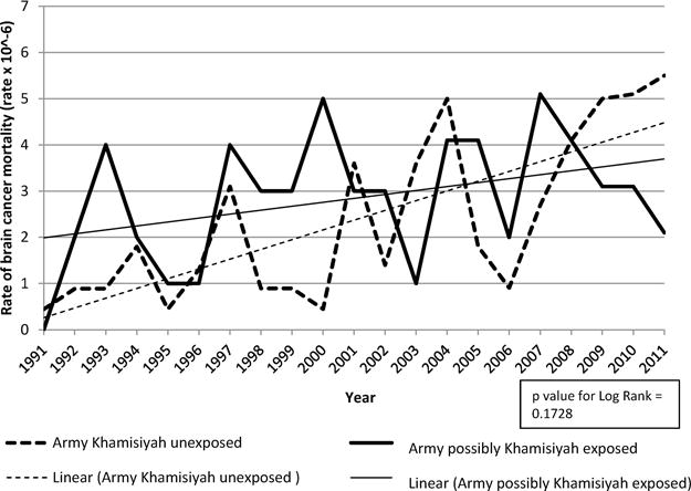
Life test hazard model: Comparison of brain cancer mortality risk among deployed U.S. Gulf War Army Veterans possibly exposed at Khamisiyah and unexposed Gulf War Army Veterans, 1991-2011, United States.
Figs. 3-6 present results from the Joinpoint regression analyses. The estimated annual percent change (APC) in rates of brain cancer mortality was 10.8% (95% confidence interval: 8.7, 12.9) for Gulf War deployed veterans, 8.0% (95% confidence interval: 6.6, 9.3) for Gulf War era veterans, 8.9% (95% confidence interval: 4.5, 13.6) for the unexposed Army group, and 2.3% (95% confidence interval: −1.3, 6.1) for the Army, possibly exposed at Khamisiyah group. The APC estimate was statistically significant and similar for all groups except the Army, possibly exposed at Khamisiyah group. Plots in all figures slope upwards, possibly due to the expected increase in brain cancer mortality that occurs with aging. This occurs in both the deployed and non-deployed groups and exposed and nonexposed groups.
Fig. 3.
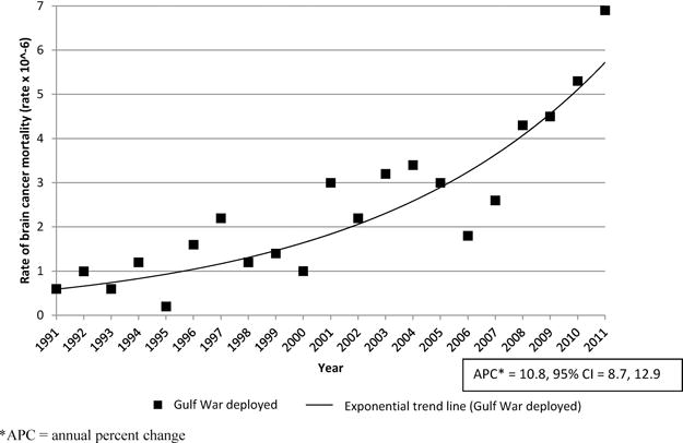
Joinpoint model for brain cancer mortality rate trend among U.S. Gulf War deployed veterans, 1991-2011, United States.
*APC = annual percent change.
Fig. 6.
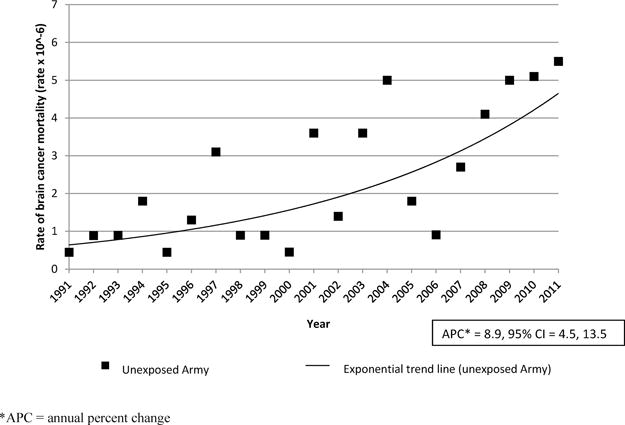
Joinpoint model for brain cancer mortality rate trends among unexposed U.S. Gulf War Army Veterans, 1991-2011, United States.
*APC = annual percent change.
4. Discussion
The objective of this study was to investigate trends and the risk of brain cancer mortality among U.S. Gulf War veterans, particularly those possibly exposed to environmental contaminants at Khamisiyah. Comparisons included aggregate and time series analyses of brain cancer mortality risk among 1) Gulf War deployed and Gulf War era veterans and 2) Army, possibly exposed at Khamisiyah versus unexposed Army veterans.
Notably, U.S. veterans in this study had lower rates of brain cancer mortality compared to U.S. adults (deployed = 0.23/10,000 person-years; non-deployed = 0.30/10,000 person-years; possible exposed at Khamisiyah = 0.28/10,000 person-years; unexposed Army = 0.23/10,000 person-years). Among U.S. men and women, the rate of brain cancer deaths was 0.43 per 10,000 per year [37].
Cox proportional hazards models using data through 2011 did not identify an increased risk of brain cancer mortality among U.S. Gulf War deployed veterans when compared to U.S. Gulf War era veterans or among U.S. Army, possibly exposed at Khamisiyah veterans (regardless of the duration of exposure) when compared to unexposed U.S. Army veterans. The results among the Army deployed exposed and unexposed groups differ from findings from previous follow-up periods through 2000 and 2004 which found increased risk for brain cancer mortality among those who were possibly exposed at Khamisiyah for two or more days compared to the unexposed group (2000: adjusted risk ratio = 3.26; 95% confidence interval: 1.33, 7.96; 2004: adjusted risk ratio = 2.71, 95% confidence interval: 1.25, 5.87) [11,12].
Life test hazard models were calculated to assess the risk of brain cancer mortality between groups over the study period. Over time, U.S. Gulf War era veterans had higher rates of brain cancer mortality when compared to U.S. Gulf War deployed veterans; however, there was no statistically significant difference in the brain cancer mortality rate between U.S. Army, possibly exposed at Khamisiyah and unexposed U.S. Army veterans. Though plots of hazard rates for these groups initially showed a gap in the rate of brain cancer mortality, this difference in risk diminished over time and eventually converged. This gap, suggesting increased risk, in the earlier years of follow up was reported in previous studies of these cohorts. The early increased risk followed by an apparent drop in later years of follow-up may suggest that an agent, such as a neoplastic initiator or promoter, could have been present and that exposure to this agent, in sufficient dose or duration, could accelerate the development of brain cancer in susceptible population members. However, results from this study suggest a time-dependent increase in risk for brain cancer that cannot be explained by known contaminants or current disease models. Specifically, this hypothesis assumes 1) between-person variability in risk for brain cancer, 2) exposure to an unknown agent that would change expected course of disease, and 3) that any effects of exposure would be limited to populations with pre-existing risk.
Results from the Joinpoint analyses indicate statistically significant increases in the risk of brain cancer mortality among the U.S. Gulf War deployed veterans, U.S. Gulf War era veterans, and unexposed U.S. Army veterans. All three groups have similar, statistically significant annual percent changes in brain cancer mortality risk, with estimates ranging from 7.9% to 10.5%. The observed trends in brain cancer mortality risk are consistent with age-based patterns of brain cancer mortality in the U.S. general population [38]. There was no significant increase in the risk of brain cancer mortality in the U.S. Army, possibly exposed at Khamisiyah group. For this group, the annual percent change was 2% and was not statistically significant. As previously discussed, the findings of earlier studies showing an increased risk coupled with an apparent absence of the normally observed age-related increase in brain cancer mortality for this group in the later years of follow-up could suggest an accelerated manifestation of disease among those who were susceptible to environmental exposures.
Importantly, the observed plot of the hazard rates for brain cancer mortality for the U.S. Army, possibly exposed at Khamisiyah group was not consistent with plots of the hazard rates in other groups. Specifically, the distribution of brain cancer mortality risk among U.S. Army, possibly exposed at Khamisiyah group over time had a higher point estimate at the first year of follow-up, lower point estimate at the final year of follow-up, and did not appear to have the same distribution across time that was observed in the other groups.
Studies on amyotrophic lateral sclerosis (ALS) among Gulf War veterans demonstrate a similar pattern of increased risk for a neurological disease among Gulf War veterans in the early years after the war, leveling off in more recent years of follow-up. Horner et al. [39] reported the relative risk of ALS for deployed Gulf War veterans to be 1.92 (95% confidence interval: 1.29, 2.84) using incidence data from 1991 to 1998. When looking at trends in ALS incidence over time, there was no evidence of differences in incidence between deployed and non-deployed Gulf War veterans until 1995, when the number of cases among the deployed more than doubled between 1995 and 1998. The authors hypothesized that Gulf War-specific neurotoxic exposures likely caused the increases in ALS after the war. It should be noted that not only are the time trends for brain cancer mortality and ALS mortality consistent, but the earlier observed risk for brain cancer mortality among possibly exposed Army Gulf War veterans is stronger through 2000 than what was reported by Horner et al. for ALS in Army deployed Gulf War veterans through 1998 (3.26 vs. 2.04) [11,39].
There are several strengths to this study. The large cohort size of approximately 1.38 million total U.S. Gulf War deployed and U.S. Gulf War era veterans allows for detection of risk for rare diseases such as brain cancer. This study extended the follow-up of previous studies of brain cancer mortality to 21 years post-war, allowing analyses of mortality trends over time. A weakness of this study is the use of the Khamisiyah plume model for exposure assessment. Although several studies have used the plume model to determine exposure [40-42], a report published by the General Accountability Office in 2004 concluded that the Department of Defense/Central Intelligence Agency 2000 Khamisiyah plume model was unsupported and recommended that Department of Defense and Department of Veteran Affairs no longer use this plume model for epidemiological studies of the Gulf War [43]. The Department of Veteran Affairs concurred with this decision but allowed for completion of studies already underway, including the work presented here. Additionally, brain cancer is rare, with a 0.6% lifetime risk of diagnosis among adult U.S. men and women [37] and the cause of the majority of brain cancers is unknown [44], which contributes to the challenges of this research question. Furthermore, this is a retrospective epidemiologic study and therefore significant findings do not indicate causation.
Results from these analyses suggest that U.S. Army veterans possibly exposed at Khamisiyah experienced different patterns of brain cancer mortality risk than the other three groups included in this study. We did not find an overall increased risk of brain cancer mortality among U.S. Gulf War deployed veterans when compared to U.S. Gulf War era veterans after 21 years of follow-up. Analyzing trends over time using the life test hazard and Joinpoint regression models provided us with a greater understanding of the overall patterns of brain cancer mortality among these cohorts. The reason for observed between group differences remains unclear and could be attributed to an early acceleration of disease among those most susceptible to developing brain cancer following environmental exposures. The observed trends in brain cancer mortality risk are consistent with age-based patterns of brain cancer mortality in the U.S. general population [38].
Fig. 4.
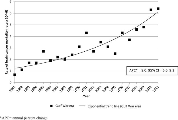
Joinpoint model for brain cancer mortality rate trend among U.S. Gulf War era veterans, 1991-2011, United States. *APC = annual percent change.
Fig. 5.
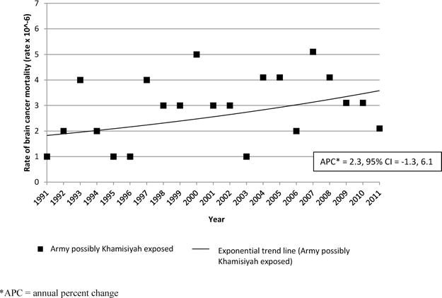
Joinpoint model for brain cancer mortality rate trend among deployed U.S. Army Gulf War Veterans possibly exposed at Khamisiyah, 1991-2011, United States.
*APC = annual percent change.
Acknowledgments
Funding
This research was supported by the Department of Veterans Affairs. This research did not receive any specific grant from funding agencies in the public, commercial, or not-for-profit sectors.
Footnotes
Conflicts of interest
None.
Authors’ contribution
Shannon Barth was responsible for data analyses and manuscript writing. Dr. Erin Dursa contributed to the review of the data analyses, manuscript writing and review. Dr. Robert Bossarte contributed to the statistical analyses plan, and manuscript writing and review. Dr. Aaron Schneiderman contributed to manuscript writing and review.
References
- 1.Winkenwerder W. Technical Report: Modeling and Risk Characterization of US Demolition at the Khamisiyah Pit, Office of the Assistant Secretary of Defense (Health Affairs) and Special Assistant to the Under Secretary of Defense (Personnel and Readiness) for Gulf War Illnesses, Medical Readiness, and Military Deployments. Department of Defense; Washington, DC: 2002. http://www.gulflink.osd.mil/khamisiyah_tech/index.htm, Published 16 April 2002 (Accessed 10 January 2016) [Google Scholar]
- 2.Winkenwerder W. Case Narrative: US Demolition Operations at Khamisiyah: Final Report, Office of the Assistant Secretary of Defense (Health Affairs) and Special Assistant to the Under Secretary of Defense (Personnel and Readiness) for Gulf War Illnesses, Medical Readiness, and Military Deployments. Department of Defense; Washington, DC: 2002. http://www.gulflink.osd.mil/khamisiyah_iii/, Published 16 April 2002 (Accessed 10 January 2016) [Google Scholar]
- 3.Proctor SP, Heaton KJ, Heeren T, et al. Effects of sarin and cycolsarin exposure during the 1991 GW on neurobehavioral functioning in US army veterans. Neurotoxicology. 2006;27(6):931–939. doi: 10.1016/j.neuro.2006.08.001. [DOI] [PubMed] [Google Scholar]
- 4.Heaton KJ, Palumbo CL, Proctor SP, et al. Quantitative magnetic resonance brain imaging in US army veterans of the 1991 GW potentially exposed to sarin and cyclosarin. Neurotoxicology. 2007;28(5):761–769. doi: 10.1016/j.neuro.2007.03.006. [DOI] [PubMed] [Google Scholar]
- 5.Kalra R, Singh SP, Razani-Boroujerdi S, et al. Subclinical doses of the nerve gas sarin impair T cell responses through the autonomic nervous system. Toxicol Appl Pharmacol. 2002;184(2):82–87. [PubMed] [Google Scholar]
- 6.Henderson RF, Barr EB, Blackwell WB, et al. Response of F344 rats to inhalation of subclinical levels of sarin: exploring potential causes of Gulf War illness. Toxicol Ind Health. 2001;17(5-10):294–297. doi: 10.1191/0748233701th105oa. [DOI] [PubMed] [Google Scholar]
- 7.Chao LL, Abadjian L, Hlavin J, Meyerhoff DJ, Weiner MW. Effects of low-level sarin and cyclosarin exposure and Gulf War illness on brain structure and function: a study at 4T. Neurotoxicology. 2011;32(6):814–822. doi: 10.1016/j.neuro.2011.06.006. [DOI] [PubMed] [Google Scholar]
- 8.Toomey R, Alpern R, Vasterling JJ, Baker DG, Reda DJ, Lyons MJ, Henderson WG, Kang HK, Eisen SA, Murphy FM. Neuropsychological functioning of U.S. Gulf War veterans 10 years after the war. J Int Neuropsychol Soc. 2009;15(5):717–729. doi: 10.1017/S1355617709990294. [DOI] [PubMed] [Google Scholar]
- 9.Chao LL, Rothlind JC, Cardenas VA, Meyerhoff DJ, Weiner MW. Effects of low-level exposure to sarin and cyclosarin during the 1991 Gulf War on brain function and brain structure in US veterans. Neurotoxicology. 2010;31(5):493–50. doi: 10.1016/j.neuro.2010.05.006. [DOI] [PMC free article] [PubMed] [Google Scholar]
- 10.Chao LL, Kriger S, Buckley S, Ng P, Mueller SG. Effects of low-level sarin and cyclosarin exposure on hippocampal subfields in Gulf War veterans. Neurotoxicology. 2014;44:263–269. doi: 10.1016/j.neuro.2014.07.003. [DOI] [PMC free article] [PubMed] [Google Scholar]
- 11.Bullman TA, Mahan CM, Kang HK, et al. Mortality in US Army GW veterans exposed to 1991 Khamisiyah chemical munitions destruction. Am J Public Health. 2005;95(8):1382–1388. doi: 10.2105/AJPH.2004.045799. [DOI] [PMC free article] [PubMed] [Google Scholar]
- 12.Barth SK, Kang HK, Bullman TA, Wallin MT. Neurological mortality among U.S. veterans of the Persian Gulf War: 13-year follow-up. Am J Ind Med. 2009;52(9):663–670. doi: 10.1002/ajim.20718. [DOI] [PubMed] [Google Scholar]
- 13.Amourette C, Lamproglou I, Barbier L, et al. Gulf War illness: effects of repeated stress and pyridostigmine treatment on blood-brain barrier permeability and cholinesterase activity in rat brain. Behav Brain Res. 2009 Nov;203(2):207–214. doi: 10.1016/j.bbr.2009.05.002. [DOI] [PubMed] [Google Scholar]
- 14.Abdullah L, Evans JE, Montague H, et al. Chronic elevation of phosphocholine containing lipids in mice exposed to Gulf War agents pyridostigmine bromide and permethrin. Neurotoxicol Teratol. 2013 Nov-Dec;40:74–84. doi: 10.1016/j.ntt.2013.10.002. [DOI] [PubMed] [Google Scholar]
- 15.Ojo JO, Abdullah L, Evans J, et al. Exposure to an organophosphate pesticide, individually or in combination with other Gulf War agents, impairs synaptic integrity and neuronal differentiation, and is accompanied by subtle microvascular injury in a mouse model of Gulf War agent exposure. Neuropathology. 2014 Apr;34(2):109–127. doi: 10.1111/neup.12061. [DOI] [PubMed] [Google Scholar]
- 16.O’Callaghan JP, Kelly KA, Locker AR, Miller DB, Lasley SM. Corticosterone primes the neuroinflammatory response to DFP in mice: potential animal model of Gulf War illness. J Neurochem. 2015 Jun;133(5):708–721. doi: 10.1111/jnc.13088. doi: http://dx.doi.org/10.1111/jnc.13088. [DOI] [PMC free article] [PubMed] [Google Scholar]
- 17.Chao LL, Reeb R, Esparza IL, Abadjian LR. Associations between the self-reported frequency of hearing chemical alarms in theater and regional brain volume in Gulf War veterans. Neurotoxicology. 2016 Mar;53:246–256. doi: 10.1016/j.neuro.2016.02.009. doi: http://dx.doi.org/10.1016/j.neuro.2016.02.009. [DOI] [PMC free article] [PubMed] [Google Scholar]
- 18.Speed HE, Blaiss CA, Kim A, et al. Delayed reduction of hippocampal synaptic transmission and spines following exposure to repeated subclinical doses of organophosphorus pesticide in adult mice. Toxicol Sci. 2012 Jan;125(1):196–208. doi: 10.1093/toxsci/kfr253. [DOI] [PMC free article] [PubMed] [Google Scholar]
- 19.Menon PM, Nasrallah HA, Reeves RR, Ali JA. Hippocampal dysfunction in Gulf War syndrome. A proton MR spectroscopy study. Brain Res. 2004 May;1009(1–2):189–194. doi: 10.1016/j.brainres.2004.02.063. [DOI] [PubMed] [Google Scholar]
- 20.Chao LL, Zhang Y, Buckley S. Effects of low-level sarin and cyclosarin exposure on white matter integrity in Gulf War veterans. Neurotoxicology. 2015 May;48:239–248. doi: 10.1016/j.neuro.2015.04.005. doi: http://dx.doi.org/10.1016/j.neuro.2015.04.005. [DOI] [PMC free article] [PubMed] [Google Scholar]
- 21.Zakirova Z, Crynen G, Hassan S, Abdullah L, Horne L, Mathura V, Crawford F, Ait-Ghezala G. A chronic longitudinal characterization of neurobehavioral and neuropathological cognitive impairment in a mouse model of gulf war agent exposure. Front Integr Neurosci. 2016 Jan;12(9):71. doi: 10.3389/fnint.2015.00071. doi: http://dx.doi.org/10.3389/fnint.2015.00071. [DOI] [PMC free article] [PubMed] [Google Scholar]
- 22.Hubbard NA, Hutchison JL, Motes MA, Shokri-Kojori E, Bennett IJ, Brigante RM, Haley RW, Rypma B. Central executive dysfunction and deferred prefrontal processing in veterans with Gulf War illness. Clin Psychol Sci. 2014 May;2(3):319–327. doi: 10.1177/2167702613506580. [DOI] [PMC free article] [PubMed] [Google Scholar]
- 23.Rostker B. Environmental Exposures Report: Oil Well Fires, Office of the Special Assistant to the Deputy Secretary of Defense for Gulf War Illnesses. Department of Defense; Washington, DC: 2000. Apr, http://www.gulflink.osd.mil/owf_ii/, Published 2 August 2000 Accessed 10 January 2016. [Google Scholar]
- 24.Cowan DN, Lange JL, Heller J, et al. A case-control study of asthma among U. S. Army GW veterans and modeled exposure to oil well fire smoke. Mil Med. 2002;167(9):777–782. [PubMed] [Google Scholar]
- 25.Lange JL, Schwartz DA, Doebbeling BN, et al. Exposures to the Kuwait oil fires and their association with asthma and bronchitis among Gulf War veterans. Environ Health Perspect. 2002;110(11):1141–1146. doi: 10.1289/ehp.021101141. [DOI] [PMC free article] [PubMed] [Google Scholar]
- 26.Kelsall HL, Sim MR, Forbes AB, et al. Respiratory health status of Australian veterans of the 1991 Gulf War and the effects of exposure to oil fire smoke and dust storms. Thorax. 2004;59(10):897–903. doi: 10.1136/thx.2003.017103. [DOI] [PMC free article] [PubMed] [Google Scholar]
- 27.Oberdorster G, Sharp Z, Atudorei V, Elder A, Gelein R, Kreyling W, Cox C. Translocation of inhaled ultrafine particles to the brain. Inhal Toxicol. 2004;16:437–445. doi: 10.1080/08958370490439597. [DOI] [PubMed] [Google Scholar]
- 28.Elder A, Gelein R, Silva V, Feikert T, Opanashuk L, Carter J, Potter R, Maynard A, Ito Y, Finkelstein J, Oberdörster G. Translocation of inhaled ultrafine manganese oxide particles to the central nervous system. Environ Health Perspect. 2006;114(8):1172–1178. doi: 10.1289/ehp.9030. [DOI] [PMC free article] [PubMed] [Google Scholar]
- 29.Calderón-Garcidueñas L, Maronpot RR, Torres-Jardon R, Henríquez-Roldán C, Schoonhoven R, Acuña-Ayala H, Villarreal-Calderón A, Nakamura J, Fernando R, Reed W, Azzarelli B, Swenberg JA. DNA damage in nasal and brain tissues of canines exposed to air pollutants is associated with evidence of chronic brain inflammation and neurodegeneration. Toxicol Pathol. 2003 Sep-Oct;31(5):524–538. doi: 10.1080/01926230390226645. [DOI] [PubMed] [Google Scholar]
- 30.Campbell A, Oldham M, Becaria A, Bondy SC, Meacher D, Sioutas C, Misra C, Mendez LB, Kleinman M. Particulate matter in polluted air may increase biomarkers of inflammation in mouse brain. Neurotoxicology. 2005;26(1):133–140. doi: 10.1016/j.neuro.2004.08.003. [DOI] [PubMed] [Google Scholar]
- 31.Donaldson K, Stone V, Borm PJA, Jimenez LA, Gilmour PS, Schins RPF, Knaapen AM, Rahmen I, Faux SP, Brown DM, MacNee W. Oxidative stress and calcium signaling in the adverse effects of environmental particles (PM10) Free Radic Biol Med. 2003;34(11):1369–1382. doi: 10.1016/s0891-5849(03)00150-3. [DOI] [PubMed] [Google Scholar]
- 32.Donaldson K, Tran L, Jimenez LA, Duffin R, Newby DE, Mills N, MacNee W, Stone V. Combustion-derived nanoparticles: a review of their toxicology following inhalation exposure. Part Fibre Toxicol. 2005 Oct;21(2):10. doi: 10.1186/1743-8977-2-10. [DOI] [PMC free article] [PubMed] [Google Scholar]
- 33.Boeglin ML, Wessels D, Henshel D. An investigation of the relationship between air emissions of volatile organic compounds and the incidence of cancer in Indiana counties. Environ Res. 2006;100(2):242–254. doi: 10.1016/j.envres.2005.04.004. [DOI] [PubMed] [Google Scholar]
- 34.Liu CC, Chen CC, Wu TN, Yang CY. Association of brain cancer with residential exposure to petrochemical air pollution in Taiwan. J Toxicol Environ Health A. 2008;71(5):310–314. doi: 10.1080/15287390701738491. [DOI] [PubMed] [Google Scholar]
- 35.Walpole RD, Rostker B. Modeling the Chemical Warfare Agent Release at the Khamisiyah Pit. US Department of Defense; Washington, DC: 2017. http://www.gulflink.osd.mil/cia_092297/, Published 4 September 1997 Accessed 10 January 2017. [Google Scholar]
- 36.Joinpoint Regression Program, Version 4.2.0. Statistical Methodology and Applications Branch, Surveillance Research Program, National Cancer Institute; Apr, 2015. [Google Scholar]
- 37.National Cancer Institute. Surveillance, Epidemiology, and End Results Program. SEER Fact Sheets: Brain Cancer and Other Nervous System Cancer. http://seer.cancer.gov/statfacts/html/brain.html (Accessed 10 January 2017)
- 38.Centers for Disease Control and Prevention, National Center for Health Statistics. Underlying Cause of Death 1999-2013 on CDC WONDER Online Database, released 2015. Data are from the Multiple Cause of Death Files, 1999-2013, as compiled from data provided by the 57 vital statistics jurisdictions through the Vital Statistics Cooperative Program. http://wonder.cdc.gov/ucd-icd10.html, (Accessed 15 October 2015)
- 39.Horner RD, Kamins KG, Feussner JR, Grambow SC, Hoff-Lindquist J, Harati Y, Mitsumoto H, Pascuzzi R, Spencer PS, Tim R, Howard D, Smith TC, Ryan MA, Coffman CJ, Kasarskis EJ. Occurrence of amyotrophic lateral sclerosis among Gulf War veterans. Neurology. 2003;61(6):742–749. doi: 10.1212/01.wnl.0000069922.32557.ca. [DOI] [PubMed] [Google Scholar]
- 40.Haley RW, Tuite JJ. Epidemiologic evidence of health effects from long-distance transit of chemical weapons fallout from bombing early in the 1991 Persian Gulf War. Neuroepidemiology. 2013;40(3):178–189. doi: 10.1159/000345124. [DOI] [PubMed] [Google Scholar]
- 41.Miranda ML, Galeano M Alicia Overstreet, Tassone E, Allen KD, Horner RD. Spatial analysis of the etiology of amyotrophic lateral sclerosisamong 1991 Gulf War veterans. Neurotoxicology. 2008;29:964–970. doi: 10.1016/j.neuro.2008.05.005. [DOI] [PubMed] [Google Scholar]
- 42.Gackstetter GD, Hooper TI, DeBakey SF, Johnson A, Nagaraj BE, Heller JM, Kang HK. Fatal motor vehicle crashes among veterans of the 1991 gulf war and exposure to munitions demolitions at Khamisiyah: a nested case-control study. Am J Ind Med. 2006;49:262–270. doi: 10.1002/ajim.20280. [DOI] [PubMed] [Google Scholar]
- 43.U.S. General Accountability Office. Report No GAO 04-821T. Washington, DC: 2004. Testimony Before the Subcommittee on National Security, Emerging Threats, and International Relations, Committee on Government Reform, House of Representatives; GULF WAR ILLNESSES DOD’s Conclusions About U.S. Troops’ Exposure Cannot Be Adequately Supported, U.S. General Accountability Office. [Google Scholar]
- 44.National Cancer Institute. A Snapshot of Brain and Central Nervous System Cancers. 2017 http://www.cancer.gov/research/progress/snapshots/brain, Published 5 November 2014 (Accessed 10 January 2017)