Abstract
Anorectal complaints are not uncommon in pediatric care, but the etiology and management can differ significantly from adults. Age is an important factor when considering etiology and management, distinguishing between infants, children, and adolescents. For all ages, malignancy is rarely a consideration, but a thorough examination of infants and children typically requires deep sedation or general anesthesia. Very little primary literature or evidence exists to guide care; so there are many opportunities for careful study to enhance our understanding beyond personal experience and historical practice patterns.
Keywords: pediatric, anal fissure, fistula-in-ano, perianal abscess, hemorrhoid, rectum
Hemorrhoid is the most common purported anorectal diagnosis for pediatric patients referred for evaluation by a surgeon. However, the incidence of hemorrhoids in the pediatric population is exceedingly low and an alternative explanation is often identified. A general concern in the evaluation of reported pediatric hemorrhoids is the identification of alternate soft tissue growths, which may have been misinterpreted as hemorrhoids, particularly rectal polyps, skin tags, and anal condylomata. While simple history and examination often elucidate the correct diagnosis, some patients require anorectoscopy. In some teens, the diagnosis can be clarified by speculum exam in the clinic. More often, discomfort or emotional distress is predictable from the patient's behavior, and consideration should be given to performing the exam under anesthesia with possible excision at the same time. This is especially true in young children but may also be the case with some teenagers.
The etiology of hemorrhoids is venous congestion/stasis. The most common cause of hemorrhoids in pediatric patients is chronic liver failure. Alternately, chronic constipation in adolescent patients can lead to hemorrhoids as a result of straining to pass stool or simply habitually sitting on a toilet for a long time. As constipation is common in young children, it is surprising that hemorrhoids are not seen more commonly in this population; this may be due to the fact that hemorrhoids develop slowly. To support this point, hemorrhoids in pediatric patients rarely develop until the teenage years.
Presentations of both internal and external hemorrhoids are similar to adults. Most patients with internal hemorrhoids will report feeling protuberant tissue only intermittently. A persistently palpable hemorrhoid indicates that it is an external hemorrhoid or Grade III-IV internal hemorrhoid. Prolapsed internal hemorrhoids can be painful and tender, as are thrombosed external hemorrhoids. In the absence of a prolapse or thrombosis, the dominant symptom is painless bleeding after stooling and/or wiping. Primary treatment consists of relieving constipation, adjusting behavior (avoiding long “dwell times” on the toilet), and observing for gradual resolution. With time, growth, and control of the above factors, limited internal hemorrhoids subside and small external hemorrhoids regress into skin tags.
Most hemorrhoids in pediatric patients resolve without operation. However, redundant tissue can be uncomfortable, causing bleeding and interference with hygiene. If these symptoms persist, hemorrhoidectomy is considered. This is performed with traditional excision techniques (i.e., open hemorrhoidectomy). There are case reports of successful use of the Ligasure technique in children. 1 There is no empiric reason to think that stapled hemorrhoidectomy and photocoagulation techniques may not eventually be extended to the adolescent population, but no cases have yet been reported. As hemorrhoidectomy is painful in adults, it is often tolerated more poorly in pediatric patients. The patient must be prepared for surgery by achieving a stable, nonconstipated state. To limit pain and risk of cicatrix, only a single column is addressed at a time.
Anal Fissures
Fissures are a common complication of constipation in children. The passage of large, firm stools can literally tear the anoderm and result in a fissure. This can be quite painful, and the pain is exacerbated by defecation. The most common symptoms of anal fissure are stools streaked with blood, pain, and/or crying with defecation. Most fissures can be easily identified in the clinic by separating the buttocks to inspect the anoderm. While fissures are most common in the posterior midline in infants, they may be found in any location (particularly anterior midline in female infants). 2 Anal pain creates a deleterious positive feedback loop, in which pain causes stool retention, stool then becomes more bulky and devoid of water, passage of this stool reinjures the fissures, and so on. This cycle of injury must be broken to allow the fissures to heal.
Most anal fissures (particularly in infancy) resolve without operation. Management centers on relief of constipation, whether by increasing water, dietary adjustments, fiber supplements, cathartic medications, or investigating other underlying causes of constipation (a lengthy discussion in itself). Softening stool consistency is preferred to using stimulants because the latter may merely serve to more forcefully push forward the same firm, large stools. Wiping with moist “baby wipes” or medicated pads may irritate the area less than dry toilet paper.
Second-line treatment involves topical agents. Topical lidocaine, calcium channel blockers, or nitroglycerin are frequently employed agents in managing adult fissures and have been reported to be efficacious in pediatric patients as well. 2 These are reasonable for application in adolescents and not as often used in younger children due to concerns of unpredictable systemic absorption, though it has been described. 3 Chronic or recurrent fissures may be treated with sphincteric botulinum toxin injection. 4 Because the effect is self-limited, this does not risk permanent incontinence. Internal sphincterotomy––while effective and definitive in adults ––is not advised in children because the sphincter complex is very thin and risk of permanent dysfunction is thus higher. 2
A particularly pediatric concern related to anal fissures is the possibility of nonaccidental trauma (formerly referred to as abuse). 5 6 Multiple fissures in different locations can be suggestive of nonaccidental penetration. History of abdominal pain may be functional abdominal pain rather than just constipation. In such patients, at least a screening should be performed for safety concerns and social risk factors for nonaccidental trauma.
Pediatric patients sometimes present with a perianal skin tag with reports from the parent that it has been there since birth. Close questioning may reveal a remote history of self-limited blood in the stools as a newborn with no diagnosis of an anal fissure previously established. The skin tag is a benign remnant of the healed fissure and does not require excision. Parents sometimes have the impression that excision is empirically required, but explanation and reassurance are all that is needed.
Abscesses
Abscesses of the backside are commonly referred to as “perirectal abscesses” by pediatricians and emergency medicine physicians seeking input from a surgeon. However, it is critically important to distinguish between perirectal abscesses, perianal abscesses, and gluteal abscesses.
True perirectal abscesses are very rare in infants and children. These are rarely visible, and though they may be suggested by history or exam in a suitable patient, diagnosis is usually based on imaging. Etiology is related to trauma, Crohn's disease, immune deficiency, or an infected mass lesion (e.g., rectal duplication or teratoma). Imaging is necessary to characterize extent and help determine the etiology. Treatment depends on the etiology, particularly if inflammatory bowel disease is suspected.
Gluteal abscesses are very common, and most of these are simple matters though they can become quite large and extend into the ischiorectal region. Drainage is the mainstay of therapy, though sometimes fluctuance can be difficult to distinguish from soft, infant gluteal fat when there is associated induration at the rim. If there is uncertainty on exam, simple bedside ultrasound can be employed to remove doubt. Initial antibiotic treatment can be helpful to decrease surrounding cellulitis, prevent extension, and sometimes even treat very small collections. When coupled with warm compresses, spontaneous drainage will frequently occur and invasive procedures are avoided. Whereas any such abscesses in adults will require drainage, in infants and children, the combination of antibiotics and warm compresses are adequate to heal many of the minor abscesses. So, many of these are handled in pediatric clinics without referral to surgeons.
When drainage is required, aggressive irrigation is performed and typically a gauze packing/wick is left in place. Such packing is usually removed the following day, and younger children seldom tolerate repacking, so a few approaches are taken to prevent reformation of an abscess: Sitz baths, continuation of antibiotics for a few days after drainage, and use of a counter-incision with a vessel loop through both incisions allow ongoing drainage from particularly large cavities.
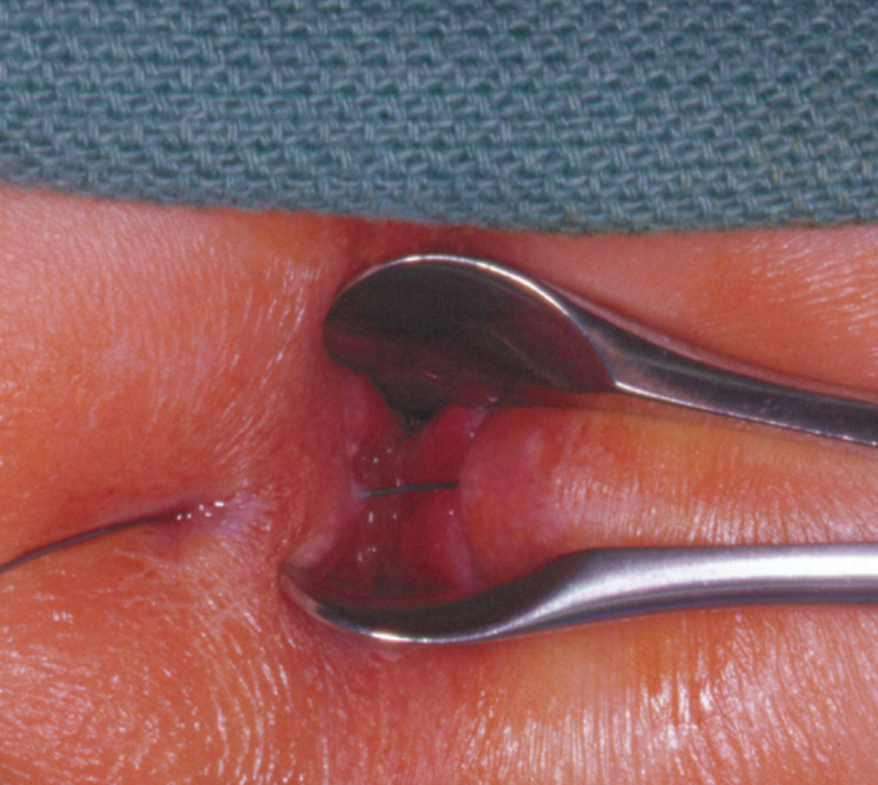
Perianal abscesses are also particularly common in infants. As the name implies, these originate immediately around the anus and most present as a small, superficial abscess. A perianal abscess originates from an infection of the anal glands that subsequently necessitates toward the perianal skin. The anal glands are located at the dentate line and can be identified by a distal transverse fold. This creates pockets termed crypts of Morgagni, into which the anal glands open. In many cases, perianal abscesses present with minimal symptoms and are simply noted by the parents during a diaper change. 7 Many can be managed adequately with warm soaks, oral antibiotics, and occasionally needle aspiration. 7 Perianal abscesses that are successfully managed without operative drainage are less likely to later present with fistula-in-ano. Less commonly, perianal abscesses are large, have associated systemic symptoms, or have failed nonoperative treatment, and thus require formal drainage to relieve symptoms and reduce likelihood of fistula formation. 8 For these, the general principles of perianal drainage apply: using superficial incisions, avoiding damage to the sphincter complex, and carefully searching for fistula-in-ano. The author contends that this is optimally done only with deep sedation or general anesthesia. In resource-limited settings, of course, compromises may be necessary. It is fairly common after drainage for these to present as recurrent abscesses at the same site. 7 9 In such cases, a fistula-in-ano should be presumed and managed as discussed later ( Fig. 1 ).
Fig. 1.

Use of a lacrimal probe through the perianal skin opening is used to demonstrate fistula-in-ano through an anal crypt. The tract is opened and curetted, and allowed to heal secondarily. (Reprinted with permission from Stites and Lund. 2 )
Operations should be performed in a setting so as to minimize discomfort and psychological distress as well as allowing the surgeon proper visualization and safe intervention. For older patients with gluteal abscesses and a very calm demeanor, this may be accomplished with topical local anesthetic and possibly an anxiolytic. Injection of a local anesthetic is typically avoided because of concern of causing tissue injury in an infected field. For any sort of perianal or transanal evaluation or intervention, the author's practice is to perform it under conscious sedation or general anesthesia to achieve a thorough and accurate assessment.
Fistula-in-Ano
Fistulae occur in infants with recurrent perianal abscesses and rarely signify underlying pathology. However, in older children and teens, these may be the first recognized manifestation of Crohn's disease. In fact, a recurrent fistula-in-ano beyond infancy may be the presenting sign of Crohn's disease and should prompt gastrointestinal tract (GI) consultation.
Drainage of perianal abscess in any age group should be accompanied by a careful examination to evaluate for fistulous connection to the anorectum. Recurrent abscesses may be caused by persistent granulation tissue in the cavity and/or development of an epithelialized tract communicating with an anal gland. This can result in a chronic draining sinus, which requires repeated debridement or excision of the tract while protecting the sphincter complex.
Searching for a fistula is very important because if it is not identified, the infection will predictably recur and an opportunity for definitive intervention would be missed. However, one must not be too aggressive in the search for a fistula as to falsely create a passage into the rectum. While identification of a fistula is important, at index operation, it is arguable whether or not to specifically address the fistula or to just incise and drain, then reevaluate for fistula persistence later.
Most pediatric perianal abscesses will not demonstrate a fistula on initial presentation, but if a fistula is identified with a first abscess, a decision must be made about whether to perform fistulotomy or simply incision and drainage with debridement. The latter is frequently sufficient, though it does have a higher likelihood of recurrence. 10 The former risks deeper injury or incontinence. Various other approaches to management of pediatric fistula-in-ano have been described. 11 12 13 However, these have not been compared with one another and management decisions do not have a solid basis in published data. Even in adults, management of fistula-in-ano is an area of ongoing evolution and debate. 14
After resolution of the acute infectious process, the site remains somewhat inflamed and continues to drain for days or even weeks. If persistent granulation tissue is observed, this can be addressed with topical silver nitrate. Repeat intervention should be reserved for those cases in which recurrent infection or chronic drainage are observed.
Conclusion
Pediatric anorectal complaints are almost always benign processes. Attention to accurate diagnosis and age-specific considerations in management will lead to the best outcomes. In infants and young children, anorectal complaints are quite common and rarely associated with an underlying pathology such as inflammatory bowel disease (IBD); in adolescence, IBD should always be considered. Mostly, the care is based on historical practice patterns and influence of mentors on surgeons' training. There is much room for research to generate objective data on which to guide the treatment.
References
- 1.Grossmann O, Soccorso G, Murthi G. Ligasure hemorrhoidectomy for symptomatic hemorrhoids: first pediatric experience. Eur J Pediatr Surg. 2015;25(04):377–380. doi: 10.1055/s-0034-1382258. [DOI] [PubMed] [Google Scholar]
- 2.Stites T, Lund D P. Common anorectal problems. Semin Pediatr Surg. 2007;16(01):71–78. doi: 10.1053/j.sempedsurg.2006.10.010. [DOI] [PubMed] [Google Scholar]
- 3.Klin B, Efrati Y, Berkovitch M, Abu-Kishk I. Anal fissure in children: a 10-year clinical experience with nifedipine gel with lidocaine. Minerva Pediatr. 2016;68(03):196–200. [PubMed] [Google Scholar]
- 4.Husberg B, Malmborg P, Strigård K. Treatment with botulinum toxin in children with chronic anal fissure. Eur J Pediatr Surg. 2009;19(05):290–292. doi: 10.1055/s-0029-1231052. [DOI] [PubMed] [Google Scholar]
- 5.Rougé-Maillart C, Houdu S, Darviot E, Buchaillet C, Baron C. Anal lesions presenting in a cohort of child gastroenterological examinations. Implications for sexual traumatic injuries. J Forensic Leg Med. 2015;32:25–29. doi: 10.1016/j.jflm.2015.02.008. [DOI] [PubMed] [Google Scholar]
- 6.Hobbs C J, Wright C M. Anal signs of child sexual abuse: a case-control study. BMC Pediatr. 2014;14:128. doi: 10.1186/1471-2431-14-128. [DOI] [PMC free article] [PubMed] [Google Scholar]
- 7.Serour F, Somekh E, Gorenstein A. Perianal abscess and fistula-in-ano in infants: a different entity? Dis Colon Rectum. 2005;48(02):359–364. doi: 10.1007/s10350-004-0844-0. [DOI] [PubMed] [Google Scholar]
- 8.Afşarlar C E, Karaman A, Tanır G et al. Perianal abscess and fistula-in-ano in children: clinical characteristic, management and outcome. Pediatr Surg Int. 2011;27(10):1063–1068. doi: 10.1007/s00383-011-2956-7. [DOI] [PubMed] [Google Scholar]
- 9.Christison-Lagay E R, Hall J F, Wales P W et al. Nonoperative management of perianal abscess in infants is associated with decreased risk for fistula formation. Pediatrics. 2007;120(03):e548–e552. doi: 10.1542/peds.2006-3092. [DOI] [PubMed] [Google Scholar]
- 10.Murthi G V, Okoye B O, Spicer R D, Cusick E L, Noblett H R. Perianal abscess in childhood. Pediatr Surg Int. 2002;18(08):689–691. doi: 10.1007/s00383-002-0761-z. [DOI] [PubMed] [Google Scholar]
- 11.Inoue M, Sugito K, Ikeda T et al. Long-term results of seton placement for fistula-in-ano in infants. J Gastrointest Surg. 2014;18(03):580–583. doi: 10.1007/s11605-013-2351-x. [DOI] [PubMed] [Google Scholar]
- 12.Osman M A, Elsharkawy M A, Othman M H. Repair of fistulae in-ano in children using image guided Histoacryl injection after failure of conservative treatment. J Pediatr Surg. 2013;48(03):614–618. doi: 10.1016/j.jpedsurg.2012.11.029. [DOI] [PubMed] [Google Scholar]
- 13.Pini Prato A, Zanaboni C, Mosconi M et al. Preliminary results of video-assisted anal fistula treatment (VAAFT) in children. Tech Coloproctol. 2016;20(05):279–285. doi: 10.1007/s10151-016-1447-1. [DOI] [PubMed] [Google Scholar]
- 14.Yassin N A, Hammond T M, Lunniss P J, Phillips R K. Ligation of the intersphincteric fistula tract in the management of anal fistula. A systematic review. Colorectal Dis. 2013;15(05):527–535. doi: 10.1111/codi.12224. [DOI] [PubMed] [Google Scholar]


