Fig. 3.
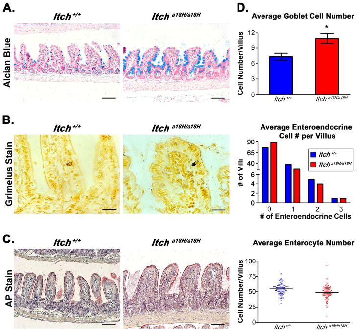
Loss of ITCH promotes goblet cell differentiation. (A) Paraffin-embedded intestinal sections stained with alcian blue, pH 2.5 and counterstained with nuclear fast red distinguish goblet cells from adult Itch+/+ and Itcha18H/a18H animals. Scale bars = 200 μm. (B) Pictured are representative Itch+/+ and Itcha18H/a18H derived paraffin-embedded sections stained with 1% silver nitrate to identify enteroendocrine cells in the small intestine. Scale bars = 200 μm. (C) Paraffin-embedded sections stained with AP highlights the brush boarder of enterocytes on the villi of Itch+/+ and Itcha18H/a18H animals. Scale bars = 200 μm. (D) Average number of goblet cells, enteroendocrine cells, and enterocytes located on the villus of Itch+/+ (n = 4) and Itcha18H/a18H (n = 4) animals. Indicated type of cells were counted on 30 villus structures from a minimum of eight different 10x fields per animal. Enteroendocrine cell counts are presented as a histogram of the total number of villus structures bearing no, one, two or three cells in Itch+/+ (n = 4) and Itcha18H/a18H (n = 4) animals. Itcha18H/a18H animals have a statistically significant increase (*p < 0.05) in goblet cells. There was no statistically significant difference in enteroendocrine or enterocyte cell number. Error bars represent SEM for goblet cell counts.

