

Abstract
Ovarian cystic teratomas constitute 10–15% of all ovarian tumours and are the most common ovarian neoplasms found in adolescence and during pregnancy. Nevertheless, ovarian cystic teratomas have also been described in patients aged 1-91 years. We report an unusual case of a benign ovarian cystic teratoma presenting as a rectal mass that was managed surgically using radical resection by a multidisciplinary team. This case report highlights the importance of preoperative investigations including colonoscopy and radiological investigations. A dedicated pelvic radiologist/pathologist and the involvement of a multidisciplinary team at the time of initial diagnosis and a gynaecologist and colorectal surgeon at the time of surgery will lead to an accurate diagnosis and the most appropriate treatment. Although rare, erosion of an ovarian dermoid into the rectum should be considered in young women who have an atypical presentation and are found to have a lesion in the rectum with biopsies indicating benign pathology.
Keywords: Ovarian cystic teratoma, Rectal mass
Ovarian cystic teratomas constitute 10–15% of all ovarian tumours and are the most common ovarian neoplasm found in adolescence and during pregnancy.1 Even though commonly found in the adolescent group, ovarian cystic teratomas have also been reported in patients aged 1–91 years.1 We describe an unusual case of a benign ovarian cystic teratoma presenting as a rectal mass that was managed surgically using radical resection by a multidisciplinary team.
Case history
A 44-year-old woman presented to the colorectal clinic with a 4-week history of weight loss and loose stools and bleeding per rectum for eight days. She had no significant past medical history. Following the initial consultation, a colonoscopy was performed.
The colonoscopy showed a polypoidal lesion, measuring 1.5cm, that was 7cm from the anal verge (Fig 1). Biopsies taken from this lesion did not show any features of malignancy but ‘strips of squamous epithelium exhibiting intense active chronic inflammation with ulceration’ were reported. Further assessment of the lesion was carried out using abdominal/transvaginal ultrasonography, computed tomography (CT) and magnetic resonance imaging (MRI). These scans showed a cystic lesion containing calcium and gas indenting into and possibly communicating with the rectosigmoid junction (Fig 2). The lesion was suspected to be originating from the left ovary.
Figure 1.

Colonoscopy view of the rectal mass
Figure 2.

Computed tomography showing the cystic mass
Following the initial investigation, the patient under-went examination under anaesthesia (EUA) with no obvious masses palpable in the rectouterine pouch and no extension to the pelvic sidewall. A further biopsy taken of the original cystic mass showed squamous epithelium with chronic inflammatory changes.
EUA was followed by a diagnostic laparoscopy, where an inflammatory mass was noted involving the left ovary and rectum. The initial laparoscopy was converted to a laparotomy and carried out as a combined procedure between a colorectal surgeon and the gynaecologist. An inflammatory mass in the pelvis, which was adherent to the left ovary, was identified. This was found to be eroding into the rectum. A left oophorectomy was performed with the removal of the cystic mass in the standard method (Fig 3). This left a 3cm defect in the rectum that was closed with polydioxanone sutures. Postoperatively, the patient made an unremarkable recovery apart from having a superficial wound infection, which was treated successfully with oral antibiotics.
Figure 3.
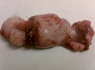
The cystic mass and left ovary following resection
Figure 4.

Haematoxylin and eosin stain of the ovary (20x magnification)
Histology of the left ovarian mass confirmed the mass to be a benign ovarian cystic teratoma with no evidence of malignancy. The patient has been followed up for a period of 18 months with no signs of recurrence.
Discussion
A cystic teratoma is a benign cystic lesion containing tissue from developmentally mature skin with hair follicles and sweat glands. Cystic teratomas can also contain pockets of sebum, blood, fat, bone, nails, teeth and cartilage.2 They constitute 10–15% of ovarian tumours and tend to occur in young women in the reproductive years. They are almost always benign as they normally contain mature tissue; less than 1% of the tumours are malignant. The rare malignant teratoma usually develops into a squamous cell carcinoma in adults whereas in babies and children it usually develops into an endodermal sinus tumour.
The most common complication of a cystic teratoma is torsion, occurring in 16% of cases.2 Other presentations include pain, discomfort and pressure symptoms. Due to the presence of a thick wall, the frequency of rupture is rare and was quoted as low as 0.7% in one series.3 Acute rupture with acute peritonitis is usually associated with pregnancy and labour.4 Intra-abdominal spillage can also result in severe chemical peritonitis and the intense inflammatory reaction can lead to fistula formation.4
In contrast to patients with acute cystic rupture and resultant peritonitis, patients with chronic involvement may present with progressive distension, anorexia, nausea, vomiting or diarrhoea.4 If rupture occurs into a hollow viscus, the situation is generally not suspected until hair, teeth or sebaceous material are passed through the orifice. Previous cases of bowel perforation due to cystic teratomas presented with rectal bleeding, hairy stools and acute abdominal pain.3
Our patient presented to the colorectal clinic with a history of diarrhoea, weight loss and rectal bleeding. Following the initial investigations, the lesion was identified correctly as originating from the left ovary. To our knowledge, the only other case report of a benign cystic teratoma presenting as a rectal mass is in a seven-year-old girl who was managed with an abdominoperineal resection.5 The management of a benign cystic teratoma depends on its symptoms and only follow-up ultrasonography is appropriate in asymptomatic patients. If associated with adnexal torsion, pain or rupture leading to chemical peritonitis, surgical treatment may become necessary.3 Our patient was treated with an oophorectomy, thereby avoiding all the morbidity associated with bowel resection and also the consequences of a permanent or a temporary stoma. The initial laparoscopy was converted to an open procedure as the nature of the disease was unsure.
Conclusions
This case report highlights the importance of preoperative investigations including colonoscopy and radiological investigations (CT and MRI) with a dedicated pelvic radiologist and pathologist. The involvement of a gynaecologist and colorectal surgeon at the time of surgery will lead to accurate diagnosis and the most appropriate treatment.
Although rare, erosion of an ovarian dermoid into the rectum should be considered in young women who have an atypical presentation and are found to have a lesion in the rectum with biopsies indicating benign pathology.
References
- 1.Williams KM, Bain CJ, Kelly MD. Laparoscopic resection of a torted ovarian dermoid cyst. World J Emerg Surg 2007; 2: 12. [DOI] [PMC free article] [PubMed] [Google Scholar]
- 2.Pantoja E, Noy MA, Axtmayer RW et al. Ovarian dermoids and their complications. Comprehensive historical review. Obstet Gynecol Surv 1975; 30: 1–20. [DOI] [PubMed] [Google Scholar]
- 3.Stern JL, Buscema J, Rosenshein NB, Woodruff JD. Spontaneous rupture of benign cystic teratomas. Obstet Gynecol 1981; 57: 363–366. [PubMed] [Google Scholar]
- 4.Peterson WF, Prevost EC, Edmunds FT et al. Benign cystic teratomas of the ovary: a clinico-statistical study of 1,007 cases with a review of the literature. Am J Obstet Gynaecol 1955; 70: 368–382. [DOI] [PubMed] [Google Scholar]
- 5.Bacon HE, Eisenberg SW. Ovarian dermoid perforating the rectum in a child: excision by abdominoperineal proctosigmoidectomy. Ann Surg 1951; 133: 408–412. [DOI] [PMC free article] [PubMed] [Google Scholar]