

Abstract
This case report describes the delayed diagnosis of inflammatory breast cancer following initial presentation with a subclavian/axillary deep vein thrombus. The relationship of thrombosis and cancer is discussed and the typical presentation of inflammatory breast cancer described. Understanding the relationship between thromboembolism and cancer is crucial to support the early diagnosis of breast cancer, which can present insidiously. The literature is reviewed, highlighting the improving prognosis of this rare condition and the current preferred treatment modalities.
Keywords: Venous thrombosis, Lymphedema, Inflammatory breast cancer
Case history
A 57-year-old woman presented to her general practitioner with several weeks of increasing right arm swelling associated with right chest and arm pain. Her co-morbidities included obesity (body mass index: 35kg/m2), type 2 diabetes, hypertension, hypercholesterolaemia and a hiatus hernia. She had stopped hormone replacement therapy five years previously.
Ultrasonography showed a right subclavian and axillary deep vein thrombosis considered spontaneous with no sign of underlying disease. Chest x-ray was normal. The patient was warfarinised and follow-up Doppler ultrasonography was arranged for three months’ time. This showed the thrombosis was resolving but the signal suggested extrinsic compression of the subclavian and axillary veins by enlarged axillary lymph nodes. Further imaging was recommended and computed tomography (CT) of the neck, chest and abdomen was requested.
Meanwhile, the patient was reviewed in primary care. She was noted to have a diffuse swelling through her right breast without a discrete lump. Her right arm still remained swollen despite the resolution of her thrombosis, suggesting ongoing lymphoedema. She was referred to our diagnostic breast clinic. The CT showed right supraclavicular and axillary lymphadenopathy. There was prominent glandular tissue in the right breast and the right latissimus dorsi muscle appeared diffusely enlarged. Liver metastases were also noted.
At the diagnostic breast clinic the patient reported painful swelling in her right breast over the previous four weeks. The skin overlying her right breast had become blotchy over the last two weeks and folliculitis has developed over her right forearm. On examination, hard fixed nodes were found in both her right supraclavicular fossa and right axilla. Her entire right breast was discoloured with irregular blotches (Fig 1). It was oedematous with peau d’orange appearance. No discrete mass was found. The right arm was extremely swollen; the patient mentioned it felt heavy and uncomfortable. The left side was entirely normal on examination. No back swelling was noted but the patient clearly recalled a prominent swelling on her back becoming apparent around this time.
Figure 1.
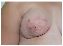
Inflammatory breast cancer at time of diagnosis
Mammography showed global oedema with gross skin thickening. No focal abnormality was demonstrated in the breast itself and appearances were reported as consistent with diffuse inflammation. Ultrasonography of the right breast showed global oedema, again with no focal abnormality. Ultrasonography of the axilla was difficult to perform as the patient was unable to raise her arm but there was evidence of abnormal adenopathy and the supraclavicular nodes were also enlarged. Overall, the appearances were felt to be most consistent with inflammatory breast carcinoma.
Biopsies were arranged once the international normalised ratio was at a safe level. Core biopsy confirmed a grade 3 invasive ductal carcinoma that was weakly oestrogen receptor/progesterone receptor positive and HER2 positive. Unfortunately, repeat CT showed metastatic disease in the liver. The patient was referred for systemic therapy. She is currently receiving weekly paclitaxel with three-weekly trastuzumab. CT after 10 weeks’ treatment suggested a partial response with a decrease in the size and number of metastatic hepatic deposits but stable appearances with the right axilla. Response to treatment can also be seen on the skin of the breast in a photograph taken three months after chemotherapy commenced (Fig 2).
Figure 2.
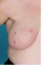
Response after 12 weeks of chemotherapy
The patient has also used the lymphoedema service for management of her arm. After an initial period of compression bandaging, she was given a set of simple exercises to maximise her range of shoulder movement. She also began wearing a compression sleeve. She has very good symptomatic relief in her arm and has had a 20% reduction in limb circumference over a 3-month period.
Discussion
Inflammatory breast cancer (IBC) represents the most virulent form of breast cancer characterised by rapid progression and relatively poor prognosis. Pleasingly, the use of neoadjuvant chemotherapy to shrink the disease prior to surgery appears to be impacting positively on patient outcomes.1 Because of its rarity, IBC is often misdiagnosed. A painful breast that appears red and swollen is the most common presentation.2 In such a scenario the differential diagnosis is extensive, and both benign and malignant diseases must be considered.3 The rapid progression of the skin changes is often the underlying clue that the symptoms represent an inflammatory malignancy.2 However, IBC may also present with symptoms distant to the breast, as in this case.
It is unusual for breast cancer to present as either unilateral arm lymphoedema or acute central venous thrombosis. A patient presenting with both these clinical problems and underlying IBC has not been described before. However, such a presentation is not unique to IBC and breast cancer recurrence, not infrequently, also presents this way. Raising awareness of the myriad of ways that breast cancer can present is obviously important as a failure to recognise the association can lead to misdiagnosis or a significant delay in diagnosis and treatment.
The pathophysiology of breast cancer presenting as lymphoedema is likely to be due to the obstruction of axillary lymphatic channels by malignant cells. In our case the body habitus of the patient may have made clinical examination of the axilla more difficult and axillary lymphadenopathy unappreciable. Careful imaging is therefore even more important when upper limb thrombus is identified to assess for possible axillary disease.
Venous thromboembolism (VTE) and cancer have a two-way clinical association: VTE may be the presenting feature of an occult cancer and patients with clinically overt cancer may develop VTE as a complication at any stage of their disease. Patients who have VTE in the absence of other risk factors have a 10% likelihood of having an underlying cancer.4
Conclusions
It therefore remains imperative for clinicians to be vigilant and consider underlying breast cancer as the possible cause of ‘idiopathic’ VTE or unilateral arm lymphoedema. Such vigilance may have led to a more timely diagnosis and earlier treatment in the patient described. Although earlier diagnosis may not have impacted on her long-term outcome, the morbidity of her lymphoedema is likely to have been reduced.
References
- 1.Dushkin H, Cristofanilli M. Inflammatory breast cancer. J Natl Compr Canc Netw 2011; 9: 233–240. [DOI] [PubMed] [Google Scholar]
- 2.Robertson FM, Bondy M, Yang W et al. . Inflammatory breast cancer: the disease, the biology, the treatment. CA Cancer J Clin 2010; 60: 351–375. [DOI] [PubMed] [Google Scholar]
- 3.Froman J, Landercasper J, Ellis R et al. . Red breast as a presenting complaint at a breast center: an institutional review. Surgery 2011; 149: 813–819. [DOI] [PubMed] [Google Scholar]
- 4.Agnelli G. Venous thromboembolism and cancer: a two-way clinical association. Thromb Haemost 1997; 78: 117–120. [PubMed] [Google Scholar]