

Abstract
Introduction
Faecal concretions or faecalomas (’stone of faeces’) are symptomatic of many well-recognised colorectal conditions. Faecalomas are usually located in the colon or rectum and associated with disorders of colorectal transit. We describe an unusual case of ’faecaloma in ano’ secondary to a chronic fissure in ano. A 67-year-old woman with a 10-year history of chronic constipation and intermittent anal discomfort presented with a painless perianal lump of similar duration.
Methods
The patient’s case notes were reviewed and a literature search was carried out.
Results
Examination under anaesthesia, sigmoidoscopy and anoscopy did not reveal any mucosal abnormality or an internal opening or any connection with the perianal lump. The mass was enucleated after incising the skin, leaving behind a cavity completely separate from the anal canal and sphincters. Histology revealed inspissated faecal material with evidence of calcification.
Conclusions
Chronic fissures may be complicated by sepsis and, rarely, a ’fissure fistula’ may develop. We suspect that this was the underlying pathogenesis of this ’faecaloma in ano’. The term faecaloma in ano befits the clinical picture.
Keywords: Colorectal surgery, Fissure in ano, Anorectal malformation
Case history
A 67-year-old woman with a 10-year history of chronic constipation and intermittent anal discomfort presented with a painless perianal lump of similar duration. She was otherwise well and denied a history of perianal discharge. On digital examination, there was a large posterior perianal lump (4cm x 3cm). At examination under anaesthesia, sigmoidoscopy and anoscopy did not reveal any mucosal abnormality or an internal opening or any connection with the perianal lump. There was no apparent increased anal tone and no acute fissure but the lower anal canal was scarred posteriorly.
The patient underwent excision of the hard but mobile lump (Fig 1). The lump appeared to contain an ‘old clot’, causing pressure necrosis of the overlying skin and some degree of extrinsic anal obstruction. The mass was enucleated after incising the skin, leaving behind a cavity with a glistening white lining completely separate from the anal canal and sphincters. Histology revealed inspissated faecal material with evidence of calcification. At the two-month follow-up visit the raw area had completely healed, digital examination was normal and her constipation improving with stool softeners.
Figure 1.
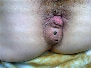
Faecaloma in ano
Discussion
Common anorectal conditions include haematomas, haemorrhoids, anal fissures and anal fistulas. This case is unusual as the patient had harboured this lump for almost a decade thinking it was associated with her tendency to constipation. Indeed, she only presented to her family physician when the overlying skin started to break down. The physician described it as a ‘large perianal external haemorrhoid’.
Faecalomas are hard, laminated masses that sometimes contain calcification and are usually located in the colon or rectum. They are associated with disorders of colorectal transit.1
Conclusions
Chronic fissures may be complicated by sepsis and, rarely, a ‘fissure fistula’ may develop that is essentially submucosal.2 We suspect that this was the underlying pathogenesis of this ‘faecaloma in ano’ despite the paucity of symptoms of local sepsis. In our case, deposition of faecal material occurred in the ‘fissure fistula’ over a long period of time and the fissure subsequently burnt itself out. The term faecaloma in ano befits the clinical picture.
References
- 1.Abella ME, Fernández AT. Large fecalomas. Dis Colon Rectum 1967; 10: 401–404. [DOI] [PubMed] [Google Scholar]
- 2.Parks AG. Pathogenesis and treatment of fistula-in-ano. Br Med J 1961; 1: 460–469. [DOI] [PMC free article] [PubMed] [Google Scholar]