



Abstract
Pharmacomechanical thrombolysis is being used increasingly for the treatment of deep vein thrombosis (DVT) and aims to reduce the severity of post-thrombotic syndrome. We report the case of a 60-year-old woman with extensive lower limb DVT that was treated using pharmacomechanical thrombolysis leading to complete recovery of her deep venous system. The prompt use of pharmacomechanical thrombolysis for the acute management of extensive DVT should be considered when treating patients with extensive DVT in order to facilitate return of normal function.
Keywords: Deep vein thrombosis, Pharmacomechanical, Thrombolysis, Thrombosis
Pharmacomechanical thrombolysis is being used increasingly for the treatment of deep vein thrombosis (DVT). It provides a more aggressive treatment of DVT than catheter directed thrombolysis and improves thrombolysis time and recanalisation rate.1 The Trellis®-8 system (Bacchus Vascular, Santa Clara, CA, US) isolates the thrombus between two balloons and breaks down the clot by rotating a guidewire in the isolated thrombosed segment in combination with thrombolytic agent.2 Previous studies have provided early clinical results demonstrating the efficacy of the Trellis®-8 system.2–4 This report demonstrates the successful deployment of the Trellis®-8 system for the treatment of acute extensive DVT and complete restoration of valvular function.
Case history
A 60-year-old woman was admitted with an acutely painful and swollen left lower limb. There was no associated chest pain or shortness of breath at presentation. She had several risk factors for DVT: recent long haul travel by aeroplane and train in addition to a recent subtotal colectomy and ileostomy formation for an exacerbation of Crohn’s disease from which she had made a good recovery. There was no underlying malignancy, nor was she taking any regular medication.
At presentation the left leg was markedly swollen and comparatively cooler with impalpable left popliteal and posterior tibial pulses. However, ankle brachial pressure indices were normal (>1.0) with triphasic signals.
Initial investigation with duplex ultrasonography established DVT and complete occlusion extending from the left posterior tibial vein to the common iliac vein. Computed tomography venography was carried out, confirming acute DVT of the left lower limb vessels (Fig 1). No abnormality was seen in the pelvic viscera.
Figure 1.
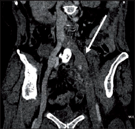
Computed tomography venography curved coronal reformat image showing left iliac system deep vein thrombosis (arrow)
An intravenous heparin infusion was commenced immediately for anticoagulation and the patient was given full length thromboembolic deterrent stockings. Four days after presentation, she attended the interventional radiology department for pharmacothrombolysis. Initially, a retrievable inferior vena cava (IVC) filter (OptEase™; Cordis, Bridgewater, NJ, US) was placed via the right common femoral vein. This was followed by ultrasonography guided insertion of a 4Fr thrombolysis catheter into the posterior tibial vein to treat the below-knee DVT. Via a 10Fr sheath inserted under ultrasonography guidance into the thrombosed popliteal vein, the Trellis®-8 procedure was performed from the IVC to the popliteal vein (Fig 2). Subsequent clearance of the clot revealed a severe stenosis at the left common iliac vein origin (May–Thurner syndrome), across which a bare metal self-expanding stent (Luminexx™; Bard, Covington, GA, US) was placed with a satisfactory result.
Figure 2.
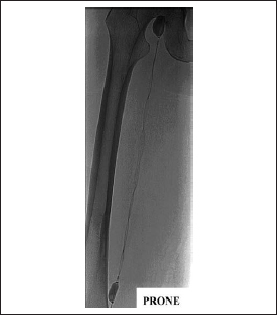
Trellis®-8 device in situ from left common iliac vein to popliteal vein
Due to some residual clots below the knee in the region not amenable to Trellis®-8, thrombolysis was continued for 16 hours via the posterior tibial vein catheter from the ankle and the popliteal vein. Post-thrombolysis venography demonstrated excellent thrombus resolution. The deep venous system was free of thrombus from the IVC to the calf except for a minor residual clot in a small deep venous branch vessel of the posterior tibial vein (Fig 3). There was good flow throughout the deep venous system and thrombolysis was stopped. The IVC filter was removed two days later. All intervention including filter removal was completed within four days.
Figure 3.

Venography after Trellis®-8 and 16-hour thrombolysis showing thrombus resolution and patent common iliac vein stent
Prior to discharge, warfarin was commenced for six months. This was felt to be an appropriate duration given the multiple risk factors for DVT.
At the follow-up appointments at three and six months, the patient’s lower limb venous system duplex scan revealed no evidence of DVT or scarring throughout the left deep venous system. Furthermore, all deep vein valves were competent. The patient is now symptom free, off anticoagulation and has fully returned to her usual quality of life with no evidence of post-thrombotic syndrome.
Discussion
Prompt treatment of extensive DVT using the Trellis®-8 system in combination with catheter directed thrombolysis and venous stenting can be used successfully to treat extensive lower limb DVT. More importantly, it can achieve complete return of normal valve function. Our patient had treatment and complete thrombus resolution within five days of the onset of symptoms. This led to a vastly improved outcome. Previous Trellis®-8 cases in our department have not had such complete resolution due to longer symptom duration. Consequently, we believe early treatment provides better results.
Conclusions
Results from large scale trials (eg ATTRACT)5 are awaited but this case supports the need for consideration of more aggressive DVT management.
References
- 1.Wittens C, Strijkers R. Thrombolytic Management of DVT. In: Davies AH, ed. Vascular and Endovascular Surgery Highlights 2010–11. Abingdon: Health Press; 2011; pp41–45. [Google Scholar]
- 2.O’Sullivan GJ, Lohan DG, Gough N et al. Pharmacomechanical thrombectomy of acute deep vein thrombosis with the Trellis-8 isolated thrombolysis catheter. J Vasc Interv Radiol 2007; 18: 715–724. [DOI] [PubMed] [Google Scholar]
- 3.Arko FR, Davis CM, Murphy EH et al. Aggressive percutaneous mechanical thrombectomy of deep venous thrombosis: early clinical results. Arch Surg 2007; 142: 513–518. [DOI] [PubMed] [Google Scholar]
- 4.Rao AS, Konig G, Leers SA et al. Pharmacomechanical thrombectomy for iliofemoral deep vein thrombosis: an alternative in patients with contraindications to thrombolysis. J Vasc Surg 2009; 50: 1,092–1,098. [DOI] [PubMed] [Google Scholar]
- 5.The ATTRACT Study. Washington University School of Medicine. http://www.attract.wustl.edu/ (cited January 2012).