



Abstract
Background
Few data exist on the genetic variants underlying the risk of steroid-sensitive nephrotic syndrome (SSNS) in children. The objectives of this study were to evaluate HLA-DQA1 and APOL1 variants as risk factors for SSNS in African-American children and use classical HLA types and amino acid inference to refine the HLA-DQA1 association.
Study Design
Case-control study
Setting & Participants
African-American children with SSNS or steroid-resistant nephrotic syndrome (SRNS) were enrolled from Duke University and centers participating in the Midwest Pediatric Nephrology Consortium.
Factor
Genetic variants in HLA-DQA1 (C34Y [rs1129740]; F41S [rs1071630]) and APOL1 high risk alleles
Outcomes
SSNS and SRNS
Measurements
Direct sequencing for the HLA-DQA1 and APOL1 variants in 115 African-American children (65 with SSNS and 50 with SRNS). Imputation of classical HLA alleles and amino acids was done in 363 South Asian children.
Results
The two HLA-DQA1 variants were significantly associated with SSNS in African-American children (C34Y: p=5.7 × 10⁻¹¹, OR=3.53, 95% CI=2.33–5.42; F41S: p=1.2 × 10⁻¹³, OR=4.08, 95% CI=2.70–6.28), but not with SRNS (C34Y: p=0.6; F41S: p=0.2). APOL1 high risk variants were not associated with SSNS (p=0.5) but showed significant associations with SRNS (p=1.04 × 10⁻⁷, OR=4.17, 95% CI=2.23–7.64). HLA-DQA1*0201, HLA-DQB1*0201, and HLA-DRB1*0701 were the classical HLA-alleles with the most significant associations with SSNS risk. The most significantly associated amino acid positions were HLA-DQα1 56 and 76 (both p=2.8 × 10⁻⁷). Conditional analysis revealed that these variants most likely account for the observed association.
Limitations
Modest sample size and limited statistical power to detect small-to-moderate effect sizes. Children studied may not be representative of all African-American children in United States.
Conclusions
HLA-DQA1 is a risk locus for SSNS, but not SRNS, in African-American children, consistent with its role in SSNS risk in children of European, Asian and African ancestries. There is little evidence of a significant role for the APOL1 high-risk alleles in childhood SSNS in African-American children. Refinement of the HLA-DQA1 association identified the critical classical HLA types and amino acids of the HLA-DQα1 molecule.
Keywords: nephrotic syndrome; genetics; childhood; African American; corticosteroids; steroid sensitivity; SSNS, SRNS, risk loci; HLA-DQA1; APOL1; renal disease; nonmodifiable risk factor; ethnic disparities; pediatric kidney disease
Nephrotic syndrome is an important cause of kidney disease in the pediatric population1. Most cases of nephrotic syndrome in children are responsive to corticosteroid therapy and are therefore referred to as steroid-sensitive nephrotic syndrome (SSNS) while a small proportion (<20%) are steroid resistant and referred to as steroid-resistant nephrotic syndrome (SRNS)2. The pattern of response to corticosteroids is the single most important predictor of outcome; the majority of children with SRNS will progress to end stage kidney disease (ESKD)2.
The pathogenesis of SSNS is not completely known; however, there are clinical and epidemiologic data to suggest that the disease may be due to dysregulation of the immune system, leading to effects on the podocyte and other components of the glomerular filtration barrier3. Epidemiological studies have established that there is significant ethnic disparity in the prevalence and clinical course of SSNS4–6. The incidence is higher in Asian children than other ethnicities; African-American (AA) and Hispanic children tend to have a more protracted course4–6. However, it is unclear if these observations are due to differences in genetic risk factors, environmental factors or gene-environment interactions. A recent study used an extreme phenotype, exome array association approach to identify genetic risk factors for SSNS7. Starting from a discovery sample of South East Asian children, the study identified four exome-wide significant variants in or around HLA-DQA1 and HLA-DQB17. Two of these variants (HLA-DQA1 C34Y and F41S [a substitution of cysteine by tyrosine at amino acid 34 and of phenylalanine by serine at amino acid 41, respectively]) were replicated in children of European ancestry, establishing a robust genetic association for SSNS7. The role of this risk locus in other populations is unknown.
In the present study, we aimed to further replicate and refine the SSNS HLA-DQA1 locus. We test the association between the HLA-DQA1 locus and SSNS in African-American children and confirm the original association. To fine map the associated loci and identify potentially functional variants, we conduct imputation of classical HLA alleles and amino acids using a population-appropriate reference and test their association with SSNS. Moreover, we undertake a set of conditional analyses and refinement of amino acid associations, and deduce their impact on three-dimensional structure of HLA-DQA1.
APOL1 variants are associated with a variety of chronic kidney diseases in populations of African ancestry8–18. Hence, we examine whether high risk APOL1 variants are associated with SSNS in populations of African ancestry.
Methods
Study Participants
For the genetic association studies of HLA-DQA1 and APOL1, we enrolled 65 African-American children with SSNS and 50 African-American children with SRNS, making a total of 115 African-American patients.
Replication Study in African-American Children With SSNS
The sample of 65 African-American children with SSNS was enrolled as part of an ongoing study of nephrotic syndrome at Duke University and the Midwest Pediatric Nephrology Consortium (MWPNC). The children were enrolled from major tertiary medical centers. The inclusion criteria were African-American ethnicity by report, age at onset of disease of 2 to 10 years, diagnosis of nephrotic syndrome (defined as proteinuria >40 mg/m2/h, hypoalbuminemia, and edema) and complete remission following 8 to 12 weeks of corticosteroid treatment. Children with secondary nephrotic syndrome were excluded. IRB approval was obtained from all participating centers. The parents and children provided informed consent and assent, respectively. Data collection included demographic information (age, gender, race and ethnicity), family history of nephrotic syndrome or other kidney disease, age at onset of nephrotic syndrome, therapies and clinical outcome. DNA was extracted from blood or saliva samples collected on enrolment.
Genotyping for HLA-DQA1 C34Y (corresponding to reference single-nucleotide polymorphism identification number [rs]1129740) and F41S (rs1071630) was done by direct sequencing (see primers in Table S1). For controls, we used data from the NHLBI GO Exome Sequencing Project (ESP) African-American sample (n=2303) accessed through the Exome Variant Server (http://evs.gs.washington.edu/EVS/).19 While the samples in the ESP were ascertained for various phenotypes, the population frequency of SSNS (1 in 16,000) means that the chances of misclassification are negligible as it is unlikely that there is more than one case in the control dataset. Association tests with SSNS were done under an additive genetic model.
In view of the role of APOL1 variants in a range of kidney disorders in populations of African ancestry8–18, in this sample of 115 African-American children we tested the hypothesis that the known APOL1 risk variants are associated with SSNS or SRNS. The samples were genotyped for the APOL1 G1 and G2 alleles by direct sequencing. Control data was obtained from published figures based on 5,543 African-Americans from the BioMe biobank of the Institute of Personalized Medicine, Icahn School of Medicine at Mount Sinai, New York20. This is one of the largest samples of African-Americans that were genotyped for the APOL1 G1 and G2 high risk alleles. High risk for APOL1 alleles is defined by having the genotype G1 or G2 in the recessive state (i.e. G1/G1, G1/G2 or G2/G2). Since G1 and G2 are derived from two genomic positions (G1 is rs73885319 [or rs60910145 with which it is usually in complete linkage disequilibrium]); G2 is a 6-bp [base pair] indel [insertion-deletion], rs71785313), they are better described as diplotypes. However, we have retained their description as “alleles” to be consistent with the literature. Association tests were conducted to test the association between APOL1 high risk alleles (G1/G1, G1/G2, G2/G2) and SSNS, as well as between the APOL1 high risk alleles and SRNS.
Evaluating the Role of HLA-DQA1 in SRNS
To test the hypothesis that HLA-DQA1 variants are also associated with SRNS, we identified 50 African-American children who had a diagnosis of SRNS. They were enrolled at Duke University and participating centers in the Midwest Pediatric Nephrology Consortium (MWPNC). Ascertainment, enrolment and sample collection were done the same way as with the children with SSNS. The DNA samples were sequenced for the two polymorphisms as for the samples with SSNS and association tests were done using the same methods.
Imputation and Association Tests With Classical HLA Alleles and Amino Acids
Imputation of classical four-digit HLA alleles and amino acids was done using exome array data generated on 214 South Asian children with SSNS and 149 controls as previously described7. We focus on four-digit (rather than two-digit) HLA alleles because four-digit HLA alleles correspond to specific HLA molecules whereas two-digit HLA alleles represent allele groups or groups of similar HLA molecules. Therefore, the higher specificity and resolution of 4-digit alleles facilitates further analyses in terms of amino acid residues and functional domains of the protein. Imputation was done with SNP2HLA21 using the Pan Asian reference panel,22 which was developed for South East Asian and South Asian populations. A total number of 115 four-digit HLA alleles, 76 two-digit HLA-alleles and 896 amino acid positions were successfully imputed. Imputed HLA alleles and amino acid positions were filtered for frequency ≥ 1% and r2 ≥ 0.9 before analysis. Logistic regression was used to test association between the imputed dosages of the variants and SSNS. For testing association at amino acid positions with three or more possible states, we used a multi-df omnibus test constructed by testing a model with all amino acid alleles of a given position, fitting individual effects of each allele and evaluating significance by testing the deviance of the alternative model compared to the null model. Therefore, the omnibus test of association tests all residues at the amino acid position simultaneously instead of the usual “one-vs-others” tests. Conditional analyses were conducted by including the most significant variant in the regression model and testing for residual association in the other variants.
Evaluation of Significant Amino Acids on Three-Dimensional Structure of HLA-DQA1
The entry with identifier 4OZI (S2 protein complex consisting of HLA-DQ-α1 chain, HLA-DQ-β1 chain, T-cell receptor S2 α chain, T-cell receptor S2 α chain and deaminated α1-gliadin peptide) was retrieved from the Research Collaboratory for Structural Bioinformatics (RCSB) Protein Data Bank and used as the reference three-dimensional structure of HLA-DQα1. The structure was visualized and manipulated using UCSF Chimera 23–24.
Results
Clinical Characteristics of Study Population
We identified 115 African-American patients with childhood-onset nephrotic syndrome: 65 with SSNS and 50 with SRNS. Children with SSNS were younger at presentation compared with children with SRNS (SSNS: mean age, of 5.4 ± 3.6 [standard deviation] years; SRNS: mean age, 10.4 ± 5.0 years). There was male preponderance in both groups (SSNS: 68.3%; SRNS: 56.5%). Kidney biopsy was performed in 20 of 65 patients with SSNS. The majority (12 of 20) had minimal change disease (MCD) while the remaining 8 children with SSNS had focal segmental glomerulosclerosis (FSGS; n=3), IgM nephropathy (n=1), membranoproliferative glomerulonephritis (MPGN; n=1), C1Q nephropathy (n=1) or focal glomerular scarring (n=1). We have information about kidney biopsy for 37 of 50 of children with SRNS. The majority (32 of 37 [85%]) of the SRNS patients had FSGS while the histological diagnosis in the others was MCD in three children and membranous nephropathy in two children.
HLA-DQA1 Associations With SSNS in African-American Children
The two HLA-DQA1 variants are significantly associated with SSNS in African-American children (rs1129740 [C34Y]: p=5.7 × 10⁻¹¹; rs1071630 [F41S]: p=1.2 × 10⁻¹³) (Table 1). Carriers of the risk variant of C34Y had 3.5 times the risk of SSNS (OR, 3.53; 95% CI, 2.33–5.42) while carriers of the risk variant for F41S had approximately four-fold the risk of SSNS (OR, 4.08; 95% CI, 2.70–6.28) compared to non-carriers.
Table 1.
Association of HLA-DQA1 risk variants with SSNS compared with SRNS in African-American children
| rs1129740 (A) | rs1071630 (G) | |
|---|---|---|
| SSNS | ||
| Frequency risk allele in cases (n=65) | 0.721 | 0.720 |
| Frequency risk allele in controls (n=2203) | 0.424 | 0.388 |
| Association p value | 5.7 ×10⁻¹¹ | 1.2 ×10⁻¹³ |
| OR (95% CI) | 3.53 (2.33–5.42) | 4.08 (2.70–6.28) |
| SRNS | ||
| Frequency risk allele in cases (n=50) | 0.452 | 0.452 |
| Frequency risk allele in controls (n=2203) | 0.424 | 0.388 |
| Association p value | 0.6 | 0.2 |
| OR (95% CI) | 1.12 (0.71–1.77) | 1.30 (0.82–2.05) |
OR = Odds ratio; CI = confidence interval; rs, reference single-nucleotide polymorphism identification number; SRNS, steroid-resistant nephrotic syndrome; SSNS, steroid-sensitive nephrotic syndrome; A, adenine; G, guanine
HLA-DQA1 Associations With SRNS in African-American Children
Neither of the two HLA-DQA1 variants showed significant association with SRNS in African-American children (rs1129740 [C34Y]: p=0.6; rs1071630 [F41S]: p=0.233) (Table 1).
Association of APOL1 High-Risk Alleles With SSNS and SRNS in African-American Children
To determine the role of APOL1 locus as a risk factor for SSNS, we tested for association between the APOL1 high risk genotype (G1/G1, G1/G2, G2/G2) with SSNS under a recessive model. There was no significant association with SSNS (frequency 16.9% versus 13.8% in controls) (Table 2). Conversely, there was a significant association between the APOL1 high-risk alleles and SRNS (frequency 40% versus 13.8% in controls; p = 1.0 × 10⁻⁷) with a four-fold difference in risk. This indicates that APOL1 is a risk locus for SRNS but not for SSNS. The distribution of APOL1 high risk alleles is found in Table S2.
Table 2.
Frequency of APOL1 risk alleles in SSNS and SRNS in African-American children
| High risk-APOL1 genotype: G1/G1 or G1/G2 or G2/G2 | Low-risk APOL1 genotype: G1 or G2 or G0/G0 | Fisher exact p | OR (95% CI) | |
|---|---|---|---|---|
| SSNS | 11/65 (16.9%) | 54/65 (83.1%) | 0.5 | 1.27 (0.60–2.8) |
| SRNS | 20/50 (40.0%) | 30/50 (60.0%) | 1.04 × 10⁻⁷ | 4.17 (2.23–7.64) |
| Population controls* | 751/5453 (13.8%) | 4702/5453 (86.2%) | – | – |
Derived from Zhang et al19 and based on 5,543 African-Americans from the BioMe biobank of the Institute of Personalized Medicine, Icahn School of Medicine at Mount Sinai, New York.
OR = odds ratio; CI = confidence interval; rs, reference single-nucleotide polymorphism identification number; SRNS, steroid-resistant nephrotic syndrome; SSNS, steroid-sensitive nephrotic syndrome
Classical HLA Allele and Amino Acid Association With SSNS in Asian Children
Association tests with classical HLA alleles in 363 South Asian children (214 with SSNS, 149 controls) showed that the most significantly associated four-digit HLA alleles were HLA-DRB1*07:01 (p=4.6 × 10⁻⁶), HLA-DQA1*02:01 (p=5.5 × 10⁻⁶), HLA-DQB1*02:01 (p=1.0 × 10⁻⁵) and HLA-DQA1*01:01 (p=3.3 × 10⁻⁴) (Table 3). A longer list of top-scoring 2-digit and 4-digit alleles are shown in tables a and b of Item S1. However, the strongest associations in a test of all variants (single-nucleotide polymorphisms [SNPs], classical HLA types, amino acid positions) were at four HLA-DQA1 amino acid positions (Table 3), including HLA-DQA1 position 56 (omnibus p=2.0 × 10⁻⁷), position 76 (omnibus p=2.0 × 10⁻⁷) and position 69 (omnibus p=8.1 × 10⁻⁷). The most significant amino acid residues at each of these positions are shown in Table 4 (a more comprehensive list is presented in Table S5). The amino acids (at positions 34 and 41) coded for by the original discovery hits (rs1129740 and rs1071630, respectively) showed significance as expected (logistic model p=2.5 × 10⁻⁶ [they have the same p value because they are completely correlated]). However, conditioning on these two amino acid positions left residual association on HLA-DQA1 positions 56 (p=8.5 × 10⁻³) and 76 (p=8.5 × 10⁻³). Conversely, conditioning on HLA-DQA1 position 56 and position 76 left no residual association in the other remaining markers, including C34Y and F41S (p = 0.9 for both amino acid positions).
Table 3.
Most significant 4-digit HLA alleles associated with SSNS in South Asian children
| Classical HLA 4-digit allele | Frequency in cases | Frequency in controls | INFO | OR | SE | P |
|---|---|---|---|---|---|---|
| HLA-DRB1*0701 | 0.3406 | 0.1915 | 0.8779 | 2.510 | 0.201 | 4.6 × 10⁻⁶ |
| HLA-DQA1*0201 | 0.3383 | 0.1907 | 0.8789 | 2.492 | 0.201 | 5.5 × 10⁻⁶ |
| HLA-DQB1*0201 | 0.3644 | 0.2120 | 0.9493 | 2.282 | 0.187 | 1.0 × 10⁻⁶ |
| HLA-DQA1*0101 | 0.1167 | 0.2173 | 0.9910 | 0.465 | 0.213 | 3.3 × 10⁻⁴ |
OR, odds ratio; SE, standard error; SSNS, steroid-sensitive nephrotic syndrome; INFO, ______
Table 4.
Most significant association tests of HLA amino acid positions with SSNS in South Asian children
| Frequency | |||||||
|---|---|---|---|---|---|---|---|
| Protein (AA position) | Omnibus P | Most significant AA residue at position | SSNS cases | Controls | OR | SE | P |
| DQA1 (56) | 2.8 × 10⁻⁷ | deletion | 0.5098 | 0.3091 | 2.376 | 0.166 | 2.0 × 10⁻⁷ |
| DQA1 (76) | 2.8 × 10⁻⁷ | L | 0.5098 | 0.3091 | 2.376 | 0.166 | 2.0 × 10⁻⁷ |
| DQA1 (−16) | 3.0 × 10⁻⁷ | M | 0.6543 | 0.4529 | 2.267 | 0.160 | 3.0 × 10⁻⁷ |
| DQA1 (69) | 8.1 × 10⁻⁷ | L | 0.6543 | 0.4529 | 2.267 | 0.160 | 3.0 × 10⁻⁷ |
| DRB1 (73) | 1.3 × 10⁻⁶ | G | 0.4245 | 0.2444 | 2.333 | 0.175 | 1.3 × 10⁻⁶ |
| DQA1 (129) | 1.3 × 10⁻⁶ | Q | 0.2285 | 0.3967 | 0.423 | 0.178 | 1.3 × 10⁻⁶ |
| DQA1 (218) | 2.5 × 10⁻⁶ | Q | 0.3273 | 0.5099 | 0.473 | 0.159 | 2.5 × 10⁻⁶ |
Note: AA, amino acid; OR = Odds ratio; SE = Standard error; SSNS, steroid-sensitive nephrotic syndrome
The location of the amino acid positions of interest on the three-dimensional model of HLA-DQA1 are shown in Figure 1. The two HLA-DQA1 positions 56 and 76 are in the extracellular topological domain of the DQ-α1 chain. An examination of the S2 protein complex’s pentameric structure (consisting of HLA-DQ-α1 chain, HLA-DQ-β1 chain, T-cell receptor S2 α chain, T-cell receptor S2 α chain and deaminated α1-gliadin peptide) (Molecular Modeling Database [MMDB] ID:119261) showed that the HLA-DQα1 residues in amino positions 56 and 76 are in the antigen-binding pocket of the protein and come into close contact with bound peptides (for example, they are within 3 Angstroms of the bound deaminated α1-gliadin peptide in the specified structure). Since binding of peptides derived from exogenous antigens is a key process in antigen presentation by class II major histocompatibility complex proteins, variation in the amino acid residues at these positions are likely to have important functional consequences as has indeed been previously reported.
Figure 1. Amino acid associations of HLA-DQA1 with SSNS (model based on Protein Data Bank [PDB] entry 4OZI).
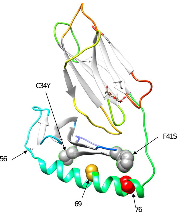
Figure showing model of HLA-DQA1 molecule and indicating the most significant amino acid positions in this study as well as the amino acid variants (C34Y, F41S) encoded by the original SNP association study.
Relationship With Reported Loci for Celiac Disease/Gluten Sensitivity
We noted the overlap of some of our findings with reported HLA associations with celiac disease and/or non-celiac gluten sensitivity25–27. Therefore, we examined this issue further in our dataset. The most commonly reported SNP and one of the strongest associations with celiac disease is HLA-DQA1 SNP rs218766825–26. This SNP, which is not in strong linkage disequilibrium with the lead SNP for SSNS (rs1129740; r2=0.05), does not show strong association with SSNS in our dataset (allelic p = 0.08). For classical HLA alleles, some of the alleles that have been associated with celiac disease27–28 also show association with SSNS in this study. Most patients with celiac disease carry DQA1*05 and DQB1*02 alleles (HLA-DQ2.5 heterodimers); these classical alleles show significant association with SSNS in this study: DQA1*05 (p = 2.6 × 10⁻³) and DQB1*02 (p=1.0 × 10⁻⁵). One of the specific 4-digit HLA-alleles implicated in celiac disease – (DQB1*0201) is one of the most strongly associated classical HLA-alleles in this study (Table 2)27. HLA-DQ2.5 (i.e. co-carriage of DQA1*05 and DQB1*02) was associated with a roughly two-fold increase in risk of SSNS (frequency in SSNS: 10.1% versus 5.5% in controls; OR, 2.1; p=2.3 × 10⁻²). On the other hand, DQB1*03:02 (the most important allele encoding DQ8 heterodimers found in most DQ2.5-negative celiac patients)29 does not show significant association with SSNS (p=0.3) (Table S6). The frequency of DQ8 did not differ significantly between SSNS cases and controls (frequency in cases 9.4% versus 11.1% in controls; p=0.4).
Discussion
The HLA-DQA1 association was the first locus for SSNS reported in a population-based study of the sporadic form of SSNS7. The discovery was made in children of South Asian ancestry and was replicated in children of European ancestry7. In the present study, we extended these findings to children of African-American ancestry and confirmed a strong association of the two HLA-DQA1 variants with SSNS. Our findings imply that HLA-DQA1-SSNS association is present in several populations and not population-specific. Our findings of the more severe manifestations of nephrotic syndrome in AA children, namely SRNS5,30, showed that the two HLA-DQA1 SSNS-associated variants are not associated with SRNS.
For individuals of African ancestry, the APOL1 G1 and G2 risk alleles are of special importance with regards to several renal phenotypes associated with this locus, including but not limited to end-stage kidney disease, HIV nephropathy and hypertension-attributable nephropathy8–16. This association has been consistently demonstrated in populations of African ancestry and with effect sizes that are large and comparable to what is often seen in HLA associations. Given the African specificity of this genetic risk factor, we tested a cohort of African-American children with SSNS in the present study for known APOL1 risk variants and demonstrated that APOL1 G1 and G2 alleles show limited evidence of association with childhood SSNS although more data is needed given the limited sample size in this study. Conversely, the APOL1 high risk alleles show significant association with SRNS, with an estimated four-fold increased risk. This is an important finding in several respects. The finding is consistent with the reported association of APOL1 with FSGS8,17 given that FSGS is the predominant histological type associated with clinical SRNS in this study. Taken together with the findings on HLA-DQA1 association, the finding provides genetic evidence to support the notion that SSNS and SRNS are distinct entities and not likely to be on the same continuum of clinical kidney disease in children. While the differences in age, histological types, and clinical course are well-established, there are little or no data on differences at the biomarker and genetic level. This study demonstrates that genetic variation also differentiates SSNS and SRNS, with SSNS displaying increased risk with HLA-DQA1 but no association with APOL1 whereas SRNS shows the opposite pattern of genetic association. If the findings from the present study is confirmed in a larger cohort of African-American children, genotyping for both HLA-DQA1 and APOL1 risk alleles may help in predicting pattern of therapy response and therefore tailoring of immunosuppression.
Recently, the German Chronic Kidney Disease (GCKD) study—a study primarily of adults—reported that the SSNS risk rs1129740 was nominally associated with MCD (OR, 1.64; p = 0.01) in a genome-wide association study (GWAS)31. Given that MCD is often the most common histological type associated with SSNS, the GCKD findings provides additional support for the role of this variant in SSNS. Interestingly, the GCKD also found significant association between this SNP and membranous nephropathy with similar effect sizes to what was found with SSNS in the discovery report and with FSGS (OR, 1.52; p = 0.002)7. In summary, these findings indicate a complex role for HLA-DQA1 rs1129740 in the risk of kidney disorders: in children it is associated with SSNS risk but not risk of SRNS; in adults it is associated with multiple kidney disorders, including membranous nephropathy and MCD.
We found that the most important classical HLA alleles associated with SSNS risk are HLA-DQA1*0201, HLA-DQB1*0201, and HLA-DRB1*0701. This is consistent with the findings of previous studies, despite some limitations with the ability to do direct comparisons, given that most previous studies of SSNS typed the HLA loci at two-digit resolution. For example, a recent study of children with nephrotic syndrome from South India showed a significant association between SSNS and both DRB1*07 and DQB1*02, as well as with the haplotype DRB1*07-DQB1*0232. Interestingly, some of the classic HLA alleles found to be significantly associated with SSNS in the present study have previously been reported to be associated with celiac disease and non-celiac gluten sensitivity25–29. This is intriguing because some difficult-to-manage cases of childhood nephrotic syndrome are known to be responsive to a gluten free or hypoallergenic diet33–38. While there are no clinical or epidemiological studies that studied the association between the two conditions, we find no evidence from the literature that this is a commonly observed association among patients. This may not be unexpected in view of the low population frequency of each of these disorders. However, the HLA findings lead us to speculate that the same HLA alleles may be associated with risk of both gluten sensitivity and SSNS and/or that gluten may be one of the potential environmental triggers for SSNS. Future studies that explicitly evaluate gluten sensitivity and SSNS would provide data on this hypothesis. Since not all children with SSNS respond to a gluten-free diet, it will be interesting to explore in future studies if classical HLA type can predict the subset of children who are likely to respond to dietary modification to induce remission of SSNS or control the frequency of relapse in frequently-relapsing/steroid-dependent SSNS. Such a biomarker would be immensely useful for risk stratification in children at initial presentation of nephrotic syndrome. Other opportunities for clinical and epidemiological research in SSNS that would be beneficial include evaluating the association between SSNS and gluten sensitivity as well as better estimates of the incidence and prevalence of SSNS in various populations beyond the mostly small and outdated studies that currently exist.40,41
In the present study, specific HLA-DQα1 amino acid positions yielded the strongest association with SSNS when all markers (SNPs, classical HLA alleles, amino acids) were considered together. In particular, HLA-DQα1 positions 76, 56, 69, and -16 (the last in the signal peptide) showed strong association with SSNS, with the first two seemingly the most critical since conditioning on them yielded no residual association in other markers including the originally reported SNPs. They are also involved in peptide binding, providing further support for a functional role for these amino acids. There have been no previous studies of specific HLA amino acids being associated with SSNS risk, so there are currently no studies with which we can compare these findings. Interestingly, amino acids at positions 76 and 56 show strong association with giant cell arteritis, an autoimmune form of vasculitis39. While the omnibus tests for the two positions were highly significant in the two studies, the direction of effect for each specific amino acid residue was inconsistent. This indicates that this may just be a coincidental finding.
The present study has some limitations. The limited sample sizes mean that the study is only powered to detect large effect sizes (ORs of 3 to 4 and higher) but is underpowered to detect small-to-moderate effect sizes (OR < 2). Future studies with larger sample sizes would detect these smaller effect sizes while providing better precision around the estimates. Also, the sample of African-American children studied may not be representative of all African-American children with SSNS or SRNS, especially since we did not have a large panel of SNPs to evaluate admixture and population stratification.
In summary, we confirmed in African-American children the HLA-DQA1 association with SSNS previously reported in children of South Asian and European ancestry7. We found that APOL1 G1 and G2 alleles show strong association with SRNS but not with SSNS in African-American children. With the aid of imputation, we refined the HLA-DQA1 locus further by identifying the classical HLA alleles and amino acid positions that are most critical in the observed association. The classical HLA alleles showed some overlap with reported alleles for celiac disease and non-celiac gluten sensitivity. The findings of the present study extend current knowledge about the genetic architecture of SSNS in children.
Supplementary Material
Supplementary Material Descriptive Text for Online Delivery
Supplementary Item S1 (PDF). Top associations of 2-digit and 4-digit classical HLA alleles with SSNS in Asian children.
Supplementary Item S2 (PDF).
Supplementary Table S1 (PDF). Primers for genotyping APOL1 and HLA-DQA1 variants.
Supplementary Table S2 (PDF). Genotype distribution of APOL1 risk alleles in a cohort of African American children with nephrotic syndrome.
Supplementary Table S3 (PDF). Top associations of amino acid variants with SSNS in Asian children.
Supplementary Table S4 (PDF). Associations with SSNS for reported classical HLA-types encoding HLA heterodimers associated with celiac disease (DQ 2.5 and DQ8).
Acknowledgments
We thank the family members of the Duke Genetics of Kidney Disease study and the personnel of the Genomic Core of the Duke Molecular Physiology Institute (DMPI), Duke University. We also acknowledge the Nephrotic Syndrome Group and participating centers in the MWPNC. Molecular graphics and analyses were performed with the UCSF Chimera package. Chimera is developed by the Resource for Biocomputing, Visualization, and Informatics at the University of California, San Francisco (supported by National Institute of General Medical Sciences P41-GM103311).
Support: Dr Gbadegesin received funding from the National Institute of Diabetes and Digestive and Kidney Diseases (NIDDK) of the National Institutes of Health (NIH) (5R01DK098135 and 5R01DK094987). This study was supported in part by the Doris Duke Charitable Foundation DDCSD award to Dr Gbadegesin. We acknowledge support from the Renal Genomics Core (Core B) of the Duke O’Brien Center for Kidney Research supported by theNIDDK/NIH (award P30DK096493). Dr Adeyemo is supported by the Intramural Research Program of the Center for Research on Genomics and Global Health (CRGGH). The CRGGH is supported by funds from the Office of the Director, NIDDK, and National Human Genome Research Institute at the NIH (Z01HG200362). The funding agencies had no role in study design, data collection and analysis, interpretation, and writing of the manuscript, or the decision to submit it for publication.
Footnotes
Publisher's Disclaimer: This is a PDF file of an unedited manuscript that has been accepted for publication. As a service to our customers we are providing this early version of the manuscript. The manuscript will undergo copyediting, typesetting, and review of the resulting proof before it is published in its final citable form. Please note that during the production process errors may be discovered which could affect the content, and all legal disclaimers that apply to the journal pertain.
Authors’ Contributions: Research idea and study design: all authors; data acquisition: all authors; data analysis/interpretation: AAd, RG; statistical analysis: AAd, RG. Each author contributed important intellectual content during manuscript drafting or revision and accepts accountability for the overall work by ensuring that questions pertaining to the accuracy or integrity of any portion of the work are appropriately investigated and resolved.
Financial Disclosure: The authors declare that they have no other relevant financial interests.
Peer Review: Received June 12, 2017. Evaluated by three external peer reviewers and a statistician, with editorial input from an Associate Editor and the Editor-in-Chief. Accepted in revised form October 5, 2017.
References
- 1.Niaudet P. Steroid-resistant idiopathic nephrotic syndrome in children. In: Avner ED, Harmon WE, Niaudet P, editors. Pediatric Nephrology. Philadelphia: Lippincott Williams & Wilkins; 2004. pp. 557–573. [Google Scholar]
- 2.Eddy AA, Symons JM. Nephrotic syndrome in childhood. Lancet. 2003;362(9384):629–639. doi: 10.1016/S0140-6736(03)14184-0. [DOI] [PubMed] [Google Scholar]
- 3.Bierzynska A, Saleem M. Recent advances in understanding and treating nephrotic syndrome. F1000Res. 2017;6:121. doi: 10.12688/f1000research.10165.1. [DOI] [PMC free article] [PubMed] [Google Scholar]
- 4.Feehally J, Kendell NP, Swift PG, Walls J. High incidence of minimal change nephrotic syndrome in Asians. Arch Dis Child. 1985;60(11):1018–1020. doi: 10.1136/adc.60.11.1018. [DOI] [PMC free article] [PubMed] [Google Scholar]
- 5.Bonilla-Felix M, Parra C, Dajani T, et al. Changing patterns in the histopathology of idiopathic nephrotic syndrome in children. Kidney Int. 1999;55(5):1885–1890. doi: 10.1046/j.1523-1755.1999.00408.x. [DOI] [PubMed] [Google Scholar]
- 6.Banh TH, Hussain-Shamsy N, Patel V, Vasilevska-Ristovska J, et al. Ethnic Differences in Incidence and Outcomes of Childhood Nephrotic Syndrome. Clin J Am Soc Nephrol. 2016;11(10):1760–1768. doi: 10.2215/CJN.00380116. [DOI] [PMC free article] [PubMed] [Google Scholar]
- 7.Gbadegesin RA, Adeyemo A, Webb NJ, et al. HLA-DQA1 and PLCG2 Are Candidate Risk Loci for Childhood-Onset Steroid-Sensitive Nephrotic Syndrome. J Am Soc Nephrol. 2015;26(7):1701–1710. doi: 10.1681/ASN.2014030247. [DOI] [PMC free article] [PubMed] [Google Scholar]
- 8.Genovese G, Friedman DJ, Ross MD, et al. Association of trypanolytic ApoL1 variants with kidney disease in African Americans. Science. 2010;329(5993):841–845. doi: 10.1126/science.1193032. [DOI] [PMC free article] [PubMed] [Google Scholar]
- 9.Papeta N, Kiryluk K, Patel A, et al. APOL1 variants increase risk for FSGS and HIVAN but not IgA nephropathy. J Am Soc Nephrol. 2011;22(11):1991–1996. doi: 10.1681/ASN.2011040434. [DOI] [PMC free article] [PubMed] [Google Scholar]
- 10.Kopp JB, Nelson GW, Sampath K, et al. APOL1 genetic variants in focal segmental glomerulosclerosis and HIV-associated nephropathy. J Am Soc Nephrol. 2011;22(11):2129–2137. doi: 10.1681/ASN.2011040388. [DOI] [PMC free article] [PubMed] [Google Scholar]
- 11.Ashley-Koch AE, Okocha EC, Garrett ME, et al. MYH9 and APOL1 are both associated with sickle cell disease nephropathy. Br J Haematol. 2011;155(3):386–394. doi: 10.1111/j.1365-2141.2011.08832.x. [DOI] [PMC free article] [PubMed] [Google Scholar]
- 12.Lipkowitz MS, Freedman BI, Langefeld CD, et al. Apolipoprotein L1 gene variants associate with hypertension-attributed nephropathy and the rate of kidney function decline in African Americans. Kidney Int. 2013;83(1):114–120. doi: 10.1038/ki.2012.263. [DOI] [PMC free article] [PubMed] [Google Scholar]
- 13.Parsa A, Kao WH, Xie D, et al. APOL1 risk variants, race, and progression of chronic kidney disease. N Engl J Med. 2013;369(23):2183–2196. doi: 10.1056/NEJMoa1310345. [DOI] [PMC free article] [PubMed] [Google Scholar]
- 14.Freedman BI, Langefeld CD, Andringa KK, et al. End-stage renal disease in African Americans with lupus nephritis is associated with APOL1. Arthritis Rheumatol. 2014;66(2):390–396. doi: 10.1002/art.38220. [DOI] [PMC free article] [PubMed] [Google Scholar]
- 15.Freedman BI, Langefeld CD, Lu L, et al. APOL1 associations with nephropathy, atherosclerosis, and all-cause mortality in African Americans with type 2 diabetes. Kidney Int. 2015;87(1):176–181. doi: 10.1038/ki.2014.255. [DOI] [PMC free article] [PubMed] [Google Scholar]
- 16.Carney EF. Chronic kidney disease: Mechanisms of APOL1-associated renal disease. Nat Rev Nephrol. 2017;13(2):62. doi: 10.1038/nrneph.2016.175. [DOI] [PubMed] [Google Scholar]
- 17.Sampson MG, Robertson CC, Martini S, et al. Integrative Genomics Identifies Novel Associations with APOL1 Risk Genotypes in Black NEPTUNE Subjects. J Am Soc Nephrol. 2016;27(3):814–823. doi: 10.1681/ASN.2014111131. [DOI] [PMC free article] [PubMed] [Google Scholar]
- 18.Ng DK, Robertson CC, Woroniecki RP, et al. APOL1-associated glomerular disease among African-American children: a collaboration of the Chronic Kidney Disease in Children (CKiD) and Nephrotic Syndrome Study Network (NEPTUNE) cohorts. Nephrol Dial Transplant. 2016 Apr 27; doi: 10.1093/ndt/gfw061. pii: gfw061. [DOI] [PMC free article] [PubMed] [Google Scholar]
- 19.Exome Variant Server, NHLBI GO Exome Sequencing Project (ESP) Seattle, WA: URL: http://evs.gs.washington.edu/EVS/. Accessed: October 2, 2017. [Google Scholar]
- 20.Zhang J, Fedick A, Wasserman S, et al. Analytical Validation of a Personalized Medicine APOL1 Genotyping Assay for Nondiabetic Chronic Kidney Disease Risk Assessment. J Mol Diagn. 2016;18(2):260–266. doi: 10.1016/j.jmoldx.2015.11.003. [DOI] [PMC free article] [PubMed] [Google Scholar]
- 21.Jia X, Han B, Onengut-Gumuscu S, et al. Imputing amino acid polymorphisms in human leukocyte antigens. PloS one. 2013;8(6):e64683. doi: 10.1371/journal.pone.0064683. [DOI] [PMC free article] [PubMed] [Google Scholar]
- 22.Pillai NE, Okada Y, Saw WY, et al. Predicting HLA alleles from high-resolution SNP data in three Southeast Asian populations. Hum Mol Genet. 2014;23(16):4443–4451. doi: 10.1093/hmg/ddu149. [DOI] [PubMed] [Google Scholar]
- 23.Pettersen EF, Goddard TD, Huang CC, et al. UCSF Chimera–a visualization system for exploratory research and analysis. J Comput Chem. 2004;25(13):1605–1612. doi: 10.1002/jcc.20084. [DOI] [PubMed] [Google Scholar]
- 24.Wang Y, Geer LY, Chappey C, Kans JA, Bryant SH. Cn3D: sequence and structure views for Entrez. Trends Biochem Sci. 2000;25(6):300–302. doi: 10.1016/s0968-0004(00)01561-9. [DOI] [PubMed] [Google Scholar]
- 25.van Heel DA, Franke L, Hunt KA, et al. A genome-wide association study for celiac disease identifies risk variants in the region harboring IL2 and IL21. Nat Genet. 2007;39(7):827–829. doi: 10.1038/ng2058. [DOI] [PMC free article] [PubMed] [Google Scholar]
- 26.Dubois PC, Trynka G, Franke L, et al. Multiple common variants for celiac disease influencing immune gene expression. Nat Genet. 2010;42(4):295–302. doi: 10.1038/ng.543. [DOI] [PMC free article] [PubMed] [Google Scholar]
- 27.Escudero-Hernández C, Peña AS, Bernardo D. Immunogenetic Pathogenesis of celiac Disease and Non-celiac Gluten Sensitivity. Curr Gastroenterol Rep. 2016;18(7):36. doi: 10.1007/s11894-016-0512-2. [DOI] [PubMed] [Google Scholar]
- 28.Megiorni F, Pizzuti A. HLA-DQA1 and HLA-DQB1 in Celiac disease predisposition: practical implications of the HLA molecular typing. J Biomed Sci. 2012;19:88. doi: 10.1186/1423-0127-19-88. [DOI] [PMC free article] [PubMed] [Google Scholar]
- 29.Lundin KE, Scott H, Hansen T, et al. Gliadin-specific, HLA-DQ(alpha 1*0501,beta 1*0201) restricted T cells isolated from the small intestinal mucosa of celiac disease patients. J Exp Med. 1993;178(1):187–196. doi: 10.1084/jem.178.1.187. [DOI] [PMC free article] [PubMed] [Google Scholar]
- 30.Kim JS, Bellew CA, Silverstein DM, Aviles DH, Boineau FG, Vehaskari VM. High incidence of initial and late steroid resistance in childhood nephrotic syndrome. Kidney Int. 2005;68(3):1275–81. doi: 10.1111/j.1523-1755.2005.00524.x. [DOI] [PubMed] [Google Scholar]
- 31.Sekula P, Li Y, Stanescu HC, Wuttke M, et al. Genetic risk variants for membranous nephropathy: extension of and association with other chronic kidney disease aetiologies. Nephrol Dial Transplant. 2017;32(2):325–332. doi: 10.1093/ndt/gfw001. [DOI] [PMC free article] [PubMed] [Google Scholar]
- 32.Ramanathan AS, Senguttuvan P, Chinniah R, et al. Association of HLA-DR/DQ alleles and haplotypes with nephrotic syndrome. Nephrology (Carlton) 2016;21(9):745–752. doi: 10.1111/nep.12669. [DOI] [PubMed] [Google Scholar]
- 33.Sandberg DH, Bernstein CW, McIntosh RM, Carr R, Strauss J. Severe steroid-responsive nephrosis associated with hypersensitivity. Lancet. 1977;1(8008):388–391. doi: 10.1016/s0140-6736(77)92603-4. [DOI] [PubMed] [Google Scholar]
- 34.Lagrue G, Laurent J, Rostoker G. Food allergy and idiopathic nephrotic syndrome. Kidney Int Suppl. 1989;27:S147–151. [PubMed] [Google Scholar]
- 35.Laurent J, Lagrue G. Dietary manipulation for idiopathic nephrotic syndrome. A new approach to therapy. Allergy. 1989;44(8):599–603. doi: 10.1111/j.1398-9995.1989.tb04207.x. [DOI] [PubMed] [Google Scholar]
- 36.Laurent J, Rostoker G, Robeva R, Bruneau C, Lagrue G. Is adult idiopathic nephrotic syndrome food allergy? Value of oligoantigenic diets. Nephron. 1987;47(1):7–11. doi: 10.1159/000184448. [DOI] [PubMed] [Google Scholar]
- 37.Lemley KV, Faul C, Schramm K, et al. The Effect of a Gluten-Free Diet in Children With Difficult-to-Manage Nephrotic Syndrome. Pediatrics. 2016;138(1):e20154528. doi: 10.1542/peds.2015-4528. [DOI] [PMC free article] [PubMed] [Google Scholar]
- 38.Uy N, Graf L, Lemley KV, Kaskel F. Effects of gluten-free, dairy-free diet on childhood nephrotic syndrome and gut microbiota. Pediatr Res. 2015;77(1–2):252–255. doi: 10.1038/pr.2014.159. [DOI] [PMC free article] [PubMed] [Google Scholar]
- 39.Carmona FD, Mackie SL, Martín JE, et al. A large-scale genetic analysis reveals a strong contribution of the HLA class II region to giant cell arteritis susceptibility. Am J Hum Genet. 2015;96(4):565–580. doi: 10.1016/j.ajhg.2015.02.009. [DOI] [PMC free article] [PubMed] [Google Scholar]
- 40.El Bakkali L, Rodrigues Pereira R, Kuik DJ, et al. Nephrotic syndrome in The Netherlands: a population-based cohort study and a review of the literature. Pediatr Nephrol. 2011;26(8):1241–6. doi: 10.1007/s00467-011-1851-8. [DOI] [PMC free article] [PubMed] [Google Scholar]
- 41.Sharples PM, Poulton J, White RH. Steroid responsive nephrotic syndrome is more common in Asians. Arch Dis Child. 1985;60(11):1014–7. doi: 10.1136/adc.60.11.1014. [DOI] [PMC free article] [PubMed] [Google Scholar]
Associated Data
This section collects any data citations, data availability statements, or supplementary materials included in this article.
Supplementary Materials
Supplementary Material Descriptive Text for Online Delivery
Supplementary Item S1 (PDF). Top associations of 2-digit and 4-digit classical HLA alleles with SSNS in Asian children.
Supplementary Item S2 (PDF).
Supplementary Table S1 (PDF). Primers for genotyping APOL1 and HLA-DQA1 variants.
Supplementary Table S2 (PDF). Genotype distribution of APOL1 risk alleles in a cohort of African American children with nephrotic syndrome.
Supplementary Table S3 (PDF). Top associations of amino acid variants with SSNS in Asian children.
Supplementary Table S4 (PDF). Associations with SSNS for reported classical HLA-types encoding HLA heterodimers associated with celiac disease (DQ 2.5 and DQ8).