Abstract
Background
The purpose of this study was to evaluate the prognostic impact of Positron Emission Tomography Response Criteria in Solid Tumors (PERCIST) and Response Evaluation Criteria in Solid Tumors (RECIST) and of pre- and post-treatment maximum Standard Uptake Value (SUVmax) in regards to survival and tumor control for patients treated for early-stage non-small cell lung cancer (ES-NSCLC) with stereotactic body radiotherapy (SBRT).
Methods
This is a retrospective review of patients with ES-NSCLC treated at our institution using SBRT. Lobar, locoregional, and distant failures were evaluated based on PERCIST/RECIST and clinical course. Univariate analysis of the Kaplan-Meier curves for overall survival (OS), progression free survival (PFS), lobar control (LC), locoregional control (LRC), and distant control (DC) was conducted using the log-rank test. Pre- and post-treatment SUVmax were evaluated using cutoffs of < 5 and ≥ 5, < 4 and ≥ 4, and < 3 and ≥ 3. ∆SUVmax was also evaluated at various cutoffs. Cox regression analysis was conducted to evaluate survival outcomes based on age, gender, pre-treatment gross tumor volume (GTV), longest tumor dimension on imaging, and Charlson Comorbidity Index (CCI).
Results
This study included 95 patients (53 female, 42 male), median age 75. Lung SBRT was delivered in 3–5 fractions to a total of 48–60 Gy, with a BEDα/β = 10Gy of at least 100 Gy. Median OS and PFS from the end of SBRT was 15.4 and 11.9 months, respectively. On univariate analysis, PERCIST/RECIST response correlated with PFS (p = 0.039), LC (p = 0.007), and LRC (p = 0.015) but not OS (p = 0.21) or DC (p = 0.94). Pre-treatment SUVmax and post-treatment SUVmax with cutoff values of < 5 and ≥ 5, < 4 and ≥ 4, and < 3 and ≥ 3 did not predict for OS, PFS, LC, LRC, or DC. ∆SUVmax did not predict for OS, PFS, LC, LRC, or DC. On multivariate analysis, pre-treatment GTV ≥ 30 cm3 was significantly associated with worse survival outcomes when accounting for other confounding variables.
Conclusions
PERCIST/RECIST response is associated with improved LC and PFS in patients treated for ES-NSCLC with SBRT. In contrast, pre- and post-treatment SUVmax is not predictive of disease control or survival.
Keywords: NSCLC, Stereotactic body radiotherapy, PERCIST/RECIST, SUVmax
Background
Lung cancer is globally the leading cause of death for men and the second leading cause of death for women, with an estimated 1.8 million new cases every year accounting for nearly 13% of all cancer diagnoses [1, 2], with non-small cell lung cancer (NSCLC) accounting for 80–85% of cases. The American Cancer Society estimates that lung cancer in the United States will cause more than 155,000 deaths in 2017 [3]. For patients with early-stage NSCLC (stages IA, IB, IIA), the 5-year survival rate is 49%, 45%, and 30%, respectively [3]. As such, novel diagnostic and interventional approaches have the potential to improve survival rates of patients with NSCLC.
Due to medical comorbidities often related to heavy cigarette use, 25% of early-stage NSCLC (ES-NSCLC) patients are inoperable at presentation [4]. As a result, stereotactic body radiation therapy (SBRT) has emerged as a viable treatment method capable of displaying high local control rates [4]. Overall survival (OS) associated with SBRT has been shown to correlate with the development of distant metastases, emphasizing the need for predictive identification of tumors that demonstrate a potential for both local and distant recurrence [5].
[18F]-fluoro-2-deoxy-glucose positron emission tomography with computed tomography (FDG PET/CT) is often used for tumor staging and post-treatment evaluation in early-stage NSCLC. Maximum standardized uptake value (SUVmax) provides a quantitative approximation of tumor glucose metabolism [5]. Although SUVmax has been consistently demonstrated to be predictive of overall survival for surgically treated NSCLC patients [6], existing research is less consistent on the prognostic value of both pre- and post-treatment SUVmax with regard to OS for patients receiving SBRT for NSCLC. Several studies have demonstrated an association between pre-treatment SUVmax and OS [7–9], while others have not shown a similar correlation [10–12]. Similarly, post-treatment FDG PET/CT is often used to evaluate tumor response, but interpretation of these findings can be difficult due to FDG uptake at the tumor site caused by radiation-induced pneumonitis, inflammation, and fibrosis [13, 14]. In addition, SUVmax has been demonstrated to persist [10] or even increase [15] at the conclusion of SBRT, even without evidence of local, regional, or distant failure, possibly due to radiation-induced pneumonitis and fibrosis. As such, the FDG uptake in these situations does not provide clear evidence of metabolic tumor activity. Based on this uncertainty in the literature, the current retrospective study examines the prognostic impact of pre- and post-SBRT SUVmax, as well as PERCIST/RECIST to assess for potential correlation to clinical disease control.
Methods
This single institution retrospective review utilized a large cohort of patients receiving relatively consistent FDG PET/CT assessments in conjunction with SBRT for early stage non-small lung cancer (ES-NSCLC). The study population consisted of all patients treated for T1-2aN0M0 NSCLC with SBRT. Tumor stage was determined according to the American Joint Committee on Cancer, 7th edition [16]. The cohort also included patients presenting with pathology suspicious of cancer on biopsy accompanied by clinical history and imaging that was consistent with ES-NSCLC. For all included patients, SBRT was the preferred modality after consensus recommendation provided by a multidisciplinary team of oncologists and cardiothoracic surgeons. Tumor size, tumor histology, smoking status, and smoking pack-years were obtained for each patient. Patients were excluded from the study if they were previously treated with thoracic radiotherapy, presented with simultaneous lung cancers, or had inconclusive, non-suspicious pathology on biopsy. The Institutional Review Board at East Carolina University approved this retrospective review (UMCIRB-15-000410).
SBRT was delivered using the CyberKnife® Robotic Radiosurgery System. Biologically Equivalent Dose (BED) was calculated for each patient assuming an α/β ratio of 10 Gy. All patients were treated in 3–5 fractions to a total of 48–60 Gy with a BEDα/β = 10Gy of at least 100 Gy (range 100–151.2). The majority of patients were treated with fiducial tracking with Synchrony® System for tumor motion tracking. Spine tracking was utilized when the tumor was located adjacent to the spine and respiratory motion was deemed negligible. A small number of patients who could not have fiducials placed were treated with Xsight® Lung Tracking System.
The SUVmax is a central component of Positron Emission Tomography Response Criteria in Solid Tumors (PERCIST) (17]. PERCIST 1.0 is based on the percentage change seen in SUV markers in pre- and post-treatment PET scans, which yields four classifications of tumor metabolism in response to therapy: complete metabolic response (CMR) – complete resolution of FDG uptake within the measurable target lesion such that it is less than mean liver activity and indistinguishable from surrounding background blood-pool levels with no new FDG-avid lesions; partial metabolic response (PMR) – reduction of a minimum of 30% in target tumor SUV; stable metabolic disease (SMD) – disease other than CMR, PMR, or progressive metabolic disease (PMD); and PMD – 30% increase in FDG SUV or beginning of new FDG-avid lesions typical of cancer [17, 18]. Similarly, Response Evaluation Criteria in Solid Tumors (RECIST) 1.1 compares pre- and post-treatment tumor dimensions to classify tumor foci changes into four categories: complete remission (CR) – absence of tumor foci for at least 4 weeks; partial response (PR) – minimum 30% decline in tumor diameter that lasts a minimum of 4 weeks; stable disease (SD) – tumor response that does not meet PR or progressive disease criteria; and progressive disease (PD) – absolute increase in total tumor diameters of at least 5 mm [17]. In the current study, PERCIST/RECIST values were obtained on subsequent FDG PET/CT scans at follow up appointments. Best-measured radiographic assessment was determined for each patient based on the follow-up FDG PET/CT scan that demonstrated the most robust tumor response to therapy regardless of previous or subsequent scans. PERCIST was used whenever possible, while RECIST was used when PERCIST could not be obtained.
Lobar control (LC), locoregional control (LRC), and distant failures (DF) were evaluated based in part on PERCIST/RECIST and confirmed by clinical or pathologic evidence of progression as the patients were followed in the clinic over time. This study defines LC as the absence of recurrence of tumor within the treated lobe, LRC as the absence of recurrence of tumor within the treated lobe or lymph node basins, and DF as the recurrence of disease outside of the treated lung or in the contralateral lung. LC was repeatedly assessed by subsequent scans in order to assess for true control of disease.
LC, LRC, overall-, progression free-, distant progression free-, and distant metastasis-free survival were estimated by the Kaplan-Meier method. Progression free survival is defined as an absence of clinical evidence of lobar failure, locoregional failure, or death. The log-rank test was used to conduct univariate comparison of survival curves to determine whether SUVmax and PERCIST/RECIST criteria influenced outcomes. Pre- and post-treatment SUVmax were evaluated using cutoffs of < 5 and ≥ 5, < 4 and ≥ 4, and < 3 and ≥ 3. PFS and OS were calculated from the final SBRT treatment day. ∆SUVmax, as defined by the change from pre-treatment to post-treatment SUVmax, was evaluated at various cutoffs. BED was also analyzed to determine whether BED cutoffs of 100 Gy versus > 100 Gy, or of < 110 Gy versus > 110 Gy, were predictive of PERCIST/RECIST criteria.
Univariate logistic regression analyses were conducted to determine whether age, gender, pre-treatment gross tumor volume (GTV), longest tumor dimension on imaging, or Charlson Comorbidity Index (CCI) were predictive of OS, PFS, LC, LRC, or DC. Each factor was assessed at various cutoffs, including the median value of the factor. Cox regression analysis was then performed on each dichotomous variable that demonstrated statistical significance (p < 0.05). All statistical calculations were performed using the MedCalc Statistical Software version 15.6.1 [19].
Results
The current study identified 95 patients with ES-NSCLC who underwent SBRT between April 27, 2009 and April 8, 2015. Patient demographics and tumor characteristics are shown in Table 1. Treatment characteristics are shown in Table 2, while treatment responses are shown in Table 3. Of the total 95 patients, 86 patients had a pre-SBRT PET/CT with a reported pre-treatment SUVmax. Sixty-one patients had a reported pre-treatment SUVmax ≥ 5. Eighty-four patients had post-treatment imaging that allowed for RECIST to be evaluated, while 71 patients had a reported post-treatment FDG PET/CT where SUV and PERCIST could be evaluated.
Table 1.
Patient & tumor characteristics
| Gender | |
| Female | 53 (66%) |
| Male | 42 (44%) |
| Age at Treatment Outset | |
| Median (years) | 75 |
| Range (years) | 51–92 |
| Smoking History | |
| Former | 59 (62%) |
| Current | 29 (31%) |
| Never | 7 (7%) |
| Pack-Years Smoking | |
| Median (pack-years) | 50 |
| Range (pack-years) | 0.75–210 |
| AJCC Stage | |
| IA | 67 (71%) |
| IB | 27 (28%) |
| IIA | 1 (1%) |
| Gross Tumor Volume | |
| Median (cm3) | 9.4 |
| Range (cm3) | 1.3–193.5 |
| Longest Tumor Dimension | |
| Median (cm) | 2.3 |
| Range (cm) | 1.0–5.4 |
| Charlson Comorbidity Index (non-age factored) | |
| Median | 4 |
| Range | 2–8 |
| NSCLC Histology | |
| Squamous | 29 (31%) |
| Adenocarcinoma | 35 (37%) |
| Bronchial Alveolar Cell | 2 (2%) |
| Poorly Differentiated | 4 (4%) |
| Atypical | 25 (26%) |
Table 2.
Treatment characteristics
| Fraction/Dose | |
| 48 Gy/4f | 14 (15%) |
| 50 Gy/4f | 5 (5%) |
| 50 Gy/5f | 54 (57%) |
| 54 Gy/3f | 3 (3%) |
| 60 Gy/5f | 19 (20%) |
| BED-Gy | |
| Median | 100 |
| Range | 100–151.2 |
Table 3.
Patient responses
| Pre-Treatment PET/CT (SUV) | |
| Patients | 86 (91%) |
| Median SUV | 8.4 |
| Range (SUV) | 1.5–31.9 |
| Post-Treatment PET/CT (SUV) | |
| Patients | 71 (75%) |
| Median SUV | 3.2 |
| Range (SUV) | 1.0–25.5 |
| Time to Post-Treatment PET/CT | |
| Median (months) | 3.1 |
| Range (months) | 1.6–26.3 |
| PERCIST/RECIST Response | |
| Patients | 84 (88%) |
| CMR/CR | 69 (82%) |
| PMR/PR | 14 (17%) |
| SMD/SD | 1 (1%) |
| PMD/PD | N/A |
| Recurrence | |
| Patients w/ Recurrence | 21 (22%) |
| Lobar Failure | 8 (8%) |
| Regional Failure | 3 (3%) |
| Locoregional Failure | 4 (4%) |
| Distant Failure | 6 (6%) |
| Overall Survival | |
| Median (months) | 15.3 |
| Range (months) | 0.85–70.7 |
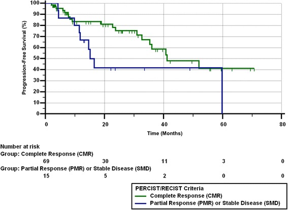
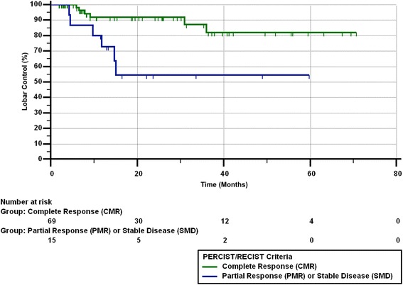
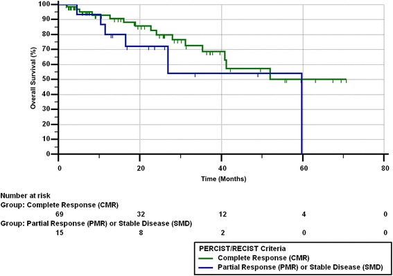
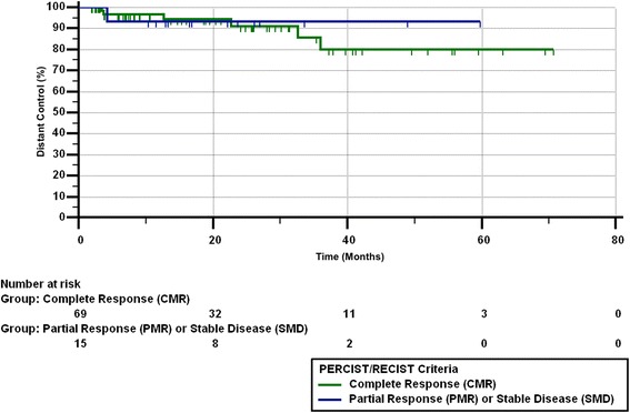
Of the 14 patients with a best response of PMR/PR on imaging, 6 had eventual lobar failure as confirmed by clinical course. Of those 6 patients, 1 had locoregional failure, 1 had lobar and distant failure, and 3 died due to lung cancer. Of the remaining 8 patients with PMR/PR, 3 are deceased from other causes and the remaining 5 are alive without disease. Median PFS from the end of SBRT was 11.9 months (range 0.59–70.7 months). Twenty-seven patients (28%) died by the end of the study. Figures 1, 2, 3 and 4 show the progression-free survival, lobar control rates, overall survival, and distant control as differentiated by PERCIST/RECIST criteria.
Fig. 1.

Progression-free survival differentiated by PERCIST/RECIST criteria
Fig. 2.

Lobar control rates differentiated by PERCIST/RECIST criteria
Fig. 3.

Overall survival differentiated by PERCIST/RECIST criteria
Fig. 4.

Distant control differentiated by PERCIST/RECIST criteria
On univariate analysis, pre-treatment SUVmax and post-treatment SUVmax with cutoff values of < 5 and ≥ 5, < 4 and ≥ 4, or < 3 and ≥ 3 did not predict for OS, PFS, LC, LRC, or DF. Complete response was predictive of PFS (p = 0.039), LC (p = 0.007), and LRC (p = 0.015), but did not correlate with OS (p = 0.21) or DF (p = 0.94). BED of 100 Gy versus > 100 Gy, or of < 110 Gy versus > 110 Gy did not predict for PERCIST/RECIST response.
Sixty four patients completed both a pre- and post-treatment SUVmax, allowing for calculation of ∆ SUVmax. Median ∆ SUVmax was − 5.1 (range = − 26.2 to + 5.9), with a negative value indicating a decrease in SUVmax from pre- to post-treatment evaluation. 53 patients (83%) demonstrated a reduction in SUVmax after treatment, while 11 patients (17%) demonstrated an increase in SUVmax. ∆ SUVmax was not predictive for OS, PFS, LC, LRC, or DF at any cutoff.
Of the 95 patients in the treatment cohort, 11 patients (11.6%) demonstrated a pre-treatment GTV ≥ 30 cm3, which was predictive for OS (p < 0.001), PFS (p = 0.004), LC (p = 0.037), and DF (p = 0.015). However, this factor was not predictive for LRC (p = 0.29). 5 patients (5.3%) demonstrated a non-age factored Charlson Comorbidity Index (CCI) ≥ 8, which was predictive for PFS (p = 0.021) and LC (p = 0.003) but not for OS (p = 0.11), DF (p = 0.63), or LRC (p = 0.71). Age, gender, and longest tumor dimension were not predictive for OS, PFS, LC, LRC, or DF at any cutoff.
Multivariate analysis demonstrated that GTV ≥ 30 cm3 was still predictive for OS (p = < 0.001), PFS (p = 0.001), LC (p = 0.020), and DF (p = 0.006) when accounting for age, gender, and non-age corrected CCI. Similarly, non-age factored CCI was still predictive for LC (p = 0.010) when accounting for age, gender, and longest tumor dimension. However, non-age factored CCI was not predictive for PFS (p = 0.17) when accounting for age, gender, and longest tumor dimension.
Discussion
SBRT has emerged as a viable treatment option for patients with medically inoperable NSCLC. Several recent studies have demonstrated clinical outcomes following SBRT as similar to those following lobectomy with systematic lymph node dissection [20, 21]. SBRT has also been associated with local control (LC) rates greater than 90% [22, 23], particularly when delivered with the target planning volume receiving a BED greater than 100 Gy [20]. In a large cohort (n = 676), long-term follow-up study of patients treated for ES-NSCLC with SBRT, Senthi et al. found that recurrence was relatively uncommon, with distant failure (DF) being the most frequent and local failure (LF) the least frequent [24]. Data from that study indicated that 12% of patients had DF, 6% of patients had locoregional failure (LRF), and 4% had LF [24]. These findings are consistent with several other patient cohorts in which DF was the most common recurrence pattern. In a patient cohort of 132, Bollineni et al. reported 13% DF and 3.6% LF [14]. In a patient cohort of 95, Horne et al. reported 15.8% DF, 10.5% LRF, and 8.4% LF [25]. In a patient cohort of 72, Burdick et al. reported 26.4% DF and 4.2% LF [5]. In a patient cohort of 57, Satoh et al. reported 30% DF, 21% LRF, and 30% LF [12].
In contrast, LF was the most common recurrence in our cohort with 8% of the patients demonstrating this finding while 6% of our patients showed DF. These disparate findings may be partially explained by our shorter follow-up period (median = 15.3 months) when compared to the follow-up timeframes of the other studies (range = 16–27 months), as there were fewer opportunities for the development or detection of distant metastasis.
Our study defines LF as the recurrence of tumor within the treated lobe and LRF as the recurrence of tumor within the treated lobe or lymph node basins. An informal sampling of similar studies illustrated several different definitions for both local and regional failure. Burdick et al. concluded that a patient had LF when two consecutive CT scans showed increasing lesion size as confirmed by PET imaging with or without positive biopsy for carcinoma [5]. Horne et al. considered a patient to have LF if recurrence was seen within the originally involved lobe or within 2 cm of the initial primary but located outside the originally involved lobe [25]. Hoopes et al. specified regional failure as occurring with lymph nodes > 1.0 cm in the expected anatomic drainage or new PET uptake in a similar location [10]. As such, this variation between studies and institutions when defining local and regional recurrence may serve to complicate any potential comparisons regarding post-treatment tumor progression.
Recent studies regarding the prognostic value of pre- and post-treatment SUVmax for patients treated for ES-NSCLC with SBRT have reached varying conclusions. Our analysis indicates that pre- and post-treatment SUVmax is not predictive of OS, PFS, LC, LRC, or DF. Hoopes et al. reached a similar conclusion, as their data showed no correlation between pre-treatment SUVmax and OS or LC [10]. Other studies have reported that post-treatment SUVmax is not predictive for OS [5, 12] or LC [12]. In contrast, several recent studies have shown pre-treatment SUVmax to be predictive for overall survival (OS) [25], progression-free survival (PFS) [25, 26], and local control (LC) [27]. Other reports demonstrate that post-treatment SUVmax is a reliable predictor for LC [14, 28] and DF [26].
The discrepancy in findings related to SUVmax and LC may be partially explained by the presence of radiation-induced pneumonitis. This inflammation and related sequelae seen on imaging may impede adequate assessment of tumor response by clouding the distinctions between residual tumor and necrosis or fibrosis [2, 14]. Therefore, acute radiation pneumonitis may limit the effectiveness of post-treatment FDG-PET/CT by inducing early increases in SUVmax and complicating the evaluation of LC following SBRT [13].
The lack of consensus regarding the prognostic value of pre- and post-treatment SUVmax may also be influenced by the relative lack of standardization in obtaining an SUV. Marom et al. reports that variation in relative SUV cutoff values, differences in elapsed time between FDG injection and imaging, fasting duration, and blood glucose correction may cause disparity in SUV findings among different institutions [29]. This procedural variation may not allow for direct comparison between studies, as patients with higher SUVmax in the current study might have been otherwise categorized with a lower SUVmax based on differences in obtaining the pre- and post-treatment SUV.
Although our study failed to demonstrate a correlation between pre- and post-treatment SUVmax and treatment outcomes, these findings are noted in the context of a relatively large patient cohort when compared to similar studies. Of the previously cited sources confirming the predictive value of pre- and post-treatment SUVmax, two studies [27, 28] had smaller patient cohorts (n = 85, n = 82, respectively), two studies [25, 26] had patient cohorts equal to our study (n = 95), and one study [14] had a larger cohort (n = 132). By comparison, the three reports [5, 10, 12] that did not find a prognostic component to SUVmax had comparatively smaller cohort sizes (n = 73, n = 58, n = 57, respectively). Therefore, we do not believe our negative findings to be a product of inadequate sample size.
The SUVmax was evaluated as both a continuous variable and a dichotomous variable using several different cutoff points (e.g. < 3.0 and ≥3.0, < 4.0 and ≥4.0, < 5.0 and ≥5.0) to assess potential correlation with specific treatment outcomes. Satoh et al. also utilized two different cutoff points in their analysis, with one demarcation at < 2.5 and ≥2.5 as well as a separate division at < 5.0 and ≥5.0 [12]. Two studies [14, 25] also utilized a cutoff of < 5.0 and ≥5.0, one study utilized a cutoff of < 4.75 and ≥4.75 [26], and another study used a cutoff of < 6.35 and ≥6.35 [10]. Despite the relative similarity in SUVmax cutoffs, the results of these studies still demonstrate varied conclusions regarding the prognostic value of SUVmax.
Given the relative inconsistency in the literature regarding the predictive value of SUVmax using FDG-PET, recent studies have examined the use of [18F]-fluorothymidine (FLT) as an additional modality for tracking tumor response and patient outcome [30, 31]. One cohort (n = 60) of patients treated for stage I-III NSCLC demonstrated rather unique findings, as superior OS was noted in patients with stable disease on FLT-PET/CT at two-week follow-up while simultaneous FDG-PET/CT was not predictive for OS. As such, the use of FLT-PET/CT might provide a more consistent tool for predicting patient outcome and treatment response when compared to FDG-PET/CT in patients treated for NSCLC.
The retrospective nature of this study presents several inherent limitations, such as the potential for inaccuracies in the medical charts and incomplete or missing information. Several patients in our original cohort were excluded from later analysis because they were lost to follow-up or were unable to obtain approval for post-treatment PET. We recognize that this may have introduced bias. However, we believe our findings to be consistent within the defined subgroups because we were not comparing between patients who did and patients who did not receive post-treatment PET scans. Our study may also have been influenced by non-uniform patient management due to variation in treatment protocols or radiographic interpretation.
Conclusion
This study demonstrates that while PERCIST and RECIST correlate with PFS, LC, and LRC, pre- and post-treatment SUVmax, as well as ∆SUVmax, were not shown to be predictive of OS, PFS, LC, LRC, or DF in patients treated for ES-NSCLC with SBRT. The results regarding pre- and post-treatment SUVmax in our study stand in contrast to the results of other recent studies that showed a significant correlation between SUVmax and those outcomes. As such, further research regarding the interpretation of pre- and post-treatment SBRT CT/PET scans is needed. Utilizing other SUVmax cutoff parameters (e.g. ≥6.0, ≥7.0) would also provide additional data points that might better illustrate the potential relationship between pre- and post-treatment SUVmax and specific treatment outcomes.
In addition, the prescribed BED did not correlate with PERCIST/RECIST, indicating the need for further research regarding whether underdosing of the tumor leads to partial response instead of complete response. Since only 1 of the 14 patients with partial response had distant failure, providing chemotherapy post-SBRT may not be indicated for these patients. However, because 6 of the 14 patients with partial metabolic response had local failure, additional ablative techniques, such as a wedge resection, may be beneficial for patients with PERCIST/RECIST partial response.
Acknowledgments
Not applicable
Funding
Funded in part by the 2015 Summer Scholars Research Program at The Brody School of Medicine and by the Vidant/ECU Research and Education Fund.
Availability of data and materials
The datasets generated in this study are not publically available due to concerns of privacy, but can be available from the corresponding author on reasonable request.
Abbreviations
- BED
Biological effective dose
- CMR/CR
Complete metabolic response
- DF
Distant failure
- FDG PET/CT
[18F]-fluoro-2-deoxy-glucose positron emission tomography/computed tomography
- FLT PET/CT
[18F]-fluorothymidine positron emission tomography/computed tomography
- LC
Lobar control
- LRC
Locoregional control
- NSCLC
Non small cell lung cancer
- OS
Overall survival
- PERCIST
Positron emission tomography response criteria in solid tumors
- PFS
Progression-free survival
- PMD/SD
Progressive metabolic disease
- PMR/PR
Partial metabolic response
- RECIST
Response evaluation criteria in solid tumors
- SBRT
Stereotactic body radiotherapy
- SMD/SD
Stable metabolic disease
- SUVmax
Standardized uptake value
Authors’ contributions
CP: Wrote and submitted the manuscript. TG: Assisted in data collection and performed the data analysis. CS: Assisted in data collection and preliminary analysis. BH: Assisted in patient identification and data collection and advised on technical details regarding SBRT delivery. TP: Assisted in patient identification and data collection. MB: Advised on the data analysis and review of the manuscript. HA: Assisted in data collection and review of the manuscript. PW: Advised on the data analysis and review of the manuscript. AJ: Principle investigator of the review, reviewed the final manuscript and oversaw data collection. All authors read and approved the final manuscript.
Ethics approval and consent to participate
This is an ECU IRB approved retrospective review under UMCIRB 15–000410. Patients have all consented for clinical information to be used in retrospective reviews in the study.
Consent for publication
No individual data is presented in this manuscript.
Competing interests
The authors declare that they have no competing interests.
Publisher’s Note
Springer Nature remains neutral with regard to jurisdictional claims in published maps and institutional affiliations.
Contributor Information
Cory Pierson, Email: piersonc16@students.ecu.edu.
Taras Grinchak, Email: grinchakt13@students.ecu.edu.
Casey Sokolovic, Email: cbsokolo@ncsu.edu.
Brandi Holland, Email: hollandb@ecu.edu.
Teresa Parent, Email: tparent@vidanthealth.com.
Mark Bowling, Email: bowlingm@ecu.edu.
Hyder Arastu, Email: arastuh@ecu.edu.
Paul Walker, Email: walkerp@ecu.edu.
Andrew Ju, Email: jua@ecu.edu.
References
- 1.Ahmedin J, Center MM, DeSantis C, Ward EM. Global patterns of cancer incidence and mortality rates and trends. Cancer Epidem Biomar. 2010;19:8. doi: 10.1158/1055-9965.EPI-10-0437. [DOI] [PubMed] [Google Scholar]
- 2.Na F, Wang J, Li C, Deng L, Xue J, Lu Y. Primary tumor standardized uptake value measured on F-18 fluorodeoxyglucose positron emission tomography is of prediction value for survival and local control in non-small-cell lung cancer receiving radiotherapy. J Thor Oncol. 2014;9:6. doi: 10.1097/JTO.0000000000000185. [DOI] [PMC free article] [PubMed] [Google Scholar]
- 3.Lung cancer (non-small cell). American Cancer Society. 2016. https://www.cancer.org/cancer/non-small-cell-lung-cancer. html. Accessed 21 January 2017.
- 4.Powell JW, Dexter E, Scalzetti EM, Bogart JA. Treatment advances for medically inoperable non-small-cell lung cancer: emphasis on prospective trials. Lancet Oncol. 2009;10:885–894. doi: 10.1016/S1470-2045(09)70103-2. [DOI] [PubMed] [Google Scholar]
- 5.Burdick MJ, Stephans KL, Reddy CA, Djemil T, Srinivas SM, Videtic GMM. Maximum standardized uptake value from staging FDG-PET/CT does not predict treatment outcome for early-stage non-small-cell lung cancer treated with stereotactic body radiotherapy. Int J Radiat Oncol. 2010;78:4. doi: 10.1016/j.ijrobp.2009.09.081. [DOI] [PubMed] [Google Scholar]
- 6.Paesmans M, Berghmans T, Dusart M, Garcia C, Hossein-Foucher C, Lafitte J, et al. Primary tumor standardized uptake value measured on fluorodeoxyglucose positron emission tomography is of prognostic value for survival in non-small cell lung cancer. J Thorac Oncol. 2010;5:612–619. doi: 10.1097/JTO.0b013e3181d0a4f5. [DOI] [PubMed] [Google Scholar]
- 7.Kohutek ZA, Wu AJ, Zhang Z, Foster A, Din SU, Yorke ED, et al. FDG-PET maximum standardized uptake value is prognostic for recurrence and survival after sterotactic body radiotherapy for non-small cell lung cancer. Lung Cancer. 2015;89:2. doi: 10.1016/j.lungcan.2015.05.019. [DOI] [PMC free article] [PubMed] [Google Scholar]
- 8.Sasaki R, Komaki R, Macapinlac H, Erasmus J, Allen P, Forster K, et al. [18F] Fluorodeoxyglucose uptake by positron emission tomography predicts outcome of non-small-cell lung caner. J Clin Oncol. 2005;23:6. doi: 10.1200/jco.2005.23.16_suppl.6. [DOI] [PubMed] [Google Scholar]
- 9.Vansteenkiste JF, Stroobants SG, Dupont PJ, De Leyn PR, Verbeken EK, Deneffe GJ, et al. Prognostic importance of the standardized uptake value on 18F-fluoro-2-deoxy-glucose-positron emission tomography scan in non-small-cell lung cancer: an analysis of 125 cases. J Clin Oncol. 1999;17:10. doi: 10.1200/JCO.1999.17.10.3201. [DOI] [PubMed] [Google Scholar]
- 10.Hoopes DJ, Tann M, Fletcher JW, Forquer JA, Lin P, Lo SS, et al. FDG-PET and stereotactic body radiotherapy for stage I non-small-cell lung cancer. Lung Cancer. 2007;56:229–234. doi: 10.1016/j.lungcan.2006.12.009. [DOI] [PubMed] [Google Scholar]
- 11.Clarke K, Taremi M, Dahele M, Freeman M, Fung S, Franks K, et al. Stereotactic body radiotherapy for non-small cell lung cancer. Radiother Oncol. 2012;104:62–66. doi: 10.1016/j.radonc.2012.04.019. [DOI] [PubMed] [Google Scholar]
- 12.Satoh Y, Nambu A, Onishi H, Sawada E, Tominga L, Kuriyama K, et al. Value of dual time point F-18 FDG-PET/CT imaging for the evaluation of prognosis and risk factors for recurrence in patients with stage I non-small cell lung cancer treated with stereotactic body radiotherapy. Eur J Radiol. 2011;81:3530–3534. doi: 10.1016/j.ejrad.2011.11.047. [DOI] [PubMed] [Google Scholar]
- 13.Vahdat S, Oermann EK, Collins SP, Yu X, Abedalthagafi M, DeBrito P, et al. CyberKnife radiosurgery for inoperable stage IA non-small cell lung cancer: 18F-fluorodeoxyglucose positron emission tomography/computed tomography serial tumor response assessment. J Hematol Oncol. 2010;3:6. doi: 10.1186/1756-8722-3-6. [DOI] [PMC free article] [PubMed] [Google Scholar]
- 14.Bollineni VR, Widder J, Pruim J, Langendijk JA, Wiegman WM. Residual 18F-FDG-PET uptake 12 weeks after stereotactic ablative radiotherapy for stage I non-small-cell lung cancer predicts local control. Int J Radiat Oncol. 2012;83:4. doi: 10.1016/j.ijrobp.2012.01.012. [DOI] [PubMed] [Google Scholar]
- 15.Ishimori T, Saga T, Nagata Y, Nakamoto Y, Higashi T, Mamede M, et al. 18F-FDG and 11C-methionine PET for evaluation of treatment response of lung cancer after stereotactic radiotherapy. Ann Nucl Med. 2004;18:669–674. doi: 10.1007/BF02985960. [DOI] [PubMed] [Google Scholar]
- 16.Edge SB, Byrd DR, Compton CC, Fritz AG, Greene FL, Trotti A, editors. AJCC cancer staging manual, 7th edition. France: Springer; 2010. [Google Scholar]
- 17.Wahl RL, Jacene H, Kasamon Y, Lodge MA. From RECIST to PERCIST: evolving considerations for PET response criteria in solid tumors. J Nucl Med. 2009;50(Suppl 1):S122-50. 10.2967/jnumed.108.057307. [DOI] [PMC free article] [PubMed]
- 18.Ding Q, Cheng X, Yang L, Zhang Q, Chen J, Li T, et al. PET/CT evaluation of response to chemotherapy in non-small cell lung cancer: PET response criteria in solid tumors versus response evaluation criteria in solid tumors. J Thorac Dis. 2014;6:677–683. doi: 10.3978/j.issn.2072-1439.2014.05.10. [DOI] [PMC free article] [PubMed] [Google Scholar]
- 19.MedCalc Statistical Software version 15.6.1. https://www.medcalc.org. Ostend, Belgium: MedCalc Software bvba; 2015.
- 20.Chang JY, Senan S, Paul MA, Mehran RJ, Louie AV, Balter P, et al. Stereotactic ablative radiotherapy versus lobectomy for operable stage I non-small-cell lung cancer: a pooled analysis of two randomized trials. Lancet Oncol. 2015;16:630–637. doi: 10.1016/S1470-2045(15)70168-3. [DOI] [PMC free article] [PubMed] [Google Scholar]
- 21.van den Berg LL, Klinkenberg TJ, Groen HJM, Widder J. Patterns of recurrence and survival after surgery or stereotactic radiotherapy for early stage NSCLC. J Thorac Oncol. 2015;10:5. doi: 10.1097/JTO.0000000000000483. [DOI] [PubMed] [Google Scholar]
- 22.Timmerman R, Paulus R, Galvin J, Michalski J, Straube W, Bradley J. Stereotactic body radiation therapy for inoperable early stage lung cancer. J Am Med Assoc. 2010;303:1070–76.2. doi: 10.1001/jama.2010.261. [DOI] [PMC free article] [PubMed] [Google Scholar]
- 23.Baumann P, Nyman J, Hoyer M, Wennberg B, Gagliardi G, Lax I, et al. Outcome in a prospective phase II trial of medically inoperable stage I non small cell lung cancer patients treated with stereotactic body radiotherapy. J Clin Oncol. 2009;27:3290–3296. doi: 10.1200/JCO.2008.21.5681. [DOI] [PubMed] [Google Scholar]
- 24.Senthi S, Lagerwaard FJ, Haasbeek CJA, Slotman BJ, Senan S. Patterns of disease recurrence after stereotactic ablative radiotherapy for early stage non-small-cell lung cancer: a retrospective analysis. Lancet Oncol. 2012;13:802–809. doi: 10.1016/S1470-2045(12)70242-5. [DOI] [PubMed] [Google Scholar]
- 25.Horne ZD, Clump DA, Vargo JA, Shah S, Beriwal S, Burton SA, et al. Pretreatment SUVmax predicts progression-free survival in early-stage non-small cell lung cancer treated with stereotactic body radiation therapy. Radiat Oncol. 2014;9:41. doi: 10.1186/1748-717X-9-41. [DOI] [PMC free article] [PubMed] [Google Scholar]
- 26.Clarke K, Taremi M, Dahele M, Freeman M, Fung S, Franks K, et al. Stereotactic body radiotherapy for non-small cell lung cancer: is FDG-PET a predictor of outcome? Radiother Oncol. 2012;104:62–66. doi: 10.1016/j.radonc.2012.04.019. [DOI] [PubMed] [Google Scholar]
- 27.Hamamoto Y, Sugawara Y, Inoue T, Kataoka M, Ochi T, Takahashi T, et al. Relationship between pretreatment FDG uptake and local control after stereotactic body radiotherapy in stage I non-small-cell lung cancer: the preliminary results. Jpn J Clin Oncol. 2011;41:4. doi: 10.1093/jjco/hyq249. [DOI] [PubMed] [Google Scholar]
- 28.Takeda A, Yokosuka N, Ohashi T, Kunieda E, Fujii H, Aoki Y, et al. The maximum standardized uptake value on FDG-PET is a strong predictor of local recurrence for localized non-small-cell lung cancer after stereotactic body radiotherapy. Radiother Oncol. 2011;101:291–297. doi: 10.1016/j.radonc.2011.08.008. [DOI] [PubMed] [Google Scholar]
- 29.Marom EM, Munden RF, Truong MT, Gladish GW, Podoloff DA, Mawlawi O, et al. Interobserver and intraobserver variability of standardized uptake value measurements in nonsmall-cell lung cancer. J Thorac Imag. 2006;21:205–212. doi: 10.1097/01.rti.0000213643.49664.4d. [DOI] [PubMed] [Google Scholar]
- 30.Everitt S, Ball D, Hicks RJ, Callahan J, Plumridge N, Trinh J, et al. Prospective study of serial imaging comparing fluorodeoxyglucose positron emission tomography (PET) and fluorothymidine PET during radical chemoradiation for non-small cell lung cancer: reduction of detectable proliferation associated with worse survival. Int J Radiat Oncol. 2017;99:947–955. doi: 10.1016/j.ijrobp.2017.07.035. [DOI] [PubMed] [Google Scholar]
- 31.Mileshkin L, Hicks RJ, Hughes BG, Mitchell PL, Charu V, Gitlitz BJ, et al. Changes in 18F-fluorodeoxyglucose and 18F-fluorodeoxythymidine positron emission tomography in patients with non-small cell lung cancer treated with erlotinib. Clin Cancer Res. 2011;17:3304–3315. doi: 10.1158/1078-0432.CCR-10-2763. [DOI] [PubMed] [Google Scholar]
Associated Data
This section collects any data citations, data availability statements, or supplementary materials included in this article.
Data Availability Statement
The datasets generated in this study are not publically available due to concerns of privacy, but can be available from the corresponding author on reasonable request.


