Abstract
Pseudohypoaldosteronism (PHA) Type 1 is characterized by mineralocorticoid resistance, manifesting as neonatal salt wasting, hypotension, hyperkalemia, hyponatremia, and metabolic acidosis in spite of elevated aldosterone levels and plasma renin activity. It is important to differentiate children with systemic PHA from renal PHA, as these children are likely to decompensate even with mild symptoms. Here, we report two neonates with PHA that presented to us with multiorgan involvement.
Keywords: Dehydration, hyperkalemia, hyponatremia, metabolic acidosis, newborn, pseudohypoaldosteronism
Introduction
Pseudohypoaldosteronism (PHA) Type 1 is a rare condition characterized by mineralocorticoid resistance. Despite elevated aldosterone levels and plasma renin activity, they manifest with neonatal salt wasting, hypotension, hyperkalemia, hyponatremia, and metabolic acidosis. These are often misdiagnosed as congenital adrenal hyperplasia (CAH).[1,2] PHA1 is subdivided into primary (genetic) and secondary (or transient) forms.[3] The primary form has two clinical entities of PHA. Type 1 is an autosomal recessive (AR) severe variant with multiple target organ involvement. Type 2 is an autosomal dominant (AD) less severe variant with isolated renal involvement. The secondary (transient) PHA1 has been explained in infants suffering from urinary tract malformations or urinary tract infections (UTIs) or both.[4,5] Here, we report two neonates with PHA1 who presented with severe dehydration, shock, and electrolyte imbalance.
Case Reports
Case 1
A 11-day-old girl baby, born second at term to third-degree consanguineous marriage, with birth weight of 2.8 kg was brought in. The baby had a history of lethargy and refusal to feed since day 5 of life. There was no other significant history.
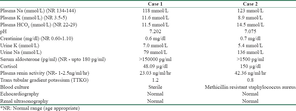
On examination, the baby portrayed features of shock and subsequently developed cardiac arrest but was revived after resuscitation. The baby was severely dehydrated (17%) with no features of virilization. Investigations revealed metabolic acidosis, hyponatremia, and hyperkalemia with electrocardiographic changes [Table 1]. Despite fludrocortisone and standard treatment including calcium gluconate, insulin infusion and K bind resin, and sodium bicarbonate infusion, hyperkalemia was refractory remaining between 9 and 11 mEq/L. The baby also developed miliaria rubra-like lesions on skin.
Table 1.
Initial workup of two babies with Pseudohypoaldosteronism

Based on the clinical and laboratory tests, a diagnosis of PHA Type 1 was made. In view of persistent hyperkalemia with hyponatremia, fludrocortisone dose was increased to 1000 mcg/day. The baby had a significant weight loss and dehydration in spite of 250 ml/kg/day of fluids. For persistent dyselectrolytemia, the baby was started on 3% NaCl by nasogastric route, initial dose of 5 mEq/kg/day and gradually increased to 20 mEq/kg/day. As her salt requirement was extremely high, sodium chloride (normal saline, 3% NaCl) was administered orally. Metabolic acidosis was corrected by sodium bicarbonate infusion. The baby showed considerable improvement in both clinical picture and weight gain, together with the ability to tolerate the salt supplementation orally. Potassium level returned to normal [Table 2] before discharge. Unfortunately, it was reported that the baby succumbed suddenly at home.
Table 2.
Discharge biochemical parameters

Case 2
An 8-day-old term boy, born second to a third-degree consanguineous marriage, with birth weight of 2.99 kg, was admitted for poor feeding and lethargy for a day. In familial history, his elder sibling had a sudden death on day 6 of his life. On examination, the baby had feeble peripheral pulses and was dehydrated with 16% weight loss. Rest of the physical examination did not yield much, and there were no signs of virilization. Initial biochemical examination revealed hyponatremia, hyperkalemia with metabolic acidosis [Table 1]. Due to features of shock, he was intubated, started on fluid bolus and inotropic support with dopamine and dobutamine. The baby was also started on insulin, calcium gluconate, and K-binding resin for hyperkalemia. With laboratory investigations [Table 1] leading to the suspicion of aldosterone insufficiency, fludrocortisone and sodium supplements were started. Renin and aldosterone were both elevated. He subsequently developed folliculitis. Vancomycin was started for methicillin-resistant Staphylococcus aureus infection.
With the lack of clinical response to treatment and persistent biochemical abnormalities, in addition to high levels of renin and aldosterone together with the history of previous sibling's unexplained sudden death, we concluded the diagnosis of PHA. DNA sequence analysis for the confirmation of the diagnosis resulted in the detection of a homozygous pathogenic mutation in the SCNN1 gene (NM_001038.5). It is a donor splice site mutation, C-1360+1G>C (r.spl), which is considered to be pathogenic because it is a mutation of the canonical splice site. No pathogenic mutations were seen in SCNN1B gene or SCNN1G genes by DNA sequence analysis of their coding region and splice sites.
With the aforementioned management process, the baby improved well and potassium levels normalized [Table 2]. The baby was brought few days later in crisis and found to have severe hyperkalemia and ventricular arrhythmias eventually suffered a cardiac arrest and could not be revived.
Genetic counseling was done for the family. In view of AR inheritance, the risk of recurrence being 25% was explained. During the next pregnancy at 13 weeks of gestation, chorionic villus sampling was done, and the fetus was not found to be homozygous for SCNN1 gene; hence, the family was reassured.
Discussion
The common causes of hyperkalemia in childhood include acute hemolysis, renal disorders, and hormonal causes. Common hormonal causes in the newborn include CAH and adrenal insufficiency and rarely PHA.[6]
Cheek and Perry in 1958 first described in an infant with severe salt-wasting syndrome, since then there have been only case reports, and rarely case series are reported in literature.[7,8]
Renal Type 1 PHA, an AD inheritance with heterogeneous mutations on the gene coding for the mineralocorticoid receptor, resulting in an absence of binding of aldosterone to the receptor. The salt loss is restricted to the kidney, and biochemically, it is characterized by salt wasting, hyponatremia, hyperkalemia, and metabolic acidosis, with elevated plasma renin and aldosterone levels. Although the treatment often involves oral salt supplementation, the clinical expression of this condition can vary widely. It typically has a mild course, followed by spontaneous remission over time.[9]
The spontaneous resolution of symptoms after infancy in patients with PHA1 can be attributed either to the change in diet from low-sodium human breast milk to higher salt diet or maturation of sodium reabsorption function of the renal tubules with increasing age.
The systemic form of Type 1 PHA, an AR disorder, is caused by mutations on the epithelial sodium channel (ENaC). In this form, symptoms are typically severe and persist into adulthood. There is a defective sodium transport in many organs containing the ENaC, including the lung, kidney, colon, sweat, and salivary glands. Consequently, sodium chloride is also elevated in sweat, saliva, and stool.[9] Miliaria rubra-like cutaneous eruptions and folliculitis present in our cases, which were typically aggravated at the time of salt-losing crisis are a characteristic feature of systemic PHA-1.
Since ENaC is expressed in the colon, sweat glands, salivary glands, and lung epithelial cells, in addition to the distal nephron, ARPHA1, which is caused by mutations in one of the three subunits of the ENaC, presents salt wasting from these various organs as well as the kidneys.[10]
Renal PHA1 results from a defect in the tubular response to aldosterone caused by inactivating mutations in the NR3C2 gene (4q31) encoding the mineralocorticoid receptor. Systemic PHA1 is caused by mutations in the genes encoding the subunits of the ENaC: alpha subunit (SCNN1A; 12p13), beta subunit (SCNN1B; 16p12.2-p12.1), or gamma subunit (SCNN1G; 16p12).[1]
The secondary (transient) form of PHA1 is due to temporary aldosterone resistance and has been described in young infants in association with UTI and/or urinary tract malformations. However, very rarely, AD PHA Type 1 in an infant with salt-wasting crisis associated with UTI and obstructive uropathy has been reported.[11] Cholelithiasis is rarely associated with PHA Type 1. Salt wasting and dehydration are assumed to be the pathogenic mechanisms leading to gallstone formation possibly beginning in fetal life.[12] Hypercalciuria leading to hypocalcemia has been previously reported to be associated with PHA.[12] Polyhydramnios is also reported in association with PHA 1.
Many studies have reported familial cases of PHA1. The symptoms of PHA1 are easily confused with the symptoms of CAH, hypoaldosteronism (HA) due to aldosterone deficiency, antenatal or infantile Bartter syndrome. At present, the main treatment for PHA1 is the intake of high doses of sodium together with ion exchange resins and dietary manipulations to reduce potassium levels.[9]
It is essential for the treating pediatrician to do genetic counseling along with molecular testing for the index baby so that the family can be benefitted during subsequent pregnancy as done in our second case. If antenatal diagnosis was not made, then genetic analysis in the immediate neonatal period with cord blood allows rapid diagnosis and management in affected baby.
Owing to the adverse effects on the respiratory tract, salivary glands, colon, and sweat glands in addition to the renal system, systemic type of PHA Type 1 is regarded to be severe. These children have increased vulnerability toward respiratory and cutaneous infection. Recurring episodes of diarrhea and failure to thrive are the other common attributes.[6]
Transtubular potassium gradient (TTKG) is a tool that is used to gauge whether the response from kidney to hyperkalemia or hypokalemia is appropriate. It is a useful indicator of aldosterone bioactivity in infants and children. It is calculated using the formula, TTKG = (urine K ÷ [urine osmolality/plasma osmolality]) ÷ plasma K. In hyperkalemia, the TTKG should be >7; lower values of which indicate HA. In hypokalemia, the TTKG should be <3; greater values indicate renal potassium wasting.
Differential diagnosis of PHA includes CAH since both involve hyperkalemia, but the latter will have hypoglycemia as well.
Probably, both the cases had unnoticed hyperkalemia and cardiac arrhythmias and thereby succumbed. Thus, these children should be monitored very closely. Moreover, educating and sensitizing the families about the need for monitoring is mandatory. Factors such as intercurrent infections and causes leading to fluid loss such as acute diarrhea/vomiting and emergencies such as surgery/trauma needing restriction/resuscitation of fluids can precipitate salt-wasting crisis, particularly hyperkalemia. Clinicians dealing with these infants should be aware of this and hence these infections and fluid loss should be treated promptly as inpatients with careful monitoring of electrolytes and fluid assessment. Wearing lifelong medical identification like MedicAlert bracelet is important for alerting health-care professionals who may be unfamiliar with the patient's rare medical condition.
Ion exchange oral resins, which act as potassium “binding agents”, are widely used in the management of hyperkalemia. The most widely used potassium binding agents are sodium polystyrene sulfonate (kayexelate) and calcium polystyrene sulfonate. However, sodium containing resin is preferred to calcium containing resin since it will correct hyponatremia and hyperkalemia simultaneously.[13] The dose of kayexelate is 1 g/kg q6 h as needed or alternatively, we could use exchange ratio of 1 mEq K+ to 1 g of resin for lower dose. Oral use is not recommended in babies <1 month old. Alternatively, rectal dose of 1 g/kg q2–6 h as needed or alternatively, use exchange ratio of 1 mEq K+ to 1 g of resin for lower dose could be administered.
Conclusion
In any neonate who presents with hyponatremia, hyperkalemia, metabolic acidosis, dehydration, shock, and failure to thrive, pediatricians should consider the possibility of PHA as it could be potentially lethal if the diagnosis is delayed. It is also important to differentiate such cases from CAH.
Financial support and sponsorship
Nil.
Conflicts of interest
There are no conflicts of interest.
References
- 1.Furgeson SB, Linas S. Mechanisms of type I and type II pseudohypoaldosteronism. J Am Soc Nephrol. 2010;21:1842–5. doi: 10.1681/ASN.2010050457. [DOI] [PubMed] [Google Scholar]
- 2.Zennaro MC, Hubert EL, Fernandes-Rosa FL. Aldosterone resistance: Structural and functional considerations and new perspectives. Mol Cell Endocrinol. 2012;350:206–15. doi: 10.1016/j.mce.2011.04.023. [DOI] [PubMed] [Google Scholar]
- 3.Hanukoglu A. Type I pseudohypoaldosteronism includes two clinically and genetically distinct entities with either renal or multiple target organ defects. J Clin Endocrinol Metab. 1991;73:936–44. doi: 10.1210/jcem-73-5-936. [DOI] [PubMed] [Google Scholar]
- 4.Schoen EJ, Bhatia S, Ray GT, Clapp W, To TT. Transient pseudohypoaldosteronism with hyponatremia-hyperkalemia in infant urinary tract infection. J Urol. 2002;167(2 Pt 1):680–2. doi: 10.1016/S0022-5347(01)69124-9. [DOI] [PubMed] [Google Scholar]
- 5.Perez-Brayfield MR, Gatti J, Smith E, Kirsch AJ. Pseudohypoaldosteronism associated with ureterocele and upper pole moiety obstruction. Urology. 2001;57:1178. doi: 10.1016/s0090-4295(01)00973-6. [DOI] [PubMed] [Google Scholar]
- 6.Rajpoot SK, Maggi C, Bhangoo A. Pseudohypoaldosteronism in a neonate presenting as life-threatening arrhythmia. Endocrinol Diabetes Metab Case Rep. 2014;2014:130077. doi: 10.1530/EDM-13-0077. [DOI] [PMC free article] [PubMed] [Google Scholar]
- 7.Cheek DB, Perry JW. A salt wasting syndrome in infancy. Arch Dis Child. 1958;33:252–6. doi: 10.1136/adc.33.169.252. [DOI] [PMC free article] [PubMed] [Google Scholar]
- 8.Kuhnle U, Nielsen MD, Tietze HU, Schroeter CH, Schlamp D, Bosson D, et al. Pseudohypoaldosteronism in eight families: Different forms of inheritance are evidence for various genetic defects. J Clin Endocrinol Metab. 1990;70:638–41. doi: 10.1210/jcem-70-3-638. [DOI] [PubMed] [Google Scholar]
- 9.Riepe FG. Clinical and molecular features of type 1 pseudohypoaldosteronism. Horm Res. 2009;72:1–9. doi: 10.1159/000224334. [DOI] [PubMed] [Google Scholar]
- 10.Kuhnle U. Pseudohypoaldosteronism: Mutation found, problem solved? Mol Cell Endocrinol. 1997;133:77–80. doi: 10.1016/s0303-7207(97)00149-4. [DOI] [PubMed] [Google Scholar]
- 11.Bowden SA, Cozzi C, Hickey SE, Thrush DL, Astbury C, Nuthakki S. Autosomal dominant pseudohypoaldosteronism type 1 in an infant with salt wasting crisis associated with urinary tract infection and obstructive uropathy. Case Rep Endocrinol 2013. 2013:524647. doi: 10.1155/2013/524647. [DOI] [PMC free article] [PubMed] [Google Scholar]
- 12.Akkurt I, Kuhnle U, Ringenberg C. Pseudohypo-aldosteronism and cholelithiasis: Coincidence or pathogenetic correlation? Eur J Pediatr. 1997;156:363–6. doi: 10.1007/s004310050615. [DOI] [PubMed] [Google Scholar]
- 13.Porter J, Kershaw M, Kirk J, Trevelyan N, Shaw NJ. The use of sodium resonium in pseudohypoaldosteronism. Arch Dis Child. 2003;88:1138–9. doi: 10.1136/adc.88.12.1138-b. [DOI] [PMC free article] [PubMed] [Google Scholar]


