

Abstract
The physis of a long bone may get ‘sandwiched’ and crushed between the metaphysis and the epiphysis if it is traumatically loaded along its long axis. Such a physeal injury may lead to complications like angular deformities and growth restrictions and hence, management of such injuries requires adequate planning and attentive execution.
Two patients with distal femoral physeal crush injury were treated using a ring fixator such that one ring had the wires passing through the epiphysis and the other through the femoral shaft. On table image intensifier controlled distraction of the crushed physis was done to bring the height of the physis similar to that of the opposite limb. Patients were followed up for more than two years clinically and radiologically. There was no clinical or radiological angular deformity of the operated limbs. MRI scans showed intact physes with no physeal bar formation in either of the two patients.
The distraction obtained by the ring fixator appears to have provided ample ‘breathing space’ to the compressed physis and that the growth potential may have been re-gained by the procedure. However, two years is a relatively short duration of follow-up and further follow-up of longer duration and in greater number of patients is needed to gauge the actual effectiveness of the technique used by us.
Keywords: Salter Harris classification, Physeal injuries, Distal femoral physis, Distraction, Ring fixator
Introduction
Management of skeletal injuries in children differs from adults. 15% of all fractures in children have physeal or epiphyseal injury components. These injuries assume significance as they can damage the growth plate and produce angular deformities or/and growth restriction.1 The impact of initial injury and clinical course unveils over the entire growth period. The first step in managing a physeal injury is classifying the injury based on clinical and radiological evidence.
The first attempt at classification of physeal injuries was done around 150 years back by Foucher. Classification by Beregnfeldt was followed by the one proposed by Aitken. But the classification by Salter and Harris,2 modified by Rang has been most widely used till date. The classification by Salter and Harris was later expanded to include variants of the physeal fractures. Peterson presented a new classification system based on an epidemiological study of 951 physeal fractures. Peterson's classification was based on the degree of damage to the physeal plate and included six types (least damage to greatest damage). This classification did not include a category similar to Salter-Harris type V. In compression injury of the physis without fracture (Salter-Harris type V), the radiographs are not suggestive of injury and pre-mature growth arrest is discovered at a later stage. Such an injury is extremely rare with only few cases reported in the literatures.3 The availability of CT and MRI scans has enabled the orthopedic surgeon to have a better understanding of physeal fracture patterns.
We are reporting two cases of physeal injury that had associated metaphyseal comminution along with a displacement of the epiphysis, which we were unable to classify into any of the available classifications and hence looked them from a different perspective.
Case report
Case 1: A 2-year-old female child presented to the emergency following a fall from height. The child had swollen lower end of right thigh and was unable to bear weight. The distal pulses were well palpable and she had no neurological deficits. Antero-posterior and lateral plain radiographs of the right femur (Fig. 1A and B) showed comminuted fracture of the distal femoral metphysis and were suggestive of a compression injury in which the distal femoral physis was sandwiched and compressed between the epiphysis and metaphysis. The affected limb was supported with a posterior plaster slab. We obtained a non-contrast CT scan of the affected limb with 2D and 3D reconstructions, which also revealed extensive comminution of the distal femoral metaphysis (Fig. 1C and D).
Fig. 1.

A: Antero-posterior radiograph of case 1 showing decreased height of physis and metaphyseal comminution. B: Lateral radiograph of case 1 showing metaphyseal comminution. C: Axial CT image of metaphysis of case 1 showing comminution. D: Sagittal CT image of case 1.
Case 2: A 4-year-old male child was brought to emergency after a roadside accident. The child had no distal neuro vascular deficits. Radiographs of the left femur and the tibia were obtained (Fig. 2) which showed a comminuted distal femoral metaphyseal fracture along with a decrease in the height of the physis. There was an undisplaced fracture of the tibia on the same side. The radiographical picture was quite similar to that of case 1 and after splinting the limb in a posterior plaster slab, CT scans were obtained. These scans confirmed the presence of significant metaphyseal comminution.
Fig. 2.

A: Antero-posterior radiograph of case 2 showing decreased height of physis and metaphyseal comminution. B: Lateral radiograph of case 2 showing comminution of metaphysis.
While the mechanism of injury was different in the two cases, they had radiographical similarity. The presence of metaphyseal comminution on both the radiographs and the CT images was a common finding. The fractures of the two cases couldn't be classified into any of the available classification systems (Salter-Harris, Ogden, Aitken and Peterson). An operative intervention aimed at decreasing the compression of the physis in both cases and stabilizing them was planned using the principle of distraction with ring fixator. The parents of the two patients were informed about the injuries and the apprehensions regarding growth disturbance. An informed consent was obtained from the parents prior to the surgical procedure.
Both the cases were operated under general anesthesia within 36 hours of admission. The whole lower limb was prepared and draped free. Under image intensifier two smooth long Kirschner's wires (K-wires) were placed into the epiphysis (Fig. 3). An Illizarov ring was attached to the wires and the wires were tensioned. Next, a similar ring was place in the femoral shaft using 3.5 mm Schanz screws for maintaining it in place. The two rings were connected using threaded connecting rods and under image intensifier controlled distraction was performed till the metaphyseal spikes fell in place and the height of distal femoral physis matched to that of the opposite non-affected limb (Fig. 4). In the second case, a unilateral external fixator was applied using 3.5 mm Schanz screws for the tibial shaft fracture. After completion of the procedure no further distraction was performed (Fig. 5). The patients were allowed assisted knee bending immediately as tolerated but weight bearing was not allowed. Patients were allowed to go home on the second postoperative day with pin track care instructions taught to the parents. Radiographs were obtained at six weeks, on which callus formation was noted at the metaphyseal areas with maintained height of the growth plate. The patients were allowed to bear weight as tolerated. Three months after surgery the fixator was removed. Patients were followed up at monthly interval for the first 6 months and then at three monthly interval for two years. Radiographs were obtained at three monthly intervals. At the latest follow-up which was 28 months after the surgery for case 1 and 24 months for case 2, both the patients had full range of motion with no discrepancy of the limb lengths. Scanograms at their latest follow-ups showed no angular deformity in either of the cases (Fig. 6). To obtain an idea about the status of the physis, non-contrast MRI of the affected physis was done, which showed no evidence of any physeal bar or bony block formation in any part of the physis (Fig. 7).
Fig. 3.
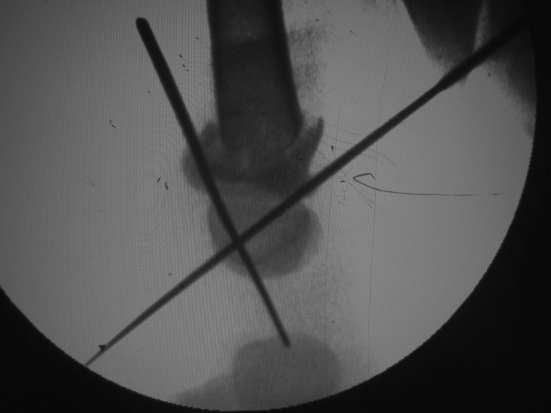
Intraoperative image intensifier picture showing the placement of Kirschner wires into the epiphysis.
Fig. 4.

A: Intraoperative image after application of distraction showing the re-alignment of metaphyseal fragments and regaining of physeal height. B: Final clinical picture after application of the fixator.
Fig. 5.

Postoperative radiograph after application of the external fixator showing well aligned metaphyseal fragments.
Fig. 6.

Scanogram of case 1 obtained after 28 months of surgery showing the distal femoral physeal and metaphyseal regions to be similar on both sides with no deformity.
Fig. 7.

A: Coronal MR image of the distal femur showing normal looking physis with no physeal bar or bony block. B: Axial MR image of the distal femoral physis region showing no physeal bar or bony block.
Discussion
Fractures of the distal femoral physis pose a challenge to the managing orthopedist as they have a notorious tendency of producing complications.4 Growth disturbance, with subsequent development of leg length discrepancy and/or angular deformities5 is the most common complication. A complication rate as high as 40% has been reported in the literature.5 The distal femoral physis, like other physis has a well-defined anatomical structure with the layer of resting cartilage (germinal layer) located towards the epiphysis and the zone of preliminary ossification towards the metaphysis.
If the Salter-Harris classification is considered, then cases with Type V injuries would have a compression of the different layers of the physis but no comminution of the metaphysis. In such an injury the physis is sandwiched between the epiphysis and metaphysis, leading to permanent damage to the germinal cells probably by decreasing the nutrition of the growth plate. Even though few investigators like Peterson et al have questioned the existence of Salter-Harris type V injuries, there are reports of such injuries.3, 5 Peterson in his classification system has described a type 1 subtype D injury in which there is metaphyseal comminution without a physeal compression and has reported the most common sites for such injuries to be distal radius, finger phalanges and metacarpals. But similar injuries in lower limb were not reported. Also, the availability of investigations like digital x-rays, CT scans with 2D and 3D reconstructions and MRI have aided in identifying injuries which could be missed on conventional radiographs. We believe that they have also brought forwards diagnostic and classification dilemmas. In the two cases presented here, comminution of the entire metaphysis was present in two orthogonal views, suggesting axial compression force transmission onto the physis. CT scans revealed the extent of comminution to be much more than that seen on the radiographs. None of the available classifications have a description of combined physeal compression and metaphyseal comminution. The two patients had an injury which could be probably considered “Salter-Harris type V equivalent” (a new term which could be proposed for such injuries). Sabharwal6 has described the use of Illizarov external fixator in the management of metadiaphyseal pediatric femur fractures. In the two cases distraction of the physis was done with ring fixators. MRI obtained after 28 months in one and 24 months in the other case was without any obvious signs of physeal bar formation, implying a good outcome. The scanograms obtained at two years follow-up showed well maintained alignment of the limb with no angular deformities. Also, the lengths of the two limbs measured clinically two years after surgery showed no limb length discrepancy. We believe that by distraction of the physis using Illizarov fixator, some “breathing space”was provided to the physis so that the nutrition of the cartilage cells was not impaired even after being subjected to crushing. The existence of unusual injuries is to be expected and needs to be searched for in patients with high velocity injuries.
There are questions which need further evaluation and answering. Whether the newer imaging modalities like CT, MRI should be included while classifying physeal injuries is one such question. Also, whether these modalities should be used for follow-ups and at what interval need to be evaluated.
There are few notable limitations. The injury pattern is rare as evidenced by only two cases reported here. The follow-up is relatively short. Also, one may argue that conservative management with casts may be an alternative way of managing these cases. Moreover, these patients need to be followed up for a longer duration to look out for any delayed growth abnormalities which may set in such cases when the child achieves the growth spurt during puberty.
Conclusion
Physeal compression injuries with metaphyseal comminution are rare among physeal injuries in children and can produce growth abnormalities. Distraction of the physis by use of Illizarov external fixator can be considered an effective way of managing such injuries.
Footnotes
Peer review under responsibility of Daping Hospital and the Research Institute of Surgery of the Third Military Medical University.
References
- 1.Basener C.J., Mehlman C.T., DiPasquale T.G. Growth disturbance after distal femoral growth plate fractures in children: a meta-analysis. J Orthop Trauma. 2009;23:663–667. doi: 10.1097/BOT.0b013e3181a4f25b. [DOI] [PubMed] [Google Scholar]
- 2.Salter R.B., Harris W.R. Injuries involving the epiphyseal plate. J Bone Jt Surg. 1963;45A:587–622. [Google Scholar]
- 3.Eid A.M., Hafez M.A. Traumatic injuries of the distal femoral physis. Retrospective study on 151 cases. Injury. 2002;33:251–255. doi: 10.1016/s0020-1383(01)00109-7. [DOI] [PubMed] [Google Scholar]
- 4.Dahl W.J., Silva S., Vanderhave K.L. Distal femoral physeal fixation: are smooth pins really safe? J Pediatr Orthop. 2014;34:134–138. doi: 10.1097/BPO.0000000000000083. [DOI] [PubMed] [Google Scholar]
- 5.Arkader A., Warner W.C., Jr., Horn B.D. Predicting the outcome of physeal fractures of the distal femur. J Pediatr Orthop. 2007;27:703–708. doi: 10.1097/BPO.0b013e3180dca0e5. [DOI] [PubMed] [Google Scholar]
- 6.Sabharwal S. Role of Ilizarov external fixator in the management of proximal/distal meta-diaphyseal pediatric femur fractures. J Orthop Trauma. 2005;19:563–569. doi: 10.1097/01.bot.0000174706.03357.26. [DOI] [PubMed] [Google Scholar]