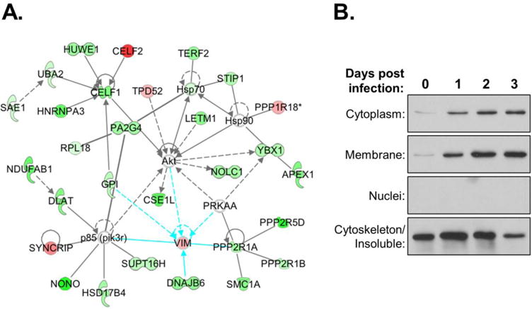
Fig. 6. VIM distribution is altered in HIV-infected cells.

(A) Protein-protein interaction network of VIM with other candidate factors in combined SWATH dataset. (B) Immunoblots of VIM in subcellular fractions of uninfected (day 0) and HIV-1 infected Jurkat cells at dpi shown. Control blots for fractionation were performed as shown in Fig. 2.
