



Abstract
Tinnitus is the perception of sound in the absence of a corresponding external acoustic stimulus, resulting in an estimated prevalence of 10% to 15% in adults. Tinnitus may be classified as pulsatile (PT) or continuous (non-PT), and may be subjective (heard only by the patient) or objective (also audible to the examiner). PT is usually related to vascular causes and is pulse synchronous (coinciding with the patient’s heartbeat). PT is much less common affecting approximately 4% of patients with tinnitus, but unlike non-PT, usually has a specific identifiable cause. We present a case of a man without previous otological disease or head trauma, with a left-ear subjective PT. MR angiography detected a left vertebro-vertebral arteriovenous fistula, which was treated by endovascular embolisation with important symptomatic relief.
Keywords: ear, nose and throat/otolaryngology; interventional radiology
Background
The main reasons to write this case report were:
Tinnitus is an important and prevalent health issue.
Most of the time, the existing treatments are scarce and with a low success rate.
Pulsatile tinnitus (PT) is rarer but usually has an identifiable cause which can be treated.
Nevertheless, tinnitus is intractable in most of the cases, sometimes we find some treatable cases with important symptomatic relief. For that, we should investigate every patient not presuming that tinnitus has no solution.
There are very few cases of vertebro-vertebral arteriovenous fistula found in the literature.
Case presentation
A 36-year-old man, bus driver, without previous relevant medical, otological or traumatic history, presented to our ear. nose and throat department with complaints of left PT, without hearing loss perception. This history had 5 years of evolution, with a slightly progressive worsening. The patient, because of his profession, had a chronic noise exposure. At physical examination, he had a normal otoscopy without tympanic pulsatility in both sides, with normal acoumetry (256 Hz), and without tinnitus variation with left carotid pressure. Cranial and neck auscultation was performed with a slight murmur at left cervical level.
Investigations
We proceeded with the audiological and immitance measurements. The pure-tone audiometry showed a symmetrical mild bilateral sensorineural hearing loss, with 15 dB of Speech Recognition Threshold (SRT) in the speech audiometry in both ears. He had stapedial reflex present in both ears with a normal (type A) tympanogram. He was submitted to an auditory brainstem evoked potentials exam which revealed a symmetrical and normal electrophysiological conduction.
Since the patient has a PT and we had a high index of suspicion that there might be some kind of abnormality, we decided to proceed to the imaging study. The cervical Doppler ultrasound showed an increased diameter and accelerated flow in the left vertebral artery (VA) at the V2 segment. The MR angiography (MRA) (figures 1 and 2) presented a left vertebro-vertebral arteriovenous fistula with ectasia of the left VA from its origin to the V3 segment (where the fistulous point was located). To this fistula, there was also contribution from the ascending cervical artery.
Figure 1.

MR angiography showing left vertebro-vertebral arteriovenous fistula (white arrow).
Figure 2.

T1-weighted, axial MR. Flow void with left VVF and VA ectasia communicating by the VVF with the VV. Note the calibre diameter difference between the VA′ and VA, and the VV′ and the VV. VA, vertebral artery; VA′, normal vertebral artery; VV, vertebral vein; VV′, normal vertebral vein; VVF, vertebro-vertebral fistula.
Treatment
The patient was proposed to neuroimaging-assisted endovascular embolisation which was consented. The procedure took 4 hours and consisted in angiography coil embolisation (figures 3 and 4). There were no occurrences to register, and the patient stayed only 24 hours resting in bed and in observation at the infirmary. He resumed to regular work after 1 month without any limitation.
Figure 3.
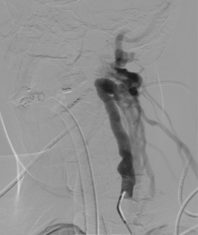
Angiography showing left vertebro-vertebral fistula, with left vertebral artery ectasia.
Figure 4.

Frontal view of the fistula. Note the circulation stop of left vertebral artery related to the turbulence of the fistula point (*).
Outcome and follow-up
More than a year has passed after the procedure, and the patient was always well, without any complaint derived from the endovascular embolisation, and, more importantly, without any sense of tinnitus or physical limitation as well as a normal head and neck auscultation without any kind of murmur.
Discussion
Tinnitus is a frequent and heterogeneous disorder, resulting in most cases from neuronal changes occurring in the central nervous system as a reaction to neural auditory deprivation.1 Nevertheless, it is essential to know the nature of the tinnitus: PT versus non-PT,2 3 and if it is objective (heard also by the physician) and subjective (only heard by the patient).3 PT is usually unilateral4 and frequently it is possible to identify an underlying cause.5 It is defined as the patient perception of an auditory sensation coinciding with heartbeat.3 Pathophysiologically, there are two possible causes5: changes in the laminar blood flow resulting in local turbulence which is transmitted to inner ear or normal blood flow that is perceived more intensely in the inner ear, as a result of increased bone conduction or higher sound conduction in the middle ear leading to loss of normal masking effect of external sounds.
PT is much less common than non-PT,1 2 6 and overall, intracranial vascular or non-vascular entities are much more common than the cervical causes of PT.2 3 6 PT has a long list of possible differential diagnosis.5–7 Usually there is an underlying vascular cause, but it is also possible due to a non-vascular cause. Vascular causes can be arterial, venous or arteriovenous.2 3 6–8 Atherosclerosis plaques, aneurysms of internal carotid artery or VT, arterial dissection and fibromuscular dysplasia are the leading causes of arterial PT.2 3 Arterial atherosclerosis stenosis is one of the most common causes of PT.2 7 Idiopathic intracranial hypertension, previously known as pseudotumour cerebri or benign intracranial hypertension, is one of the most frequent venous PT, followed by venous malformations like high jugular bulb or transverse-sigmoid sinus aneurysm.2 3 6 9 Dural arteriovenous fistula and malformations, highly vascularised skull base tumours are common arteriovenous causes for PT.2 3 Some non-vascular causes can be related to glomus tumour,2 3 5 6 8 9 histiocytosis X,8 Paget’s disease,8 9 endolymphatic sac tumour,2 high cardiac output (with an underlying anaemia)2 3 8 9 and some middle-ear conductive hearing loss entity such as middle-ear effusion and otosclerosis.5 9 There is a specific cause of non-vascular PT that may be intermittently pulsatile, but not exactly synchronised with the heartbeat, usually related with myoclonic contractions of middle-ear muscles (tensor tympani and stapedial) and palatine muscles like tensor veli palatini, levator veli palatini or salpingopharyngeus.7 9 These spasmodic contractions can range between 10 and 240/min and may be confused with the arterial pulse and mimic PT.7 In these cases, all the physical examination, audiological study and neuroimaging is completely normal.7
There are many possible approaches and guidelines to study a patient with PT. There is no consensus about one specific protocol; therefore, it is more important to choose one to fully study our patient to increase the diagnosis rate as well as to understand if our protocol needs to be improved. The first and essential step is to get a good anamnesis and understand if the patient really has a PT.5–7 9 Then we proceed to physical examination with: (1) otoscopy (Is it normal or is there any retrotympanic mass?); (2) Careful auscultation of head and neck region should be performed in a completely quiet environment (Can we hear some turbulence? It should be reinforced that some intracranial vascular pathologies are noticed at cranial auscultation.); (3) cervical palpation (Is there any PT modification after jugular and carotid compression?); (4) Can we hear the PT or is only heard by the patient? (Is the PT objective or subjective?)3 5–7 9 After physical examination, it is crucial to get to all patients a pure-tone audiometry, impedance audiometry and auditory brainstem evoked potentials (this one specially if there is a notorious interaural difference).6 7 9
After this first approach, we can narrow the possibilities. A high percentage of patients with PT will have a normal otoscopy,7 but if a violaceous retrotympanic mass is present, the three most frequent diagnosis are6 7 9: glomus tumour, high jugular bulb or an ectopic carotid artery. When we find an abnormal otoscopy, we should get a CT of the temporal bone, complemented with angiography (CTA).6 7 9 CTA demonstrates the arterial and venous anatomy of the neck and intracranial circulation and shows with detail the temporal bone, middle-ear cavity and skull base.2 5 9 If the otoscopy is normal, we must try to explore the effect of cervical palpation and auscultation. If we suspect of an arterial PT, we should get a CTA as an initial approach.9 If a venous PT is the main hypothesis, an MR complemented with the angiography should be done. If arteriovenous fistula is suspected, MRA should be the first imaging study because it is more sensitive than CTA2 and can give more detail of the brain and its vessels.3 If more detail of temporal bone is needed, we can add a CT.2 5 9 An angiography is optional for those cases with normal MRA and a high suspicion of arteriovenous fistula.7
Some protocols include, as a first step, a cervical Doppler ultrasound.7 9 Actually, it is an operator-dependent innocuous exam and can detect one of the most frequent causes of PT, carotid atherosclerosis; but we really feel that may be a redundant exam and in most of the time does not achieve the diagnosis, and even if something is abnormal, we will need another imaging study such as MRA or CTA. In our clinical case we performed a cervical Doppler ultrasound, but as we stated before, we needed to proceed to MRA to achieve the precise diagnosis. So, we think that Doppler ultrasound may be not essential, but if in particular cases if no further imaging study is available, or even, if it is much faster to obtain this exam, it may be legitimate to choose it as a first step. In our multidisciplinary team, and after some years of expertise, if we have to choose one essential imaging study we would select MRA because it is more sensitive than CTA, gives us more detailed information, and if we need more temporal bone information, we can easily add a CT. This current opinion is in line with Dietz et al,10 as they consider MRA as the first radiological study ordered in patients with PT.
Apart from our clinical case, there are very few cases of vertebro-vertebral fistulas, especially causing PT. The most common cause of this entity is a penetrating neck injury, blunt cervical trauma or some iatrogenic form of trauma such as percutaneous puncture of a carotid artery or VA for a diagnostic angiography.11 Even more rare is a vertebro-vertebral fistula created by a spontaneous arterial dissection with a resulting pseudoaneurysm.9 12 Vertebro-vertebral fistulas have been reported in association with connective tissue diseases including fibromuscular dysplasia, neurofibromatosis, Ehler-Danlos syndrome and Marfan’s syndrome.12 In our patient, we could not identify a plausible cause to this fistula. Symptoms are usually progressive and related to fistula compression of spinal cord,11 tinnitus is really unusual. To date, there are no guidelines on the best treatment modality to vertebro-vertebral fistula, but endovascular coiling seems to have advantages when compared with surgical approach.11 Periprocedural and postprocedural ischaemic or thromboembolic issues and vessel injuries are the main possible complications that may occur.11 Nevertheless, in expert and trained hands, endovascular techniques were safe without any registered iatrogenic complication, as stated by Briganti et al.11 In our clinical case, we referred to neuroradiology endovascular coil embolisation with important symptomatic relief and PT resolution. It is important to note that the physical and audiological examination was completely normal, and, only the high clinical suspicion led us to other examinations to demonstrate a very rare treatable cause of tinnitus.
Learning points.
Tinnitus is a very common complaint in ear, nose and throat practice. Only a minority will have a pulsatile tinnitus.
All pulsatile tinnitus must be investigated to rule out some treatable cause.
After a normal physical examination and audiometric investigation, the next step is the imaging study. MR angiography is a sensitive exam to study pulsatile tinnitus.
Vertebro-vertebral fistula is a very rare entity that may cause pulsatile tinnitus.
It is essential to cowork in a multidisciplinary team, especially, with an experienced neuroradiology department to achieve the diagnosis and, if it is the case, fully treat your patient.
Footnotes
Contributors: MSB is the medical doctor of the patient, drafted the initial manuscript, made revisions. JA and JR made the neuroradiological exam, performed the selection of the images, coworked in the images description and revised the manuscript. LD critically reviewed the manuscript. All authors approved the final manuscript as submitted.
Competing interests: None declared.
Patient consent: Obtained.
Provenance and peer review: Not commissioned; externally peer reviewed.
References
- 1.Langguth B, Kreuzer PM, Kleinjung T, et al. Tinnitus: causes and clinical management. Lancet Neurol 2013;12:920–30. 10.1016/S1474-4422(13)70160-1 [DOI] [PubMed] [Google Scholar]
- 2.Madani G, Connor SE. Imaging in pulsatile tinnitus. Clin Radiol 2009;64:319–28. 10.1016/j.crad.2008.08.014 [DOI] [PubMed] [Google Scholar]
- 3.Hofmann E, Behr R, Neumann-Haefelin T, et al. Pulsatile tinnitus: imaging and differential diagnosis. Dtsch Arztebl Int 2013;110:451–8. 10.3238/arztebl.2013.0451 [DOI] [PMC free article] [PubMed] [Google Scholar]
- 4.Levine RA, Nam EC, Melcher J. Somatosensory pulsatile tinnitus syndrome: somatic testing identifies a pulsatile tinnitus subtype that implicates the somatosensory system. Trends Amplif 2008;12:242–53. 10.1177/1084713808321185 [DOI] [PMC free article] [PubMed] [Google Scholar]
- 5.Mattox DE, Hudgins P. Algorithm for evaluation of pulsatile tinnitus. Acta Otolaryngol 2008;128:427–31. 10.1080/00016480701840106 [DOI] [PubMed] [Google Scholar]
- 6.Sismanis A. Pulsatile tinnitus. Otolaryngol Clin North Am 2003;36:389–402. 10.1016/S0030-6665(02)00169-X [DOI] [PubMed] [Google Scholar]
- 7.Herraiz C, Aparicio JM. [Diagnostic clues in pulsatile tinnitus (somatosounds)]. Acta Otorrinolaringol Esp 2007;58:426–33. [PubMed] [Google Scholar]
- 8.Shin EJ, Lalwani AK, Dowd CF. Role of angiography in the evaluation of patients with pulsatile tinnitus. Laryngoscope 2000;110:1916–20. 10.1097/00005537-200011000-00028 [DOI] [PubMed] [Google Scholar]
- 9.Sismanis A. Pulsatile tinnitus: contemporary assessment and management. Curr Opin Otolaryngol Head Neck Surg 2011;19:348–57. 10.1097/MOO.0b013e3283493fd8 [DOI] [PubMed] [Google Scholar]
- 10.Dietz RR, Davis WL, Harnsberger HR, et al. MR imaging and MR angiography in the evaluation of pulsatile tinnitus. AJNR Am J Neuroradiol 1994;15:879–89. [PMC free article] [PubMed] [Google Scholar]
- 11.Briganti F, Tedeschi E, Leone G, et al. Endovascular treatment of vertebro-vertebral arteriovenous fistula. A report of three cases and literature review. Neuroradiol J 2013;26:339–46. [DOI] [PMC free article] [PubMed] [Google Scholar]
- 12.Honarmand AR, Ansari SA, Alden TD, et al. Endovascular management of pediatric high-flow vertebro-vertebral fistula with reversed basilar artery flow. Interventional Neuroradiology 2013;19:215–21. 10.1177/159101991301900211 [DOI] [PMC free article] [PubMed] [Google Scholar]