Figure 1.
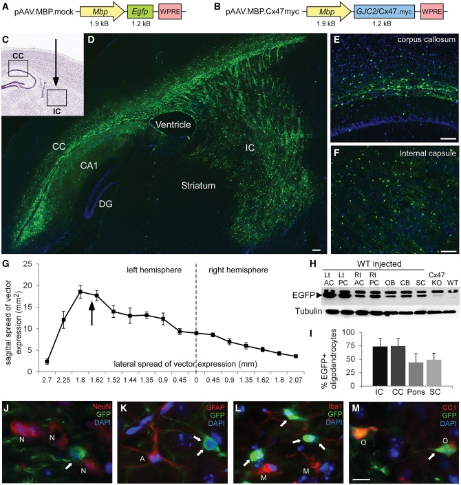
AAV vectors and EGFP expression in the CNS of AAV.MBP.EGFP injected mice. Schematic diagrams of the AAV.MBP.EGFP (mock) (A) and AAV.MBP.Cx47myc (B) vector plasmids used in this study. Injection of the mock vector in the internal capsule (IC) of wild-type (WT) postnatal Day 10 (P10) mice (indicated by arrow in the diagram in C) resulted in widespread expression in the internal capsule as well as the corpus callosum (CC). Composition of low magnification images from a sagittal section in D, as well as higher magnification images of the corpus callosum (E) and internal capsule (F) demonstrate the extend of AAV-derived reporter gene EGFP expression in oligodendrocytes (green). Cell nuclei are counterstained with DAPI (blue). (G) Quantification of expression volume in both hemispheres from sagittal brain sections taken from n = 5 injected mice. Arrow shows the level of the vector injection in the internal capsule of the left hemisphere. (H) Immunoblot analysis of EGFP expression in lysates from different CNS areas of an AAV.MBP.EGFP-injected wild-type mouse, including left (Lt) and right (Rt) anterior and posterior cerebrum (AC and PC), olfactory bulb (OB), cerebellum (CB), and spinal cord (SC) shows widespread expression with higher levels in the injected left hemisphere. A brain sample from Cx47 knockout mouse (transgenically expressing EGFP) shows lower levels of EGFP. A non-injected wild-type mouse brain is shown as negative control. A non-specific band is present (marked with asterisk) above the specific band for EGFP. Tubulin blot is shown underneath as loading control. (I) Quantification of the percentage of EGFP expressing oligodendrocytes in the corpus callosum, internal capsule, ventral pons (corticospinal tract area) and cervical spinal cord (SC) anteriolateral white matter of n = 5 mice shows high expression ratios in all areas, although lower in pons and spinal cord. (J–M) Immunostaining with cell markers as indicated confirms that AAV.MBP.EGFP expression (arrows) is not present in NeuN+ neurons (N), in GFAP+ astrocytes (A), or in Iba1+ microglia (M), but is localized only in CC1+ oligodendrocytes (O). DG = dentate gyrus.

