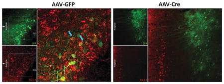
The image illustrates the elimination of Cav1.2 protein, encoded by the cacna1c gene, in the mouse prefrontal cortex (PFC) using adenoassociated viral (AAV) vector-expressing Cre recombinase (AAV-Cre). Cacna1c was focally eliminated in the PFC via bilateral stereotactic delivery of AAV-Cre (right panel) into the PFC of floxed cacna1c mice.1 AAV-expressing green fluorescent protein (AAV-GFP) was used as a control, as shown in the left panel. Double immunohistochemical analysis was used to visualize GFP (green; top left image) and Cav1.2 (red; bottom left image) protein using anti-GFP and anti-Cav1.2 (ref. 2) antibodies, respectively. The larger left image displays co-localization of GFP and Cav1.2 (blue arrows), indicating that AAV-GFP did not alter levels of Cav1.2.
The right panel shows loss of Cav1.2 protein selectively in the PFC after delivery of AAV-Cre. As AAV-Cre also expresses GFP, viral spread can be readily visualized through immunohistochemical detection of GFP (green; top right image). Co-labeling with anti-Cav1.2 antibody identifies Cav1.2 protein (red; bottom right image). The larger image in the right panel shows the absence of Cav1.2 labeling in regions expressing GFP (green), indicating focal deletion of cacna1c by AAV-Cre. c.c., corpus callosum.
For more information on this topic, please refer to the article by Lee et al., on pages 1054–1055.
References
- 1.Moosmang S, Haider N, Klugbauer N, Adelsberger H, Langwieser N, Müller J, et al. J Neurosci. 2005;25:9883–9892. doi: 10.1523/JNEUROSCI.1531-05.2005. [DOI] [PMC free article] [PubMed] [Google Scholar]
- 2.Tippens AL, Pare JF, Langwieser N, Moosmang S, Milner TA, Smith Y, et al. J Comp Neurol. 2008;506:569–583. doi: 10.1002/cne.21567. [DOI] [PubMed] [Google Scholar]


