Abstract
The purpose of this study was to determine the characteristics of the pain tolerance threshold (PTT) induced by electrical stimulation of the alveolar ridge. A total of 100 healthy volunteers studying or working at Nihon University School of Dentistry at Matsudo and patients from Nihon University School of Dentistry at Matsudo Affiliated Hospital, including 51 men (58.7 ± 17.6 years old) and 49 women (60.7 ± 17.1 years old), participated in this study. The volunteers were enrolled after obtaining written informed consent. PTT measurements were obtained using a Neurometer CPT/C® device to deliver electrical stimulation around the left greater palatine foramen at frequencies of 5 and 250 Hz. When the stimulus could no longer be tolerated, the participant released a button to automatically discontinue the stimulus. After the distribution of the PTT values was analyzed, the influence of gender, age, and Eichner index on PTT was analyzed. The Eichner index values were divided into three categories: group A (four supporting zones), group B (less than four supporting zones but with anterior tooth contact), and group C (no occlusal contact). The PTT values did not show a normal distribution. There were no significant differences in PTT between men and women. PTT was significantly associated with age (P = 0.017) at 5 Hz in men. There were no significant differences in PTT among the Eichner index groups. The characteristics of the PTT of the alveolar ridge are as follows: (1) age and PTT at 5 Hz are significantly associated with men but not with women, and (2) the Eichner index has no influence on the PTT.
Keywords: Electric stimulation, mouth mucosa, pain threshold
Introduction
The number of people requiring complete dentures is predicted to increase over the next 20 years because of aging of the population (Douglass et al., 2002; Carlsson and Omar, 2010). As the population ages, the number of denture users with advanced atrophic and thin alveolar ridge mucosa will increase. For these individuals, the nerves underlying the dentures might be subject to severe compression from occlusal force during mastication.
The standard textbook on complete dentures suggests the necessity of relief for the incisive and posterior palatine foramina in long‐term denture wearers to prevent the impingement of the nerves and vessels passing through these foramina (Swenson and Boucher, 1970). Although this suggestion was based on concerns that compression by dentures may induce functional changes to nerves and vessels located underneath the dentures, no concrete evidence of a relationship between functional changes in the nerves and compression from dentures has been reported.
We have investigated the influence of denture‐induced compression on sensory nerve responses to stimulation by measuring the current perception threshold (CPT) (Ito et al., 2012; Kimoto et al., 2013). Our previous study compared the CPT values of dentulous subjects with those of complete denture wearers and showed that complete denture wearers experience asymptomatic hypoesthesia that primarily affects the nasopalatine and greater palatine nerves and increases asymptomatic hypoesthesia in the following order: dentulous individual < partial denture wearer < complete denture wearer.
As an extension of the previous studies, we became interested in pain threshold. The pain threshold of the oral mucosa has been investigated using a number of methods. Margarida et al. (1962) measured the thermal pain threshold of the hard palate. Komiyama et al. (2008) investigated the filament‐prick pain detection threshold of the maxillary gingiva using Semmes–Weinstein monofilaments. Tanaka et al. (2004) investigated the effects of denture wearing and bite force on the pressure pain threshold of edentulous oral mucosa using an electronic pressure algometer. Isobe et al. (2013) clarified the relationship between the properties of the palatal mucosa and the pressure pain threshold of the denture‐supporting area, also using an algometer. Because these studies measured the response from intraoral nociceptors, sensory nerve fibers could not be directly stimulated and evaluated. To our knowledge, no study had investigated the oral pain tolerance threshold (PTT) induced by direct electrical stimulation of sensory nerve fibers.
Based on this background, we measured the PPT using an electrical stimulus applied to the oral mucosa via an electro‐diagnostic device that can stimulate almost every sensory nerve, including the A‐delta fibers and C fibers that respond to mechanical stimuli related to compression by dentures (Ito et al., 2012). In a previous study, we verified the reliability of an electro‐diagnostic device (Nakashima et al., 2014). Thus, we conducted the current study to evaluate the characteristics of the PTT in the oral mucosa, particularly the alveolar ridge.
Materials and Methods
Participants
This study was approved by the Human Ethics Committee of Nihon University School of Dentistry at Matsudo. The study protocol was in accordance with the Declaration of Helsinki. The participants were 100 volunteers studying or working at Nihon University School of Dentistry at Matsudo and patients at Nihon University School of Dentistry at Matsudo Affiliated Hospital, including 51 men (mean ± standard deviation [range]: 58.7 ± 17.6 [24–86] years old) and 49 women (60.7 ± 17.1 [26–86] years old). The participants were enrolled only after they gave written informed consent. Individuals were excluded if they (1) had general health problems that could affect the measurement of nerve activity (e.g., diabetes mellitus, trigeminal neuralgia, or postherpetic neuralgia); (2) experienced neuropathic complaints involving the maxillary alveolar ridge; (3) wore pacemakers; (4) showed obvious cognitive impairment; or (5) could not understand written or spoken Japanese.
Pain tolerance threshold test
Measurements of the PTT were conducted according to the protocol used in our previous report, which confirmed the reliability of the device used in this study (Nakashima et al., 2014). Each participant was seated comfortably on a dental chair in a quiet room throughout the testing. Prior to administering the actual PTT test, the operator exposed the participants to each of the two frequencies (5 and 250 Hz) used to assess the PTT, allowing them to experience the frequencies prior to the actual test and negating the startle reflex. The PTT measurements were obtained using a Neurometer CPT/C® device (Neurotron Inc., Baltimore, MD) to deliver electrical stimulation around the left greater palatine foramen at frequencies of 5 and 250 Hz (Fig. 1A). The frequencies of 5 and 250 Hz stimulate A‐delta and C fibers, respectively.
Figure 1.
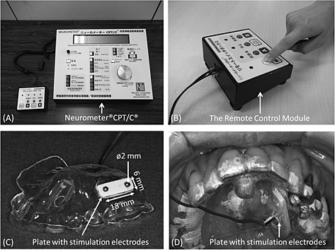
Pain tolerance threshold measuring system (A‐D). The participants press and hold the button on the remote box and release it when they can no longer tolerate the electrical stimulus (B). Thermoforming discs of 1 mm in diameter are used to ensure contact between the mucosa and the stimulation electrodes (C, D).
The participants were instructed to press and hold the “test cycle” button on a remote box (Fig. 1B). The stimulus was applied in an ascending “staircase” fashion (Raj et al., 2001). When the stimulus could no longer be tolerated, the participant released the button to automatically discontinue the stimulus. The stimulation would automatically stop if the maximum output intensity (9.99 mA) was reached. After each individual PTT measurement and before the next, the targeted area was checked using a dental mirror, and the participants were asked whether they still felt any residual irritation from the stimulus they had just received. The maximum amount of electrical stimulation that could be tolerated was recorded as the PTT.
To ensure contact between the mucosa and the stimulation electrodes, a measurement apparatus with thermoforming discs of 1 mm in diameter (Erkodur®; Erkodent, Pfalzgrafenweiler, Germany) was used for each participant. The stimulation apparatus consisted of plates (18 × 9 × 6 mm) bearing stimulation electrodes (diameter, 2 mm) mounted on a removable intraoral appliance (Fig. 1C, D).
Oral conditions
The Eichner index, which was previously validated in relation to oral function, was used to evaluate the oral condition (Ikebe et al., 2010). The Eichner index classifies patients into six groups based on the distribution of the occlusal contact in the premolar and molar regions. A maximum of four supporting zones can exist, each of which must have at least one tooth in contact with an antagonist in order to be counted. In this study, the participants were divided into groups designated as A, B, and C. Group A had four supporting zones, group B had less than four supporting zones but had anterior tooth contact, and group C had no occlusal contact. Group B was divided into four subgroups: B1 (three supporting zones), B2 (two supporting zones), B3 (one supporting zone), and B4 (anterior tooth contact but no supporting zones). Similarly, group C was divided into three subgroups: C1 (no occlusal contact, only a few remaining maxillary and/or mandibular teeth), C2 (no occlusal contact, either the maxilla or mandible was completely edentulous), and C3 (no occlusal contact, both maxilla and mandible were completely edentulous).
Statistical analyses
The normality of the PTT was tested using the Kolmogorov–Smirnov test. As a result of the Kolmogorov–Smirnov test, the Spearman rank correlation was chosen to test whether the PTT was related to age. The Mann–Whitney U‐test was used to test for differences in the PTT between men and women. All statistical analyses were performed using IBM® spss® Statistics 21 (IBM, Armonk, NY). A P‐value of <0.05 was considered statistically significant.
Results
Descriptive statistics of pain tolerance threshold
The mean PTT at 250 Hz was 145.3 ± 152.2 10−2 mA for the men in this study; the median was 80.0 10−2 mA (95% confidence interval [CI] 102.5–188.1 × 10⁻² mA). The mean PTT at 250 Hz was 152.2 ± 166.9 × 10⁻² mA for the women; the median was 80.0 10−2 mA (95% CI 104.3–200.2 10−2 mA). The mean PTT at 5 Hz was 247.5 ± 242.7 × 10⁻² mA for the men; the median was 140.0 × 10⁻² mA (95% CI 179.2–315.7 × 10⁻² mA). The mean PTT at 5 Hz was 214.1 ± 174.9 10−2 mA for the women; the median was 160.0 10−2 mA (95% CI 163.9–264.3 × 10⁻² mA). The PTT data were not normally distributed; instead, the distribution was skewed to the right (as shown in the histogram in Fig. 2), meaning that most participants had low PTT values but a few had high values. Thus, the null hypothesis that the PTT would be normally distributed was rejected (Kolmogorov–Smirnov test P < 0.05).
Figure 2.
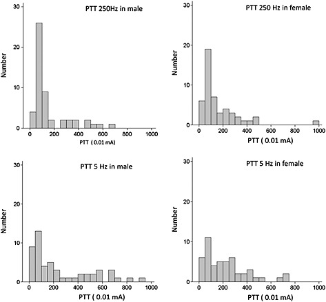
Histogram of pain tolerance threshold (PTT) at each frequency. The PTT was not normally distributed; the distribution was skewed to the right.
Pain tolerance threshold and gender
The PTT values did not differ significantly between men and women at either 250 or 5 Hz (Mann–Whitney U‐test, P > 0.05; Fig. 3).
Figure 3.
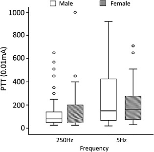
Gender differences in pain tolerance threshold (PTT). There were no significant differences in the PTT between men and women at 250 or 5 Hz.
Pain tolerance threshold and age
No significant association between age and PTT at 250 Hz was found for either gender. As shown in Fig. 4A, the regression line for the men had a negative slope, indicating a decrease in the PTT with increasing age; this was not the case for the women.
Figure 4.
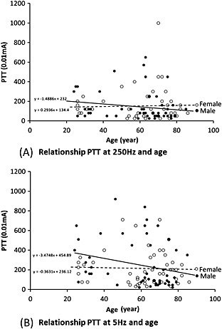
Scatter charts with the regression line between pain tolerance threshold (PTT) and age. (A) At 250 Hz, no significant association between age, and PTT was found in men or women. (B) At 5 Hz, a significant association between age and PTT at 5 Hz was found in men but not in women.
At 5 Hz, age and PTT were significantly associated with the men in this study (correlation coefficient = −0.33, P = 0.02) but not with the women (P > 0.05). As shown in Figure 4B, the regression line for the men had a negative slope, indicating a decrease in the PTT with increasing age; however, this was not the case for the women.
Pain tolerance threshold and oral conditions
At 250 Hz, the mean PTT in Eichner group A was 158.2 ± 122.7 × 10⁻² mA; the median was 120.0 × 10⁻² mA (95% CI 112.3–204.0 × 10⁻² mA). In Eichner B group, the mean PTT was 127.5 ± 124.1 × 10⁻² mA; the median was 80.0 10−2 mA (95% CI 81.2–173.9 10−2 mA). In Eichner group C, the mean PTT was 157.5 ± 202.2 × 10⁻² mA; the median was 80.0 × 10⁻² mA (95% CI 92.8–222.2 × 10⁻² mA).
At 5 Hz, the mean PTT in Eichner group A was 265.3 ± 212.3 × 10⁻² mA; the median was 202.5 × 10⁻² mA (95% CI 186.1–344.6 × 10⁻² mA). In Eichner group B, the mean PTT was 208.0 ± 216.1 × 10⁻² mA; the median was 97.5 10−2 mA (95% CI 127.3–288.7 × 10⁻² mA). In Eichner group C, the mean PTT was 222.8 ± 210.4 10−2 mA; the median was 140.0 × 10⁻² mA (95% CI 155.5–290.1 × 10⁻² mA).
There were no significant differences in the PTT among the Eichner groups (Kruskal–Wallis test, P > 0.05; Fig. 5).
Figure 5.
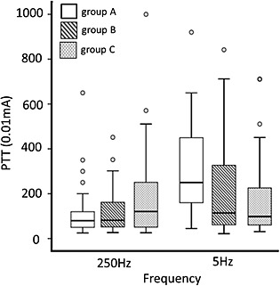
Prosthodontic conditions and pain tolerance threshold (PTT). There was no significant difference in the PTT among different oral condition categories based on the Eichner index.
Discussion
The findings of the present study were the following: (1) there was no difference between genders in the PTT at 250 or 5 Hz; (2) age was associated with the PTT only at 5 Hz in men; (3) oral condition evaluated by the Eichner index was not significantly associated with the PTT; and (4) the PTT values were not normally distributed.
The present study did not show gender‐based differences in the PTT. The question of gender‐based differences in pain tolerance remains controversial. There have been many reports that women exhibit greater tolerance to painful experimental nociceptive stimuli than men (Fillingim et al., 2009; Riley et al., 1998). However, a recent systematic review found that previous laboratory research has not demonstrated a clear and consistent pattern of greater pain sensitivity in women (Racine et al., 2012). Nevertheless, a simple comparison of our results with those of previous studies is impossible because the protocol and target organs of previous studies differed from ours. Gender differences in tolerance to experimentally induced pain may be affected by biological factors such as blood pressure, gonadal hormones, and body size, as well as by psychosocial factors (Bartley and Fillingim, 2013). The mechanisms underlying these differences have yet to be fully explained.
It is widely believed that the PTT decreases with increasing age, which means that older individuals have a lower tolerance for pain. However, in the present study, a relationship between age and PTT was found only for men and only at 5 Hz, with older men having a lower PTT than younger ones. The PTT of men did present a decreasing trend with age at 250 Hz, but the association was not significant. The lack of statistically significant results may be a result of the small sample size. On the contrary, a relationship between age and PTT was not found for women, and the regression slope was nearly flat, suggesting this relationship would not be clinically meaningful even in a large sample. Previous studies differ regarding the relationship between age and the PTT; some studies report that there is no age‐related change (Lucantoni et al., 1997; Mylius et al., 2008), whereas others report that the PTT decreases with age (Neri and Agazzani, 1984; Tucker et al., 1989), and one study found that the PTT increases with age (Kemp et al., 2014). Further studies with larger samples will be needed to address this question.
Contrary to our expectations, there were no significant differences in the PTT among the Eichner index groups. The occlusal support between maxillary and mandibular teeth decreases in the order of group A > group B > group C, and poor denture stability producing pain tends to increase in the same order. Clinically, group C denture wearers are more likely to be exposed to daily pain than group A or B denture wearers because of poor denture stability. Denture wearers are able to adapt to daily pain in the order of group C > group B > group A as a result of habituation that occurs as an adaptive response to repeated painful stimulation in patients living with more pain (Bingel et al., 2007; Nickel et al., 2014; Okayasu et al., 2012). However, in the present study, there was no significant difference in the PTT among Eichner index groups. Contrary to this result, the CPT of the alveolar ridge, which we previously reported by using the same device, significantly increases in the following order: dentulous individual < partial denture wearer < complete denture wearer. The Eichner index categorizes dentulous individuals, partial denture wearers, and complete denture wearers into groups A, B, and C based on the degree of the occlusal support. Therefore, we predicted that the PTT would also differ significantly among Eichner index groups. The difference in the results between the CPT and the PTT might be the result of different measurement procedures. Because the electrical stimulation increases until the participant releases the button when the stimulus can no longer be tolerated, the PTT might be partially influenced by the participant's patience. On the other hand, the CPT is measured at the first feeling of electrical stimulation. Therefore, the PTT should be calculated cautiously after adjusting for personality and emotions, which are known to affect the perception of pain (Shiomi, 1978; Aoki et al., 2010). Unfortunately, we did not measure the personality and emotions. This is one of the study's limitations.
The pain experienced by denture wearers is very complex. The skewed histogram plot of the PTT values showed that individuals had a wide range of pain tolerance. This large intersubject variance partially supported the complexity of the pain experience. In the clinical setting, some denture wearers complaining of severe pain do not exhibit an inflamed mucosa, while others reporting no pain or only slight pain exhibit severely inflamed mucosa. This diversity is a result of the subjective nature of the pain experience. Considering that the procedure used in the present study can evaluate directly sensory nerve A‐delta fibers corresponding to pressure pain, we believe that PTT values would assist in objectively understanding certain aspects of the pain experienced by denture wearers after denture fitting. To our knowledge, there are no studies on the relationship between the PTT of the oral mucosa and the clinical course of pain. Further studies will be needed to address this question.
Conclusion
Within the limitations of the study, the following characteristics of the PTT of the alveolar ridge are shown: (1) age and PTT at 5 Hz are significantly associated with men but not with women, and (2) the Eichner index has no influence on the PTT.
Conflict of Interest
None declared.
Acknowledgments
This study was conducted in accordance with the Declaration of Helsinki, and each subject received oral and written information about the study and provided informed consent. The study protocol was reviewed and approved by the Human Ethics Committee of Nihon University School of Dentistry at Matsudo (EC 13‐12‐003‐2). This research was carried out without external funding.
Nakashima, Y. , Kimoto, S. , Ogawa, T. , Furuse, N. , Ono, M. , and Kawai, Y. (2015) Characteristics of the pain tolerance threshold induced by electrical stimulation of the alveolar ridge. Clinical and Experimental Dental Research, 1: 80–86. doi: 10.1002/cre2.14.
References
- Aoki, J. , Ikeda, K. , Murayama, O. , Yoshihara, E. , Ogai, Y. , Iwahashi, K. , 2010. The association between personality, pain threshold and a single nucleotide polymorphism (rs3813034) in the 3′‐untranslated region of the serotonin transporter gene (SLC6A4). J. Clin. Neurosci. 17(5), 574–8. [DOI] [PubMed] [Google Scholar]
- Bartley, E.J. , Fillingim, R.B. , 2013. Sex differences in pain: a brief review of clinical and experimental findings. Br. J. Anaesth. 111(1), 52–8. [DOI] [PMC free article] [PubMed] [Google Scholar]
- Bingel, U. , Schoell, E. , Herken, W. , Buchel, C. , May, A. , 2007. Habituation to painful stimulation involves the antinociceptive system. Pain 131(1‐2), 21–30. [DOI] [PubMed] [Google Scholar]
- Carlsson, G.E. , Omar, R. , 2010. The future of complete dentures in oral rehabilitation. A critical review. J. Oral Rehabil. 37(2), 143–56. [DOI] [PubMed] [Google Scholar]
- Douglass, C.W. , Shih, A. , Ostry, L. , 2002. Will there be a need for complete dentures in the United States in 2020? J. Prosthet. Dent. 87(1), 5–8. [DOI] [PubMed] [Google Scholar]
- Fillingim, R.B. , King, C.D. , Ribeiro‐Dasilva, M.C. , Rahim‐Williams, B. , Riley, 3rd., J.L. , 2009. Sex, gender, and pain: a review of recent clinical and experimental findings. J. Pain 10(5), 447–85. [DOI] [PMC free article] [PubMed] [Google Scholar]
- Ikebe, K. , Matsuda, K. , Murai, S. , Maeda, Y. , Nokubi, T. , 2010. Validation of the Eichner index in relation to occlusal force and masticatory performance. Int. J. Prosthodont. 23(6), 521–4. [PubMed] [Google Scholar]
- Isobe, A. , Sato, Y. , Kitagawa, N. , Shimodaira, O. , Hara, S. , Takeuchi, S. , 2013. Influence of denture supporting tissue properties on pressure‐pain threshold ‐ measurement in dentate subjects. J. Prosthodont. Res. 57(4), 275–83. [DOI] [PubMed] [Google Scholar]
- Ito, N. , Kimoto, S. , Kawai, Y. , 2012. Does wearing dentures change sensory nerve responses under the denture base? Gerodontology 31(1), 63–7. [DOI] [PubMed] [Google Scholar]
- Kemp, J. , Despres, O. , Pebayle, T. , Dufour, A. , 2014. Age‐related decrease in sensitivity to electrical stimulation is unrelated to skin conductance: an evoked potentials study. Clin. Neurophysiol. 125(3), 602–7. [DOI] [PubMed] [Google Scholar]
- Kimoto, S. , Ito, N. , Nakashima, Y. , Ikeguchi, N. , Yamaguchi, H. , Kawai, Y. , 2013. Maxillary sensory nerve responses induced by different types of dentures. J. Prosthodont. Res. 57(1), 42–5. [DOI] [PubMed] [Google Scholar]
- Komiyama, O. , Gracely, R.H. , Kawara, M. , Laat, A.D. , 2008. Intraoral measurement of tactile and filament‐prick pain threshold using shortened Semmes–Weinstein monofilaments. Clin. J. Pain 24(1), 16–21. [DOI] [PubMed] [Google Scholar]
- Lucantoni, C. , Marinelli, S. , Refe, A. , Tomassini, F. , Gaetti, R. , 1997. Course of pain sensitivity in aging: pathogenetical aspects of silent cardiopathy. Arch. Gerontol. Geriatr. 24(3), 281–6. [DOI] [PubMed] [Google Scholar]
- Margarida, R. , Hardy, J.D. , Hammel, H.T. , 1962. Measurement of the thermal pain threshold of the hard palate. J. Appl. Physiol. 17, 338–42. [DOI] [PubMed] [Google Scholar]
- Mylius, V. , Kunz, M. , Hennighausen, E. , Lautenbacher, S. , Schepelmann, K. , 2008. Effects of ageing on spinal motor and autonomic pain responses. Neurosci. Lett. 446(2‐3), 129–32. [DOI] [PubMed] [Google Scholar]
- Nakashima, Y. , Kimoto, S. , Kawai, Y. , 2014. Reliability of pain tolerance threshold testing by applying an electrical current stimulus to the alveolar ridge. J. Oral Rehabil. 41(8), 595–600. [DOI] [PubMed] [Google Scholar]
- Neri, M. , Agazzani, E. , 1984. Aging and right‐left asymmetry in experimental pain measurement. Pain 19(1), 43–8. [DOI] [PubMed] [Google Scholar]
- Nickel, F.T. , Ott, S. , Mohringer, S. , Saake, M. , Dorfler, A. , Seifert, F. , Maihofner, C. , 2014. Brain correlates of short‐term habituation to repetitive electrical noxious stimulation. Eur. J. Pain 18(1), 56–66. [DOI] [PubMed] [Google Scholar]
- Okayasu, I. , Komiyama, O. , Yoshida, N. , Oi, K. , De Laat, A. , 2012. Effects of chewing efforts on the sensory and pain thresholds in human facial skin: a pilot study. Arch. Oral Biol. 57(9), 1251–5. [DOI] [PubMed] [Google Scholar]
- Racine, M. , Tousignant‐Laflamme, Y. , Kloda, L.A. , Dion, D. , Dupuis, G. , Choiniere, M. , 2012. A systematic literature review of 10 years of research on sex/gender and pain perception ‐ part 2: do biopsychosocial factors alter pain sensitivity differently in women and men? Pain 153(3), 619–35. [DOI] [PubMed] [Google Scholar]
- Raj, P.P. , Chado, H.N. , Angst, M. , Heavner, J. , Dotson, R. , Brandstater, M.E. , Johnson, B. , Parris, W. , Finch, P. , Shahani, B. , Dhand, U. , Mekhail, N. , Daoud, E. , Hendler, N. , Somerville, J. , Wallace, M. , Panchal, S. , Glusman, S. , Jay, G.W. , Palliyath, S. , Longton, W. , Irving, G. , 2001. Painless electrodiagnostic current perception threshold and pain tolerance threshold values in CRPS subjects and healthy controls: a multicenter study. Pain Pract. 1(1), 53–60. [DOI] [PubMed] [Google Scholar]
- Riley, 3rd, J.L. , Robinson, M.E. , Wise, E.A. , Myers, C.D. , Fillingim, R.B. , 1998. Sex differences in the perception of noxious experimental stimuli: a meta‐analysis. Pain 74(2‐3), 181–7. [DOI] [PubMed] [Google Scholar]
- Shiomi, K. , 1978. Relations of pain threshold and pain tolerance in cold water with scores on Maudsley Personality Inventory and Manifest Anxiety Scale. Percept. Mot. Skills 47(3 Pt 2), 1155–8. [DOI] [PubMed] [Google Scholar]
- Swenson, M.G. , Boucher, C.O. , 1970. Swenson's complete dentures , Saint Louis, Mosby, 109.
- Tanaka, M. , Ogimoto, T. , Koyano, K. , Ogawa, T. , 2004. Denture wearing and strong bite force reduce pressure pain threshold of edentulous oral mucosa. J. Oral Rehabil. 31(9), 873–8. [DOI] [PubMed] [Google Scholar]
- Tucker, M.A. , Andrew, M.F. , Ogle, S.J. , Davison, J.G. , 1989. Age‐associated change in pain threshold measured by transcutaneous neuronal electrical stimulation. Age Ageing 18(4), 241–6. [DOI] [PubMed] [Google Scholar]


