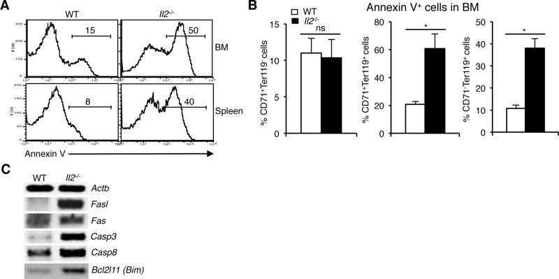
Figure 4. Enhanced NFAT signaling induces apoptosis in Il2−/− immature erythrocytes.
(A) Cell death analysis in BM and splenic Ter119+ cells from WT and Il2−/− mice as revealed by Annexin V staining. (B) Quantification of % Annexin V positive cells in BM CD71+Ter119-, CD71+Ter119+ and CD71-Ter119+ fractions from WT and Il2−/− mice. (C) Expression of apoptosis-related genes in the BM Ter119+ cells from Il2−/− mice compared to WT controls. Data in (A–C) are representative of three independent experiments. Number inside each histogram represents percent Annexin V positive population. Data in (B) are presented as mean ± s.d., ns = not significant and *p = 0.7376 or 0.0141, paired t-test.

