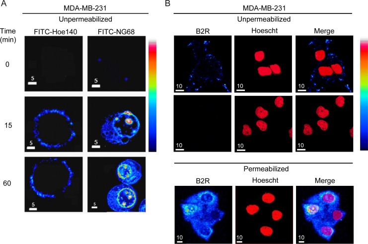
Figure 3. Binding and cellular incorporation of B2RAs in MDA-MB-231 cells.
(A) MDA-MB-231 cells were incubated with FITC-labelled peptide HOE140 or NG68 (10 µM) at 37°C for the indicated times, washed with PBS buffer, fixed with paraformaldehyde then analyzed by laser confocal microscopy. A pseudocolor fluorescence intensity scale is shown on the right; black being the lowest and white the highest value. Note the rapid cellular incorporation of FITC-NG68 peptide and its perinuclear/nucleolar localization increasing over time while FITC-HOE140 conserved an exclusive membrane distribution. No signal was detected when cells were incubated with FITC alone under the same experimental conditions. Individual midsection confocal images are representative of three replicates. (B) Immunocytochemical localization of B2R in MDA-MB-231 cells was performed in unpermeabilized (upper panels) and permeabilized (bottom panels) conditions using the B2R antiserum AS276–83. An Alexa Fluor 568-conjugated goat anti-rabbit IgG antibody was used as a secondary antibody. Nuclei stained with Hoescht 33342 (middle panels, arbitrary colors). Merge in right panels. Negative control: no primary antibody. Pseudocolor fluorescence intensity scale as in A. Representative confocal middle z-sections are shown.

