Abstract
We previously showed that metabolic activation of the environmental and tobacco smoke constituent dibenzo[a,l]pyrene (DB[a,l]P) to its active fjord region diol epoxide (DB[a,l]PDE) is required to induce DNA damage, mutagenesis and squamous cell carcinoma (SCC) in the mouse oral cavity. In contrast to procarcinogens which were employed previously to induce SCC, DB[a,l]PDE does not require metabolic activation to exert its biological effects and thus, this study was initiated to examine, for the first time, whether black raspberry powder (BRB) inhibits post-metabolic processes such as DNA damage, mutagenesis and tumorigenesis. Prior to long-term chemoprevention studies we initially examined the effect of BRB (5% added to AIN-93M diet) on DNA damage in B6C3F1 mice using LC-MS/MS and on mutagenesis in the lacI gene in the mouse oral cavity. We showed that BRB inhibited DB[a,l]PDE-induced DNA damage (p<0.05) and mutagenesis (p=0.053) in the oral cavity. Tumor incidence in the oral cavity (oral mucosa and tongue) of mice fed diet containing 5% BRB was significantly (p<0.05) reduced from 93% to 66%. Specifically the incidence of benign tumor was significantly (p<0.001) reduced from 90% to 31% (62% to 28% in the oral cavity and 28% to 2% in the tongue); a non-significant reduction of malignant tumors from 52% to 45%. Our preclinical findings demonstrate for the first time that the chemopreventive efficacy of BRB can be extended to direct-acting carcinogens that do not require phase I enzymes and is not just limited to procarcinogens.
Keywords: Dibenzo[a,l]pyrene diol epoxide; DNA adducts; mutagenesis; mouse oral cavity; black raspberry
Introduction
Worldwide, head and neck cancer is the 6th most common human cancer, and oral cancer is the most common type of this disease (1,2). Cancer of the oral cavity is a deadly disease and can strip away the patient’s voice and certain basic needs in life such as eating and drinking; there are currently over 300,000 cases, and more than 145,500 deaths occurred in 2012 worldwide; and in the USA over 30,000 cases and over 6,000 deaths from the disease occur annually (2–4). In 2017, the number of new cases of oropharyngeal carcinomas in the USA is estimated to be 49,670 and the estimated deaths from the disease 9,700 (5). The most common histological type of oral cancer is oral squamous cell carcinoma (OSCC); it accounts for more than 90% of oral cancers (1,2). Early diagnosis for oral cancer has not improved over time; up to 77% of oral cancer cases are diagnosed at advanced stages (6). The conventional treatments include surgery, radiotherapy, and chemotherapy (7). However, approximately one-third of treated patients will experience local or regional recurrence and/or distant metastasis (8). The survival rate is stagnant at about 50%; it varies greatly, with the stage of disease detection (6).
The main risk factors for oral cancer are exposure to exogenous carcinogens, such as tobacco smoke, smokeless tobacco, excess alcohol and human papilloma virus (HPV). These factors are estimated to account for 90% of oral cancers (1,2). In general, avoidance of risk factors has only been partially successful – largely because of the addictive power of tobacco smoking and alcohol consumption. Although it is not clear which chemical compounds in tobacco smoke contribute to the development of human oral cancer, certain classes of chemical carcinogens such as tobacco-specific nitrosamines (TSNA) and polycyclic aromatic hydrocarbons (PAH) are recognized as potential etiological agents for oral cancer (9–11).
Our lab has focused on developing an animal model that mimics human exposure to environmental carcinogens and reflects tumor heterogeneity; in 2012 we introduced a new mouse model to study oral carcinogenesis (12). We chose to focus on the tobacco smoke constituent and the environmental pollutant, the fjord-region PAH, dibenzo[a,l]pyrene (DB[a,l]P) (12) and its ultimate diolepoxide metabolite DB[a,l]PDE (13). Both compounds were potent carcinogens and DB[a,l]PDE greatly enhanced carcinogenicity relative to the parent compound in the mouse oral cavity (12–14).
Treating cancers including OSCC at late stages, even with recent advances in targeting therapies continues to be a major challenge, and thus prevention remains a desirable approach. Numerous sources of phytochemicals have been proposed (15), and one that has shown promise in inhibiting carcinogenesis including head and neck cancers is freeze-dried black raspberry (BRB) (14–19). In a previous study BRB has been shown to inhibit dimethylbenz[a]anthracene (DMBA)-induced oral cancer in the hamster cheek pouch (20). In that study inhibition could have resulted from inhibiting initiation or post initiation processes. For initiation, metabolic activation of DMBA (and also other oral procarcinogens, such as 4-nitroquinoline-N-oxide) is essential to induce DNA damage, mutagenesis and carcinogenesis. Therefore, the chemopreventive effect of black raspberry on DBMA-induced oral cancer could be due to factors affecting initiation, such as inhibition of phase I, induction of phase II enzymes, and enhancement of DNA repair capacity of bulky lesions derived from DMBA. Additionally post-initiation processes such as promotion and progression could have been affected by BRB. In recent in vitro studies carried out in our lab (21) we showed that black raspberry extract enhanced repair of DB[a,l]P induced bulky DNA lesions. We also showed that metabolic activation of DB[a,l]P to its active fjord-region diol epoxide DB[a,l]PDE is required to induce DNA damage, mutagenesis and squamous cell carcinoma in the mouse oral cavity (13,22). In contrast, to DMBA and 4-NQO, DB[a,l]PDE does not require metabolic activation to exert its biological effects and thus this study was initiated to examine, for the first time, whether BRB inhibits post-metabolic processes such as DNA damage, mutagenesis and tumorigenesis.
Materials and Methods
Chemicals
(±)-anti-DB[a,l]PDE was prepared according to a published method by our group (23). Structural characterization of this carcinogen was based on NMR and high-resolution mass spectral data and its purity (≥99%) was determined by HPLC.
Animals
The selection of species, strain and sex of the animals used in the present study is based on our previous report (13). Thus, female B6C3F1 mice (6–8 weeks of age, Jackson Laboratories, Bar Harbor, ME) were used to examine the effects of BRB on DNA adducts formation and tumorigenesis induced by DB[a,l]PDE in the oral cavity; Big Blue™ C57BL/6 (lacI) mice (Bioreliance, Rockville, MD) were used to examine the effect of BRB on mutagenesis induced by DB[a,l]PDE. Mice were quarantined for 1 week; then they were transferred to the bioassay laboratory. All mice were kept on a 12-h light:12-h dark cycle, maintained at 50% relative humidity and 21 ± 2°C. Water and food were provided ad libitum. The bioassay was carried out in accordance with the NIH Guide for the Care and Use of Laboratory Animals and was approved by Institutional Animal Care and Use Committee.
Effect of BRB on DNA Adducts Induced by DB[a,l]PDE in Oral Tissues of Mice
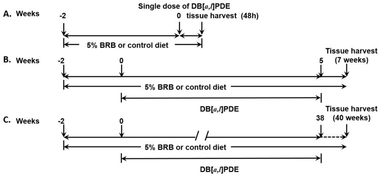
Two groups of B6C3F1 mice (6 mice/group) were fed AIN-93M diet (5% corn oil) containing BRB (5%) or control diet (AIN-93M) starting 2 weeks prior to the administration of a single dose of DB[a,l]PDE (6 nmol/mouse) by topical application into the oral cavity of mice (Scheme IA). On the basis of previous preclinical studies (reviewed in 14) 5% BRB was the most protective and thus such level was used in the present investigation. The carcinogen dose was selected based on our previous studies (13,24,25). Mice were sacrificed 48 hours after DB[a,l]PDE treatment, and tissues were removed for DNA isolation.
Scheme I.
Experimental Protocol to Determine the Effects of BRB on DB[a,l]PDE-lnduced DNA adducts (A) Mutagenesis (B) and Tumorigenesis (C) in Oral Tissues of Mice.
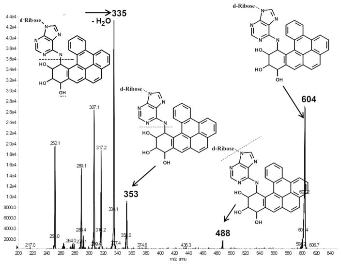
We employed our previously published method for the analysis of the major deoxyadenosine adducts by LC-MS/MS (21,24,25). Briefly, we isolated DNA from oral tissues using the Qiagen genomic DNA isolation procedure. The level of DNA was quantified by a NanoDrop ND-1000 spectrophotometer (NanoDrop Technologies, Wilmington, DE). Prior to enzymatic digestion, 150 pg of the internal standard [15N5]-anti-trans-DB[a,l]PDE-dA adduct was added to ~60 μg DNA. Subsequently, in the presence of 10 μL of 1M MgCl2/mg DNA and DNAse I (0.2 mg/mg DNA) DNA was hydrolyzed at 37 °C for 1.5 h, followed by snake venom phosphodiesterase (0.08 unit/mg DNA) and alkaline phosphatase (2 units/mg DNA). An aliquot of the DNA hydrolysate was taken for analysis of dA by HPLC. The remaining supernatant was partially purified on an Oasis HLB column (1 cm3, 30 mg, Waters Ltd.). Then, the analysis was carried out on an API 3200™ LC/MS/MS triple quadrupole mass spectrometer interfaced with an Agilent 1200 series HPLC using an Agilent extend-C18 5 μm 4.6 × 150 mm column. Adducts were monitored in multiple reaction monitoring (MRM) mode, and the MS/MS transitions of m/z 604→ m/z 335, and m/z 609→ m/z 335 were monitored for targeted adducts and the internal standard (Figure 1), respectively.
Fig. 1.
A representative mass spectra of (±)-anti-DB[a,l]PDE-dA adducts.
The Effect of BRB on DB[a,l]PDE-Induced Mutagenesis in Oral Tissues of Mice
Two groups of Big Blue™ C57BL/6 (lacI) mice (10/group) received 3 nmol (3 times weekly for 5 weeks) of (±)-anti-DB[a,l]PDE in DMSO by topical application as depicted in Scheme IB for 5 weeks. The dose was selected based on our previous report (13). One group of mice were fed AIN-93M control diet, and the other group were fed AIN-93M diet containing 5% BRB starting 2 weeks prior to carcinogen treatment and continued until termination. Mice were sacrificed 2 weeks after the cessation of carcinogen administration. The oral tissues were excised and stored at −80°C until isolation of DNA. The palate and pharynx were pooled and homogenized together to give a mixture designated oral tissue. Each tissue was gently homogenized by hand in a microcentrifuge tube using a Teflon pestle in three volumes of 10 mM Tris-HCl (pH, 8.0), 10 mM EDTA, 150 mM NaCl per gram tissue weight (w/v). SDS and protease K were added to this homogenate to obtain final concentrations of 10 and 1 mg/ml, respectively. The mixture was incubated for 3 hr at 50°C, and then for 30 min at 37°C with 0.1 mg/ml RNase A. After incubation one-third volume of 6M ammonium acetate (pH 7.4) was added, the mixture was gently mixed and then centrifuged at 14,000 rpm in an Eppendorf microfuge. The supernatant was carefully removed, leaving a small volume behind, to avoid transfer of any of the precipitate. To the supernatant an equal volume of isopropyl alcohol was added at room temperature to precipitate DNA. The supernatant was removed, the DNA was washed once with 70% ethanol, suspended in 10 mM Tris-HCl (pH, 8.0), 1 mM EDTA and left overnight at room temperature to dissolve.
Phage packaging was carried out using a homemade packaging extract prepared from bacterial strains supplied by Dr. Peter Glazer (Yale University School of Medicine, New Haven, CT); and the positive selection cII mutation assay was performed as previously described (26). At least three packaging reactions were carried out for each DNA sample. The mean mutant fractions of each group were compared with those from the control (vehicle-alone) group using a one-tailed t-test.
The Effect of BRB on DB[a,l]PDE-Induced Tumorigenesis in Oral Tissues of Mice
Two groups of B6C3F1 mice (30/group) at the age of 8 weeks received 3 nmol of (±)-anti-DB[a,l]PDE in DMSO, 3 times a week for 38 weeks as depicted in Scheme IC. The dose was selected based on our previous study (13). One group of mice was fed AIN-93M control diet, and the other group was fed AIN-93M diet containing 5% BRB starting two weeks prior to the initiation of carcinogen treatment. Treatments continued until termination. A third group of mice (n=20) treated only with the vehicle (DSMO) and fed 5% BRB diet was used as a negative control. Mice were weighed weekly in the first month and then biweekly until termination. During the progress of the bioassay, mice were culled from the group and sacrificed if we observed a sudden weight loss of more than 20% or a tumor size exceeding 0.5 cm in diameter. At termination, mice were sacrificed by CO2 asphyxiation and soft tissues of the oral cavity including tongue, pharynx, and other oral tissues (hard palate, buccal mucosa, and floor of mouth) were collected and fixed in 10% neutral buffered formalin. Tissues were processed in an automated Tissue-Tek VIP processor and paraffin-embedded with a Tissue-Tek TEC embedding station. Sections were cut at 6 μm for routine hematoxylin and eosin (H&E) staining. All tissues were examined by an ACVP diplomate pathologist blinded to treatment according to established criteria (27). Benign tumors were not invasive into the basement membrane and subjacent stroma. Microinvasive malignant tumors had focal limited invasion, less than a single 400x high power field deep to the basement membrane. Invasive malignant tumors were more extensively invasive.
Results
Effect of BRB on DB[a,l]PDE-Induced DNA Adducts in Oral Tissues of Mice
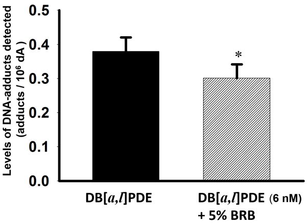
Prior to the examination of the chemopreventive effects of BRB on DB[a,l]PDE-induced tumorigenesis, we initially examined the effect of BRB on DB[a,l]PDE-induced DNA adducts in oral tissues of mice. We showed that 5% BRB in the diet significantly (p<0.05) reduced the levels of (−)-anti-trans-DB[a,l]PDE-dA in the oral cavity of mice 48hr after a single dose of 6 nmol DB[a,l]PDE (Figure 2). BRB in the diet reduced the levels of (−)-anti-trans-DB[a,l]PDE-dA adduct from 3.8 ± 0.4 to 3.0 ± 0.4 adducts/107 dA (20.7% inhibition).
Fig. 2.
Inhibition of DB[a,l]PDE-dA adduct levels by BRB in oral tissues of mice.
*p < 0.05
The Effect of BRB on DB[a,l]PDE-Induced Mutagenesis in Oral Tissues of the lacI Mice
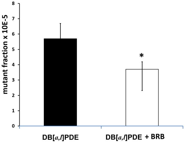
We found that 5% BRB in the diet reduced the mutation fraction induced by DB[a,l]PDE in the oral cavity of mice by about 20% (p = 0.053) (Figure 3). A background of 2.4 mutants/105 has been subtracted from the mutant fractions in Figure 3. That represents the mutant fraction in the untreated mice from our earlier study (13).
Fig. 3.
Inhibition of DB[a,l]PDE-induced mutagenesis by BRB in oral tissues of mice.
*p = 0.053
The Effect of BRB on DB[a,l]PDE-Induced Tumorigenesis in Oral Tissues of Mice
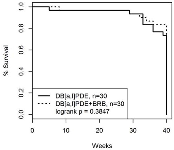
Figure 4 shows the cumulative mortality of mice during the progress of the bioassay. The probability of survival was displayed using Kaplan-Meier plot with death as an endpoint. Log-rank test was used to evaluate the difference between this pair of groups. No significant difference was observed in survival analysis in both groups of mice. All mice treated with DMSO and fed 5% BRB survived the duration of the bioassay. Body Weights (mean ± SE) of mice treated with DB[a,l]PD + 5% BRB and those treated with DB[a,l]PDE alone are provided in Table S1 and were not significantly different with the exception at week 4 and 20; however, the difference at these two time points was about 5%.
Fig. 4.
Percentage survival of B6C3F1 mice treated by topical application of DB[a,l]PDE at 3nmol three times a week for 38 weeks during the progress of the bioassay.
We found that the tumor incidence in the oral cavity (oral mucosa and tongue) of mice fed diet containing 5% BRB was reduced significantly from 93% to 66% (Table 1). Benign tumors induced by DB[a,l]PD consist of papilloma and keratoacanthoma; papilloma accounted for 94.7%. Specifically, overall benign tumor incidence was significantly reduced from 90% to 31% (62% to 28% in the oral cavity and 28% to 2% in the tongue). Malignant tumors induced by DB[a,l]PDE consist of squamous cell carcinoma, invasive anaplastic cell carcinoma and fibrosarcoma; squamous cell carcinoma accounted for 88.8%. BRB reduced malignant tumors from 52% to 45%. No tumors were found in mice treated with DMSO and fed 5% BRB diet.
Table 1.
Inhibition of Oral Tumorigenesis by BRB in DB[a,l]PDE treated mice
| Treatments | ||
|---|---|---|
| DB[a,l]PDE | DB[a,l]PDE+BRB | |
| Number of mice1 | 29 | 29 |
| Total (M + B) | 27 (93)2 | 19 (66)* |
| Benign Tumors (B) | 26 (90)3 | 9 (31)*** |
| Malignant Tumors (M) | 15 (52)4 | 13 (45) |
Mice which died before the first tumor appeared in the study or did not reach histology due to cannibalism were not counted.
Number in parentheses, percentage of mice developed tumors.
Benign tumors induced by DB[a,l]PD consist of papilloma and keratoacanthoma; papilloma accounted for 94.7%.
Malignant tumors induced by DB[a,l]PDE consist of squamous cell carcinoma, invasive anaplastic cell carcinoma and fibrosarcoma; squamous cell carcinoma accounted for 88.8%.
tumor incidence was significantly reduced compared to DBPDE treatment, p < 0.05.
tumor incidence was significantly reduced compared to DBPDE treatment, p < 0.001.
Discussion
The results of the present study clearly demonstrate the protective effects of BRB against the development of oral carcinogenesis induced by a powerful carcinogenic metabolite derived from the environmental pollutant and tobacco smoke constituent, DB[a,l]P. Numerous compounds as well as complex mixtures have been used to induce oral cancer in animal models (28). Preclinical animal models which used carcinogens found in the environment and/or tobacco smoke can mimic human exposure to such carcinogens and can recapitulate the tumor heterogeneity and can also provide a realistic platform to evaluate chemopreventive agents (29). In this study we selected DB[a,l]PDE because of 1) its remarkable carcinogenicity in the mouse oral cavity (14), and 2) to demonstrate for the first time that the efficacy of BRB can be extended to direct-acting carcinogens which do not require metabolic activation by phase I enzymes, and is not just limited to procarcinogens.
Prior to the initiation of the long-term chemopreventive bioassay it was essential to initially determine in a short-term animal bioassays whether BRB was capable of inhibiting DNA damage and consequently mutation induction by DB[a,l]PDE. Because of the positive outcomes demonstrating in vivo the protective effects of BRB on these endpoints, we then initiated the long-term bioassay to examine the efficacy of BRB against the development of oral carcinogenesis.
It was previously shown that in diet containing 5% BRB resulted in a 45% reduction in tumor incidence induced by the procarcinogen DMBA in hamster cheek pouch model, and this effect was associated with reduced formation of DNA adducts in epithelial cells of the oral mucosa (20). However, in this previous report 32P-postlabelling technique was used which may not provide accurate adduct quantification and adduct distribution remain to be assessed (20). In the present study, our LC-MS/MS method (25) was used to provide an accurate quantification and structural information on the nature of DNA adducts derived from DB[a,l]PDE. Clearly the protective effect of BRB on DNA damage is not due to inhibition of phase I enzymes but its effects on phase II and/or DNA repair enzymes could be relevant. Although we did not examine the levels of phase II or DNA repair enzymes in the present study, our results are in line with our recent in vitro cell culture study that demonstrated that BRB extract resulted in reduced DNA adducts derived from DB[a,l]PDE via enhancing DNA repair capacity which represents the integrated effects of proteins involved in the DNA repair pathways. (21,30).
This observed inhibition of DNA damage by BRB stimulated a follow-up investigation aimed at determining the effect of BRB on mutation induction by DB[a,l]PDE in vivo using Big Blue™ C57BL/6 (lacI) mice. Presumably a reduction in DNA damage by BRB would be expected to reduce mutation induction. Using lacI and lacZ rodents, we have reported that all of the known oral carcinogens are also mutagens in the mouse and/or rat oral cavity (31). As mutagenesis is considered an important step in carcinogenesis, it can represent an early biomarker in the multi-step carcinogenesis process. We previously assayed mutagenesis induced by DB[a,l]P (12) and DB[a,l]PDE (13) in the oral cavity of BGC3F1 lacI mice and showed that both are powerful mutagens in the oral cavity of mice.
In the present study we showed that BRB is capable of inhibiting mutagenesis induced by DB[a,l]PDE in the mouse oral cavity. In our previous in vitro studies, using rat oral fibroblast we showed that BRB extract was also effective at reducing mutagenesis induced by DB[a,l]PDE (21). Presumably a reduction in DNA damage by BRB as discussed above, is responsible for the reduction in mutagenesis. We also showed that BRB extract reduced oxidative stress in vitro (21). Taken together these results suggest that BRB is potentially an inhibitor of several steps in the multi-step carcinogenesis including tumor initiation by DB[a,l]PDE and thus we performed the following long-term chemoprevention bioassay.
Our results on survival and body weights demonstrated that mice tolerated 5% BRB in the diet. Clearly the ability of BRB to inhibit tumorigenesis is not limited to synthetic procarcinogens such as DMBA (20). A significant protective effect of BRB was observed against benign tumors, but the inhibition of malignant formation did not reach significance. A longer duration of the bioassay might have allowed some or all of the benign tumors to progress to malignant tumors, resulting in significant inhibition of malignancy, but this could not be possible because animals needed to be sacrificed because tumor size reached >0.5 cm in the oral cavity. In the present study dietary black raspberry powder was given 2-weeks before carcinogen administration and continued until termination of the bioassay. Thus, our design cannot dissociate the effects of berry on tumor initiation vs tumor progression. However, based on our previous studies (21,30), we showed that black raspberry enhanced DNA repair of the damage induced by DB[a,l]P, which contributes to the initiation phase of carcinogenesis.
In addition to the protective effects of BRB in preclinical animal models, the results of several clinical trials demonstrate its safety, and that it is easy to administer and can modulate critical markers of cancer in a manner consistent with prevention/treatment (14). In the oral cavity of healthy volunteers, a 10% mucoadhesive freeze-dried black raspberry gel, when treated topically on dysplastic lesions four times per day for six weeks, was found to result in a histologic regression of about 60% of the lesions (32,33). It was also found in oral cancer patients that proinflammatory and prosurvival markers were suppressed by BRB phytochemical-rich troche (34). The active components in BRB which can account for its observed effects in our oral carcinogenesis model remain to be determined. However, we recently reported on the effects of select constituents of BRB on carcinogen-induced DNA damage in cell cultures; our preliminary results indicate that the structure of the constituent is an important determining factor in inhibiting DNA damage (30). Our results provide a basis to translate our findings into future studies designed, for instance, to examine the effect of BRB on the initiation stage of carcinogenesis by assessing levels of DNA adducts derived from DB[a,l]P and benzo[a]pyrene in smokers who are considered at high risk of developing this disease.
Supplementary Material
Acknowledgments
This work was supported by the National Institutes of Health NCI grant R01-CA173465.
Footnotes
The authors declare no potential conflicts of interest
References
- 1.Tanaka T, Ishigamori R. Understanding carcinogenesis for fighting oral cancer. J Oncol. 2011;2011:603740. doi: 10.1155/2011/603740. [DOI] [PMC free article] [PubMed] [Google Scholar]
- 2.Torre LA, Bray F, Siegel RL, Ferlay J, Lortet-Tieulent J, Jemal A. Global cancer statistics, 2012. CA Cancer J Clin. 2015;65(2):87–108. doi: 10.3322/caac.21262. [DOI] [PubMed] [Google Scholar]
- 3.Parkin DM, Bray F, Ferlay J, Pisani P. Global cancer statistics, 2002. CA Cancer J Clin. 2005;55(2):74–108. doi: 10.3322/canjclin.55.2.74. [DOI] [PubMed] [Google Scholar]
- 4.Siegel RL, Miller KD, Jemal A. Cancer statistics, 2016. CA Cancer J Clin. 2016;66(1):7–30. doi: 10.3322/caac.21332. [DOI] [PubMed] [Google Scholar]
- 5.Siegel RL, Miller KD, Jemal A. Cancer Statistics, 2017. CA Cancer J Clin. 2017;67(1):7–30. doi: 10.3322/caac.21387. [DOI] [PubMed] [Google Scholar]
- 6.Groome PA, Rohland SL, Hall SF, Irish J, Mackillop WJ, O’Sullivan B. A population-based study of factors associated with early versus late stage oral cavity cancer diagnoses. Oral Oncol. 2011;47(7):642–7. doi: 10.1016/j.oraloncology.2011.04.018. [DOI] [PubMed] [Google Scholar]
- 7.Argiris A, Karamouzis MV, Raben D, Ferris RL. Head and neck cancer. Lancet. 2008;371(9625):1695–709. doi: 10.1016/s0140-6736(08)60728-x. [DOI] [PMC free article] [PubMed] [Google Scholar]
- 8.da Silva SD, Hier M, Mlynarek A, Kowalski LP, Alaoui-Jamali MA. Recurrent oral cancer: current and emerging therapeutic approaches. Front Pharmacol. 2012;3:149. doi: 10.3389/fphar.2012.00149. [DOI] [PMC free article] [PubMed] [Google Scholar]
- 9.El-Bayoumy K, Chung FL, Richie J, Jr, Reddy BS, Cohen L, Weisburger J, et al. Dietary control of cancer. Proc Soc Exp Biol Med. 1997;216(2):211–23. doi: 10.3181/00379727-216-44171. [DOI] [PubMed] [Google Scholar]
- 10.Hecht SS, Hoffmann D. The relevance of tobacco-specific nitrosamines to human cancer. Cancer Surv. 1989;8(2):273–94. [PubMed] [Google Scholar]
- 11.Ng SK, Kabat GC, Wynder EL. Oral cavity cancer in non-users of tobacco. J Natl Cancer Inst. 1993;85(9):743–5. doi: 10.1093/jnci/85.9.743. [DOI] [PubMed] [Google Scholar]
- 12.Guttenplan JB, Kosinska W, Zhao ZL, Chen KM, Aliaga C, DelTondo J, et al. Mutagenesis and carcinogenesis induced by dibenzo[a,l]pyrene in the mouse oral cavity: a potential new model for oral cancer. Int J Cancer. 2012;130(12):2783–90. doi: 10.1002/ijc.26344. [DOI] [PMC free article] [PubMed] [Google Scholar]
- 13.Chen KM, Guttenplan JB, Zhang SM, Aliaga C, Cooper TK, Sun YW, et al. Mechanisms of oral carcinogenesis induced by dibenzo[a,l]pyrene: an environmental pollutant and a tobacco smoke constituent. Int J Cancer. 2013;133(6):1300–9. doi: 10.1002/ijc.28152. [DOI] [PMC free article] [PubMed] [Google Scholar]
- 14.El-Bayoumy K, Chen KM, Zhang SM, Sun YW, Amin S, Stoner G, et al. Carcinogenesis of the Oral Cavity: Environmental Causes and Potential Prevention by Black Raspberry. Chem Res Toxicol. 2017;30(1):126–44. doi: 10.1021/acs.chemrestox.6b00306. [DOI] [PubMed] [Google Scholar]
- 15.Surh YJ. Cancer chemoprevention with dietary phytochemicals. Nat Rev Cancer. 2003;3(10):768–80. doi: 10.1038/nrc1189. [DOI] [PubMed] [Google Scholar]
- 16.Mallery SR, Tong M, Shumway BS, Curran AE, Larsen PE, Ness GM, et al. Topical application of a mucoadhesive freeze-dried black raspberry gel induces clinical and histologic regression and reduces loss of heterozygosity events in premalignant oral intraepithelial lesions: results from a multicentered, placebo-controlled clinical trial. Clin Cancer Res. 2014;20(7):1910–24. doi: 10.1158/1078-0432.ccr-13-3159. [DOI] [PMC free article] [PubMed] [Google Scholar]
- 17.Rao AV, Snyder DM. Raspberries and human health: a review. J Agric Food Chem. 2010;58(7):3871–83. doi: 10.1021/jf903484g. [DOI] [PubMed] [Google Scholar]
- 18.Stoner GD, Wang LS, Zikri N, Chen T, Hecht SS, Huang C, et al. Cancer prevention with freeze-dried berries and berry components. Semin Cancer Biol. 2007;17(5):403–10. doi: 10.1016/j.semcancer.2007.05.001. [DOI] [PMC free article] [PubMed] [Google Scholar]
- 19.Stoner GD. Foodstuffs for preventing cancer: the preclinical and clinical development of berries. Cancer Prev Res (Phila) 2009;2(3):187–94. doi: 10.1158/1940-6207.capr-08-0226. [DOI] [PMC free article] [PubMed] [Google Scholar]
- 20.Casto BC, Kresty LA, Kraly CL, Pearl DK, Knobloch TJ, Schut HA, et al. Chemoprevention of oral cancer by black raspberries. Anticancer Res. 2002;22(6c):4005–15. [PubMed] [Google Scholar]
- 21.Guttenplan JB, Chen KM, Sun YW, Kosinska W, Zhou Y, Kim SA, et al. Effects of Black Raspberry Extract and Protocatechuic Acid on Carcinogen-DNA Adducts and Mutagenesis, and Oxidative Stress in Rat and Human Oral Cells. Cancer Prev Res (Phila) 2016;9(8):704–12. doi: 10.1158/1940-6207.capr-16-0003. [DOI] [PMC free article] [PubMed] [Google Scholar]
- 22.Sun YW, El-Bayoumy K, Aliaga C, Awad AS, Gowda K, Amin S, et al. Tissue Distribution, Excretion and Pharmacokinetics of the Environmental Pollutant Dibenzo[def,p]chrysene in Mice. Chem Res Toxicol. 2015;28(7):1427–33. doi: 10.1021/acs.chemrestox.5b00097. [DOI] [PMC free article] [PubMed] [Google Scholar]
- 23.Krzeminski J, Lin JM, Amin S, Hecht SS. Synthesis of Fjord region diol epoxides as potential ultimate carcinogens of dibenzo[a,l]pyrene. Chem Res Toxicol. 1994;7(2):125–9. doi: 10.1021/tx00038a002. [DOI] [PubMed] [Google Scholar]
- 24.Zhang SM, Chen KM, Aliaga C, Sun YW, Lin JM, Sharma AK, et al. Identification and quantification of DNA adducts in the oral tissues of mice treated with the environmental carcinogen dibenzo[a,l]pyrene by HPLC-MS/MS. Chem Res Toxicol. 2011;24(8):1297–303. doi: 10.1021/tx200188j. [DOI] [PMC free article] [PubMed] [Google Scholar]
- 25.Zhang SM, Chen KM, Sun YW, Aliaga C, Lin JM, Sharma AK, et al. Simultaneous detection of deoxyadenosine and deoxyguanosine adducts in the tongue and other oral tissues of mice treated with Dibenzo[a,l]pyrene. Chem Res Toxicol. 2014;27(7):1199–206. doi: 10.1021/tx5001078. [DOI] [PMC free article] [PubMed] [Google Scholar]
- 26.Guttenplan JB, Zhao ZL, Kosinska W, Norman RG, Krzeminski J, Sun YW, et al. Comparative mutational profiles of the environmental mammary carcinogen, 6-nitrochrysene and its metabolites in a lacI mammary epithelial cell line. Carcinogenesis. 2007;28(11):2391–7. doi: 10.1093/carcin/bgm142. [DOI] [PubMed] [Google Scholar]
- 27.Leininger JR, Jokinen MP. Tumours of the oral cavity, pharynx, oesophagus and stomach. IARC Sci Publ. 1994;(111):167–93. [PubMed] [Google Scholar]
- 28.Mognetti B, Di Carlo F, Berta GN. Animal models in oral cancer research. Oral Oncol. 2006;42(5):448–60. doi: 10.1016/j.oraloncology.2005.07.014. [DOI] [PubMed] [Google Scholar]
- 29.Vitale-Cross L, Czerninski R, Amornphimoltham P, Patel V, Molinolo AA, Gutkind JS. Chemical carcinogenesis models for evaluating molecular-targeted prevention and treatment of oral cancer. Cancer Prev Res (Phila) 2009;2(5):419–22. doi: 10.1158/1940-6207.capr-09-0058. [DOI] [PubMed] [Google Scholar]
- 30.Guttenplan JB, Chen K-M, Sun Y-W, Lajara B, Shalaby N, Kosinska W, et al. Effects of Black Raspberry Extract and Berry Compounds on Repair of DNA Damage and Mutagenesis Induced by Chemical and Physical Agents in Human Oral Leukoplakia and Rat Oral Fibroblasts. Chem Res Toxicol. 2017 doi: 10.1021/acs.chemrestox.7b00242. in press. [DOI] [PMC free article] [PubMed] [Google Scholar]
- 31.von Pressentin MM, Kosinska W, Guttenplan JB. Mutagenesis induced by oral carcinogens in lacZ mouse (MutaMouse) tongue and other oral tissues. Carcinogenesis. 1999;20(11):2167–70. doi: 10.1093/carcin/20.11.2167. [DOI] [PubMed] [Google Scholar]
- 32.Shumway BS, Kresty LA, Larsen PE, Zwick JC, Lu B, Fields HW, et al. Effects of a topically applied bioadhesive berry gel on loss of heterozygosity indices in premalignant oral lesions. Clin Cancer Res. 2008;14(8):2421–30. doi: 10.1158/1078-0432.ccr-07-4096. [DOI] [PMC free article] [PubMed] [Google Scholar]
- 33.Mallery SR, Zwick JC, Pei P, Tong M, Larsen PE, Shumway BS, et al. Topical application of a bioadhesive black raspberry gel modulates gene expression and reduces cyclooxygenase 2 protein in human premalignant oral lesions. Cancer Res. 2008;68(12):4945–57. doi: 10.1158/0008-5472.can-08-0568. [DOI] [PMC free article] [PubMed] [Google Scholar]
- 34.Knobloch TJ, Uhrig LK, Pearl DK, Casto BC, Warner BM, Clinton SK, et al. Suppression of Proinflammatory and Prosurvival Biomarkers in Oral Cancer Patients Consuming a Black Raspberry Phytochemical-Rich Troche. Cancer Prev Res (Phila) 2016;9(2):159–71. doi: 10.1158/1940-6207.capr-15-0187. [DOI] [PMC free article] [PubMed] [Google Scholar]
Associated Data
This section collects any data citations, data availability statements, or supplementary materials included in this article.


