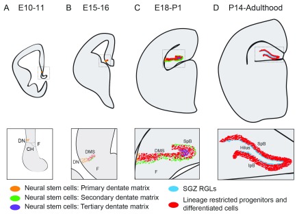
Figure 3. Development of the dentate gyrus.
( A) Around embryonic day 10–11 (E10–11), the putative dentate neuroepithelium is situated adjacent to the dentate notch (DN), a small indentation of the ventromedial ventricular wall, which in turn is placed caudal to the cortical hem (CH), neighboring the fimbria (F). These neural stem cells (NSCs) are sometimes referred to as the primary dentate matrix. ( B) During embryonic neurogenesis (E15–16), a stream of neural progenitors appears medial to the DN forming the dentate migratory stream (DMS). The DMS consists of both lineage-restricted neural progenitor cells (Tbr2 +) and NSCs (expressing Sox2, Nestin, and GFAP). The DMS leads to the dentate primordium. ( C) At about E18, the upper blade of the dentate gyrus (also called the suprapyramidal blade, or SpB) starts to form and contains post-mitotic Prox1 + neurons, as well as a layer of NSCs that are located subpially, on the upper part of the SpB blade. These NSCs are defined as the secondary dentate matrix, and the NSCs ventral to the SpB are termed the tertiary dentate matrix. ( D) At about postnatal day 14 (P14), both the SpB and the infrapyramidal blade (IpB) have formed. At this stage, the secondary and tertiary matrices are gone, and the remaining NSCs—now referred to as radial glia-like cells (RGLs)—are located in the subgranular zone (SGZ), on the border between the granule cell layer and the hilus. This structure and morphology are maintained throughout the rest of the animal’s life.

