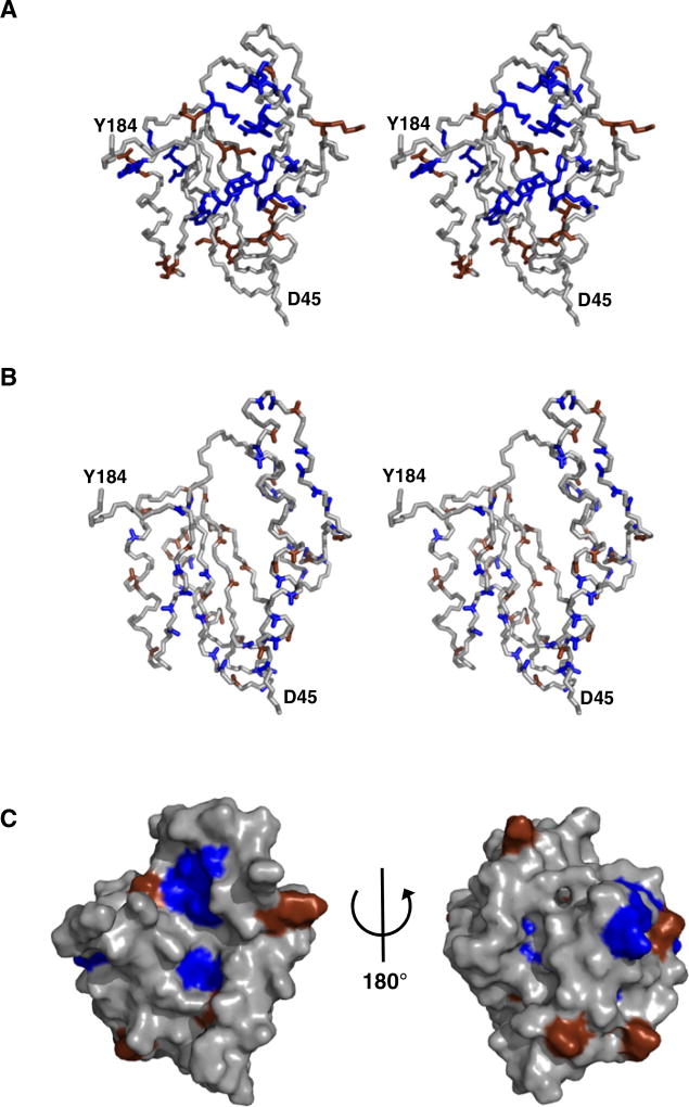
Figure 4. MESD Contains Surface-Located Hydrophobic and Acidic Residues Crucial for Function.
(A) Summary of LRP6 maturation assay. Single mutations of MESD reducing the level of mature LRP6 (blue) or not affecting the LRP6 maturation (brown) are mapped to the model structure of MESD45–184 as stereo view.
(B) The crucial hydrophobic and acidic areas are surface located, as shown by the increase of amide hydrogen T1 relaxation after addition of paramagnetic GdIII-DTPA to the sample buffer. The ratioT1Gd (III)/T1reference was lower than 0.9 for the amide hydrogens colored blue and higher than 0.9 for the brown-colored ones. Stereo view.
(C) Surface view of MESD45–184 with color coding as in (A).

