Abstract
Cytosine-guanosine deoxynucleotides (CpG) DNA can be delivered in ovo at embryo day (ED)18 for the stimulation of toll-like receptor (TLR)21 signaling pathway that ultimately protects chickens against a number of bacterial and viral infections. There is a dearth of information understanding the mechanisms of protection induced by in ovo delivered CpG DNA. The objective of this study was to determine the immune cell changes post-hatch following in ovo delivery of the TLR21 ligand, CpG DNA. In order to quantify changes of percentage of KUL01+, IgM+ B, cluster of differentiation (CD)4+ and CD8α+ cells, trachea, lung, duodenum, large intestine, spleen and bursa of Fabricius were collected on day 1 post-hatch. We found increased recruitments of KUL01+ cells, in organs of these body systems post-hatch following in ovo delivery of CpG DNA. Although IgM+ B cells, CD4+ and CD8α+ cells were increased in lungs and immune system organs, these cells were not quantifiable from the trachea, duodenum and large intestine immediately following the hatch. Furthermore, when CpG DNA is delivered in ovo and subsequently infected with infectious laryngotracheitis virus (ILTV) post-hatch on day 1, CpG DNA reduces morbidity and mortality resulting from ILTV infection. This study provides insights into the mechanisms of host responses elicited following in ovo delivery of CpG DNA in avian species.
Introduction
CpG DNA is classified into three major classes, class A, B and C based on the structural variations and their effects on peripheral blood mononuclear cell (PBMC)s [1, 2]. Class A CpG DNA mainly activates the dendritic cell (DC)s and natural killer (NK) cells mediated via interferon regulatory factor (IRF)7 signaling pathways from early endosomes leading to increasing production of type 1 interferon (IFN)s. The class B CpG DNA is a strong activator of B cells and monocytes and operates via nuclear factor (NF)-kB signaling pathway from late endosomes leading to the production of pro-inflammatory mediators. Class C CpG DNA shows the characteristics of both class A and B [3, 4] in terms of the structure and functions. Toll-like receptor (TLR)9 in mammals and TLR21 in avian species detect both bacterial and viral DNA containing unmethylated CpG motifs [5], which are generally methylated in the genomes of vertebrate [6, 7]. The frequency of CpG motifs is also negligible in vertebrate DNA, while it occurs with high frequency in microbial genomes [4] and that allow elicitation of host responses against DNA of microbial origin and not against the host origin.
Induction of innate host responses by the treatment of CpG DNA has been studied in various animal models. For example, many studies in the mouse model reported that treatment of CpG DNA significantly stimulates the recruitment of innate immune cells such as macrophages and NK cells in the respiratory and genital mucosal epithelium [8, 9] correlating with the inhibition of viral replication in the subsequent challenges with herpes simplex virus (HSV)-2 [8] and influenza virus [10] respectively. CpG DNA is also known to increase adaptive immune cells such as B cells and T cell subsets via increased cell proliferation and cell survival, which has been recorded in mammals [11–14]. In avian species, there is an indication that CpG DNA induce proliferation of B cells in vitro [15] and B cells and T cell subsets in vivo in four weeks old chickens [16].
Pre-hatch or in ovo vaccination is a major advancement in infectious disease control in chickens and it is practiced at embryo day (ED) 18. When the eggs are hatched three days following the in ovo vaccination and placed the newly hatched chickens in poultry barns, a number of vaccines have been introduced to the chicks reducing the window of susceptibility for various infectious diseases [17]. In ovo delivered CpG DNA has been shown to reduce microbial infections encountered post-hatch in chickens such as bacterial infections [18–20] and viral infections [9, 21] correlating with macrophage response in lungs. However, it is not known whether in ovo delivered CpG DNA is capable of eliciting 1) macrophage responses post-hatch in other body systems and 2) adaptive immune cells in respiratory and other body systems. In the present study, we investigated whether the prophylactic use of in ovo delivered TLR21 ligand, CpG DNA could stimulate mucosal immune responses in lungs, trachea, duodenum, large intestine, spleen and bursa of Fabricius post-hatch potentially reducing infection of infectious laryngotracheitis virus (ILTV). Our data demonstrate that in ovo delivery of CpG DNA increases recruitments of KUL01+, IgM+ B cells, CD4+ and CD8α+ cells day 1 post-hatch at variable extents. When the chickens were infected with ILTV at day 1 of age coinciding with this augmented cellular response induced by in ovo delivered CpG DNA, the ILTV induced morbidity and mortality were reduced potentially minimizing the replication of the virus indicating that in ovo delivery CpG DNA may be a prophylactic measure against ILTV infection.
Materials and methods
Animals
The Veterinary Science Animal Care Committee (VSACC) and Health Science Animal Care Committee (HSACC) have approved the use of SPF eggs, embryos, and chickens used in all our experimental procedures (animal Protocol #: AC13-0291). The sampling of chicken tissues was performed as has been approved by the institutional animal care committees. Briefly, for embryo sampling at the ED19, the egg shell was cut opened at the broader end of the egg, fetal membranes were disrupted and the embryos were decapitated. For the sampling of tissues post-hatch at day 1 and at endpoints, the chickens were euthanized using overdose of isoflurane anesthesia followed by cervical dislocation. The eggs purchased from Canadian Food Inspection Agency (CFIA), Ottawa, Canada were incubated at Health Research Innovation Center (HRIC), University of Calgary in digital incubators (Rcom Pro 20 and 50, Kingsuromax 20 and Rcom MARU Deluxe max, Autoelex Co., Ltd., GimHae, GyeongNam, Korea). During the incubation (60–70% relative humidity and 37.2–37.6°C temperature depending on the stage of the incubation) [22], the eggs were candled to select fertile eggs at ED11 for the experiments.
Virus and TLR ligand
The ILTV (strain N-71851) purchased from the American Type Culture Collection (ATCC, Manassas, Virginia, United States), was used in the studies [9, 23]. Initially, the viral titer was determined by plaque assay done using leghorn chicken hepatocarcinoma (LMH) cells [24].
The ligand for TLR21, Class B CpG DNA 2007 (5'-TCG TCG TTG TCG TTT TGT CGT T-3'), and non-CpG DNA (5'-TGC TGC TTG TGC TTT TGT GCT T-3') (Invivogen, San Diego, California, USA) were purchased from Cedarlane (Burlington, ON, Canada). These class B CpG DNA sequences, which consist of a complete phosphorothioate backbone, have been shown to induce strong monocyte and B cell responses when compared to non-CpG DNA [25, 26].
Experimental design
Characterization of cell-mediated immune responses in the embryo (ED19) lungs following in ovo delivery CpG DNA
In order to determine whether in ovo delivery of CpG DNA leads to cellular responses characterized by KUL01+, IgM+ B, CD4+ and CD8α+ cells, the lung tissues that originated from in ovo delivered CpG DNA (50 μg CpG DNA in 200 μL PBS per egg) group (n = 9), control group injected with 50 μg non-CPG DNA in 200 μL PBS in ovo (n = 4) and in ovo PBS treated group (n = 6) were collected and embedded in OCT compound (Tissue-Tek®, Sakura Finetek USA inc, Torrance, CA, USA) 24 hours following the in ovo treatments. The OCT blocks were snap frozen in dry ice and stored at -80°C before being sectioned.
Characterization of cell-mediated immune responses in multiple body systems of day 1 aged chickens following in ovo delivery CpG DNA
To study the potential cellular mediators of antimicrobial response, such as KUL01+, IgM+ B, CD4+ and CD8α+ cells elicited by CpG DNA, we delivered CpG DNA (50μg of CpG DNA diluted with 200μl of sterile PBS per egg, n = 5) in ovo at ED18 in SPF chicken eggs, while the two control groups received either 50 μg non-CPG DNA in 200 μL PBS in ovo (n = 4) or 200μl of sterile PBS per egg in ovo (n = 4). At day 1 post-hatch, the lungs, tracheas, duodenum, large intestine, spleen, and bursa of Fabricius were collected and preserved in OCT compound (Tissue-Tek®, Sakura Finetek USA inc, Torrance, CA, USA), snap frozen in dry ice and stored at -80°C before being sectioned.
Determination of antiviral response elicited by in ovo delivered CpG DNA against ILTV infection encountered post-hatch
To investigate the antiviral response elicited by in ovo delivered CpG DNA in lungs, trachea, duodenum, large intestine, spleen and bursa of Fabricius against post-hatch ILTV infection, ED18 SPF eggs were delivered in ovo either with 50μg of CpG DNA (diluted with 200μl of sterile PBS per egg, n = 8) or 50μg of non-CpG DNA in 200μl of sterile PBS per egg (n = 7). Both groups were infected with ILTV intra-tracheally at day 1 post-hatch (3x10⁴ plaque forming units or PFUs in 30μl/chicken). To facilitate the intra-tracheal ILTV infection using a pipette, the chicken tongue was gently pulled out and downwards while under isoflurane anesthetic, exposing the glottis, the opening that connects to the larynx. Then, the chickens were observed for 12 days and clinical signs and endpoints determined. The clinical signs were scored on a scale of 0–5 (No clinical signs = 0, droopy wings/ ruffled feathers and huddling together/ depression or inactive with lowered head/ loss of body weight/ mild increase of respiration = 1, moderate increase of respiration = 2 and severe increase of respiration or gasping = 3). When a birds reached a cumulative score of 5 (humane end point), the chickens were euthanized [9]. At 4 days post-infection, oropharyngeal and cloacal swabs were collected using Puritan® UniTranz-RT® Media Transport Systems (VWR, Edmonton, AB, Canada), which consisted of the sterile swabs and transport medium and ILTV genome quantified following DNA extraction. Since ILTV replicates mainly in tracheal epithelium, we preferred oropharyngeal swabs rather than tracheal swabs of 5 days old chickens in order to prevent any inadvertent injury to tracheal mucosa. When the chickens reached the humane and experimental endpoints, the lungs were collected and preserved in OCT compound (Tissue-Tek®, Sakura Finetek USA inc, Torrance, CA, USA), snap frozen in dry ice and stored at -80°C before being sectioned. Additionally, a portion of the lungs also collected in RNA Save (Biological Industries, FroggaBio, Toronto ON, Canada) and stored in -20°C before being used for DNA extraction.
In ovo delivery technique
Briefly, the egg shell was punctured through the air sac at the broader end of the eggs using a sterile 23-gauge needle after disinfecting the shell surface with 70% ethanol. Two and a half centimeters long, a 23-gauge needle was then used for in ovo delivery of compounds into the amniotic cavity by inserting the entire length of the needle perpendicularly through the punctured hole. At the end of the procedure, the holes were sealed with lacquer and eggs were placed in the incubator for further incubation.
Immunofluorescent assay
The tissues preserved in OCT were sectioned (thickness of 5 micrometer, μm) and indirect immunofluorescent assay was used to quantify KUL01+, IgM+ B cells, CD4+cells and CD8α+ cells numbers in lungs, trachea, duodenum, large intestine, spleen and bursa of Fabricius. For KUL01+, IgM + B cell, CD4+ cell and CD8α+ cell staining, 5% goat serum in TBS buffer (Trizma base: 2.42g, NaCl: 8g in 1liter (L) of distilled water, pH 7.6) was used for the purpose of blocking and incubated at room temperature for 30 minutes in a humidified chamber. Unlabeled mouse monoclonal antibody specific for chicken macrophages/monocytes/interdigitating cells, KUL01 (Southern Biotech, Birmingham, Alabama, USA), IgM (M-4, Southern Biotech, Birmingham, Alabama, USA), CD4 (CT-4, Southern Biotech, Birmingham, Alabama, USA) and CD8α (CT-8, Southern Biotech, Birmingham, Alabama, USA) were used in 1:200 dilution (except IgM which was used at 1:100 dilution) in blocking buffer and incubated for 30 minutes at the room temperature in a humidified chamber. For KUL01+, IgM and CD8α+ cell staining DyLight® 550 conjugated goat anti-mouse IgG (H+L) (Bethyl Laboratories Inc., Montgomery, TX, USA) was used in 1:500 dilution in blocking buffer as the secondary antibody and incubated for 1 hour at the room temperature in a humidified chamber. For CD4+ cell staining biotinylated Goat Anti-Mouse IgG (H+L) (Southern Biotech, Birmingham, Alabama, USA) was used in 1:250 dilution in blocking buffer and incubated for 30 minutes and then incubated with DyLight® 488 streptavidin in 15:1000 dilution for 30 minutes at the room temperature in a humidified chamber. TBS-T buffer (TBS with 0.1% Tween 20) was used as the washing buffer followed by PBS and after each step, the sections were washed 3 times (twice with TBS-T buffer and once with PBS) with an interval of 3–5 mins. Finally, the slides were mounted in Vectashield mounting medium with 4′, 6-Diamidine-2′-phenylindole dihydrochloride (DAPI) (Vector Laboratories Inc., Burlingame, CA, USA), cover slipped and edges sealed with lacquer.
DNA extraction and real time polymerase chain reaction (PCR)
From the swab samples collected at 4 days post-infection and also lungs collected from chickens that reached the end points, DNA extraction was carried out using QIAamp DNA mini kit (QIAGEN GmbH, Hilden, Germany) as per manufacturer’s guidelines and the concentration of DNA was quantified using the Nanodrop 1000 spectrophotometer at the wavelength of 260/280 nm (ThermoScientific, Wilmington, DE, USA). Two hundred nano grams (ng) (cloacal swabs, lungs) or 25 ng (oropharyngeal swabs) of the extracted DNA were used for real-time PCR. The real time PCR assay was conducted in a 96-well PCR plate (VWR, Edmonton, AB, Canada) in duplicate to quantify the protein kinase (PK) gene of ILTV in relation to β actin housekeeping gene. Briefly, the calculated copy numbers of PK gene were normalized with the copy numbers of β-actin gene and expressed for 1 x 10⁶ host cells). In order to generate a standard curve, a dilution series of ILTV plasmids (cloned into the pCR®2.1-TOPO® vector and amplified in One Shot® Escherichia coli, TOPO TA Cloning kit Top 10 (Invitrogen, Burlington, ON, Canada) were used in duplicate with copy numbers ranging from 1.56 x 10³-1.56x10¹⁰. Fast SYBR® Green Master Mix (Invitrogen, Burlington, ON, Canada) was used in 20μl of reaction volume. The detection of intercalating SYBR® Green dye was conducted in a Thermal Cycler (CFX96-C1000) (Bio-Rad Laboratories, Mississauga, ON, Canada). Five pico molar (pM) of ILTV PK (ORF2) gene specific primers (F: 5’-TAC GAT GAA GCG TTC GAC TG -3’ and R: 5’-AGG CGT GAC AGT TCC AAA GT -3’) [27, 28] or β actin primers (F: 5’-CAA CAC AGT GCT GTC TGG TGG TA-3’ and R: 5’-ATC GTA CTC CTG CTT GCT GAT CC -3’) [9, 29] were used in each reaction and gene specific plasmids were included as a positive control and DNAse/RNAse free water was included as a negative control. The optimum parameters for thermal cycling were 95°C for 20 seconds (s) of pre-incubation, 95°C for 3s of 40 amplification cycles with the final segment of 60°C for 30s. Melting curve was analyzed at 95°C for 10s, 65°C for 5s and finally 9°C for 5s. Acquisition of fluorescent signals was performed at 60°C for 30s.
Data analyses
For the quantification of numbers of KUL01+, IgM + B cells, CD4+ cells and CD8α+ cells in the examined tissues, 3–5 areas with highest DyLight® 550 (KUL01+, IgM+ B cells and CD8α+ cells) or DyLight® 488 (CD4+ cells) fluorescent signals and corresponding nuclear stained (DAPI) areas were captured under 40X magnification from each section. Then, these images were subjected to fluorescent intensity quantification using the Image-J software (National Institute of Health, Bethesda, Maryland, USA). The resultant fluorescent intensities for DyLight® 550 or DyLight® 488 positive signals were expressed relative to the total areas (as estimated by nuclear staining with DAPI) as a percentage [21].
Student’s t-test (GraphPad Prism Software 5, La Jolla, CA, USA) was used for the purpose of identifying the differences between two groups. When more than two groups are parts of an experiment, the one-way analysis of variance (ANOVA) with Bonferroni's post test for selected comparison was performed to identify the differences between groups. In addition, Log-rank test was used to identify group differences in survival percentage and Mann-Whitney U test was used to identify group differences in clinical score data for selected time points. Before being analyzed each set of data, the outlier test was conducted using the Grubbs’ test (GraphPad software Inc., La Jolla, CA, USA). The differences between groups were considered statistically significant at P ≤ 0.05.
Results
In ovo delivery of CpG DNA increases KUL01+ and CD4+ cells in the lungs pre-hatch while PBS and non-CpG DNA (control CpG DNA) induce similar minimal cellular responses
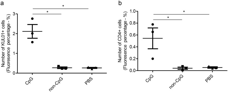
We examined pre-hatch lungs for KUL01+, IgM + B cells and CD4+ and CD8α+ cell subsets following in ovo delivery of CpG DNA, non-CpG DNA and PBS. We were able to detect and quantify only KUL01+, and CD4+ cells (Table 1). We found that in ovo delivered CpG DNA at ED18 increased the number of KUL01+ (P<0.05) and CD4+ cells (P = 0.02) in pre-hatch lungs (ED19) compared to the controls (Fig 1). The KUL01+ and CD4+ cell responses between non-CpG DNA and PBS groups were similar (P>0.05).
Table 1. The cellular responses following in ovo delivery of CpG DNA in different organs pre- and post-hatch.
| Cell types | Pre-hatch (Lungs) | Post-hatch | |||||
|---|---|---|---|---|---|---|---|
| Lungs | Trachea | Duodenum | Large intestine | Spleen | Bursa of Fabricius | ||
| KUL01+ cells | √ * | √* | √* | √ * | √ * | √ | √ * |
| CD4+ cells | √ * | √ * | - | - | - | √ * | √ * |
| CD8α+ cells | - | √ * | - | - | √ | - | √ |
| IgM+ B cells | - | √ | - | - | - | √ * | - |
* indicates significant increase in CpG DNA group at P ≤ 0.05 compared to PBS or non-CpG or both groups
√ indicates an increased trend with CpG DNA treatment and—indicates no changes/not quantifiable.
Fig 1. In ovo delivery of CpG DNA increases KUL01+ and CD4+ cell numbers in lungs pre-hatch.
SPF ED18 eggs were injected with CpG DNA (n = 9), non-CpG DNA (n = 4) or PBS (n = 6). At ED19, lungs were collected and immunofluorescent assay was performed for KUL01+ and CD4+ cells. The quantitative data for KUL01+ (a) and CD4+ cells (b) are shown. The one-way ANOVA test with Bonferroni's post test for selected comparison was performed to identify group differences and the differences were considered significant at P< 0.05. The bars represent mean ± SEM.
In ovo delivered CpG DNA increases KUL01+ cells in lungs, trachea, duodenum, large intestine and bursa of Fabricius post-hatch
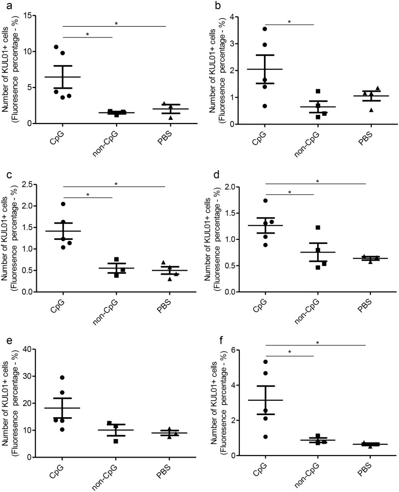
Since we observed that in ovo delivered CpG DNA induces KUL01+and CD4+ cell responses pre-hatch compared to PBS and non-CpG DNA controls in lungs, then we investigated to see whether there is similar cellular response day 1 post-hatch. First, we found that in ovo delivered CpG DNA at ED18 increased the number of KUL01+ cells post-hatch in lungs (P<0.05, Fig 2a), trachea (P<0.05, Fig 2b), duodenum (P <0.05, Fig 2c), large intestine (P<0.05, Fig 2d) and bursa of Fabricius (P<0.05, Fig 2f) when compared to non-CpG DNA delivered group. The KUL01+ cell numbers in in ovo CpG DNA delivered group was also significantly higher in lungs (P<0.05, Fig 2a), duodenum (P <0.05, Fig 2c), large intestine (P<0.05, Fig 2d) and bursa of Fabricius (P<0.05, Fig 2f) when compared to PBS control group. In the spleen, the differences of KUL01+ cell numbers between in ovo CpG DNA delivered and control groups was not significant (P>0.05, Fig 2e). The KUL01+ cell recruitment between non-CpG DNA and PBS control groups were similar (P>0.05, Fig 2). Fig 1 illustrates the KUL01+ cells responses in lungs, trachea, duodenum, large intestine, spleen and bursa of Fabricius post-hatch following in ovo CpG DNA, non-CpG DNA or PBS deliveries.
Fig 2. In ovo delivered CpG DNA increases KUL01+ cell numbers in lungs, trachea, duodenum, large intestine and bursa of Fabricius.
SPF ED18 eggs were delivered with CpG DNA (n = 5), non-CpG DNA (n = 4) or PBS (n = 4) in ovo and the eggs were incubated to hatch. At day 1 post-hatch, samples of lungs, trachea, duodenum, large intestine, spleen and bursa of Fabricius were collected in OCT, sectioned and immunofluorescent assay was performed to quantify KUL01+ cells. The quantitative data from the immunofluorescent assay of each organ is shown a) lungs, b) tracheas, c) duodenum, d) large intestine, e) spleen, f) bursa of Fabricius. The one-way ANOVA test with Bonferroni's post test for selected comparison was performed to identify group differences and the differences were considered significant at P< 0.05. The bars represent mean ± SEM.
In ovo delivered CpG DNA increases cells of adaptive immune system only in the lungs, spleen and bursa of Fabricius post-hatch
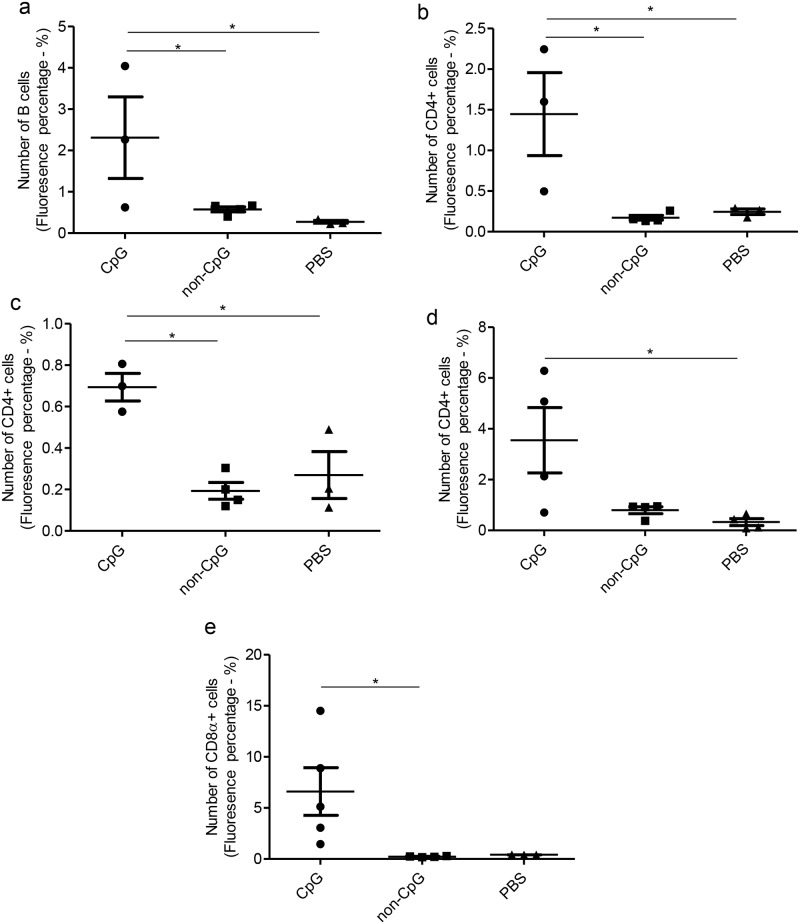
Since we observed that in ovo CpG DNA increases KUL01+ cell numbers in lungs, trachea, duodenum, large intestine and bursa of Fabricius post-hatch as an indication of enhanced innate immune response, then we investigated the potential recruitment of cells of the adaptive immune system post-hatch following in ovo CpG DNA delivery. On the contrary to the KUL01+ cells response following in ovo CpG DNA delivery, IgM + B cells were quantifiable only in lungs and spleen of the examined tissues post-hatch (Table 1). We found that in ovo delivered CpG DNA at ED18 increased the number of IgM + B cells in spleen (P<0.05) when compared to both control groups but we could not observe a difference in IgM + B cells in lungs of in ovo CpG DNA and PBS or non-CpG DNA delivered groups (P>0.05) (Fig 3a).
Fig 3. In ovo delivery of CpG DNA increases IgM+ B cells in spleen, CD4+ cells in lungs, bursa of Fabricius and spleen and CD8α+ cells in lungs post-hatch.
SPF ED18 eggs were delivered with CpG DNA (n = 5), non-CpG DNA (n = 4) or PBS (n = 4) in ovo and the eggs were incubated to hatch. At day 1 post-hatch, lungs, trachea, duodenum, large intestine, spleen and bursa of Fabricius were collected in OCT, sectioned and immunofluorescent assay was performed for the quantification of IgM + B cells, CD4+ cells and CD8α+ cells. The quantitative data from the immunofluorescent assay for IgM+ B cells in spleen (a) for CD4+ cells in lungs (b), bursa of Fabricius (c) and spleen (d) and for CD8α+ cells in lungs (e) are shown. The one-way ANOVA test with Bonferroni's post test for selected comparison was performed to identify group differences and the differences were considered significant at P< 0.05. The bars represent mean ± SEM.
Again, on the contrary to the KUL01+ cell response following in ovo CpG DNA delivery, CD4+ cells were quantifiable only in lungs, spleen and bursa of Fabricius of the examined tissues post-hatch (Table 1). We found that in ovo delivered CpG DNA at ED18 increased the number of CD4+ cells in lungs (P<0.05, Fig 3b), bursa of Fabricius (P<0.05, Fig 3c) and spleen (P<0.05, Fig 3d) of in ovo CpG DNA delivered group when compared to PBS or non-CpG DNA delivered controls.
The CD8α+ cells were quantifiable only in lungs, large intestine and bursa of Fabricius of the examined tissues post-hatch (Table 1). We found that in ovo delivered CpG DNA at ED18 increased the number of CD8α+ cells in lungs when compared to the in ovo non-CpG DNA delivered control group (P<0.05, Fig 3e). However, we could not observe a difference in CD8α+ cells in large intestine (P>0.05) and bursa of Fabricius (P>0.05) of in ovo CpG DNA and PBS or non-CpG DNA delivered groups.
In ovo delivered CpG DNA induces protective host response against ILTV infection encountered post-hatch
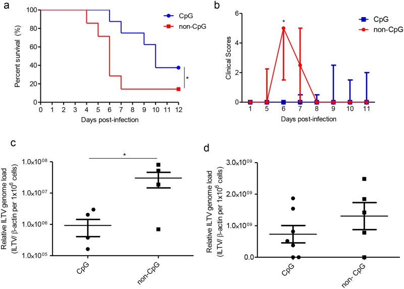
Since we observed that in ovo delivery of CpG DNA increases recruitments of KUL01+, IgM+ B cells, CD4+ and CD8α+ cells day 1 post-hatch in lungs, trachea, duodenum, large intestine, spleen and bursa of Fabricius, we then investigated to determine if CpG DNA administration influences disease presentation and viral replication after ILTV inoculation at day 1 post-hatch. We found that in ovo delivered CpG DNA at ED18 significantly reduced; 1) mortality (P≤0.05, Fig 4a), 2) clinical scores at 6 days post-infection (P<0.05, Fig 4b) and 3) cloacal excretion of the ILTV genome (P<0.05, Fig 4c) but not the oropharyngeal excretion of the ILTV genome (P>0.05, Fig 4d) at 4 days post-infection associated with post-hatch ILTV infection.
Fig 4. In ovo delivered CpG DNA induces protective host response against ILTV infection encountered post-hatch.
SPF ED18 eggs were delivered with CpG DNA (n = 8) or non-CpG DNA (n = 7) in ovo, the eggs were incubated to hatch and infected with ILTV intra-tracheally at day 1 post-hatch. The chickens were observed for 12 days. At 4 days post-infection, oropharyngeal and cloacal swabs were collected and ILTV genome loads quantified. a) Survival percentage, b) clinical scores, c) ILTV genome loads in cloacal swabs and d) ILTV genome loads in oropharyngeal swabs. Log-rank test was used to identify differences in survival percentages, Mann-Whitney U test was used to identify differences in clinical score data and student's t-test was used to identify differences in ILTV genome loads. The differences were considered significant at P< 0.05. The bars represent median ± inter quartile range in Fig. 4b and mean ± SEM in Fig. 4a, c and d.
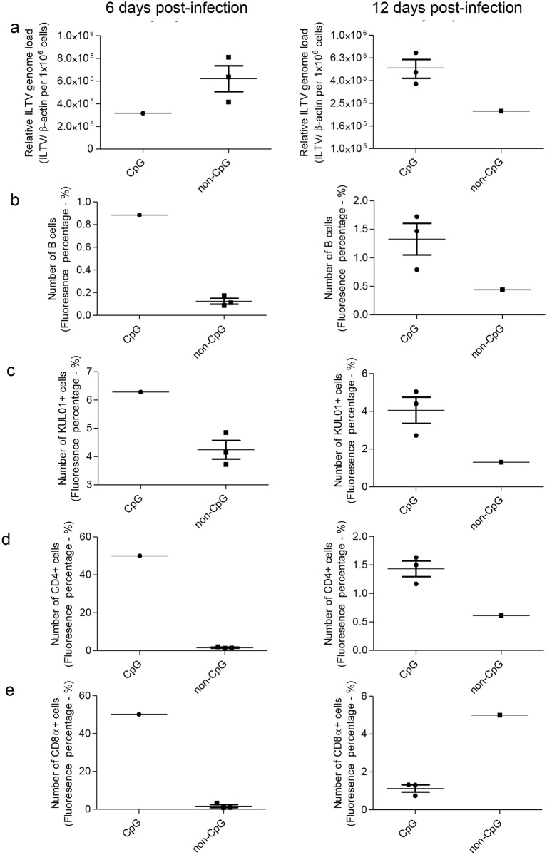
The chickens that reached the selected humane and experimental end points were evaluated for their ILTV genome loads in lungs. Although there is a trend of increased ILTV genome load in lungs at 6 days post-infection in in ovo non-CpG DNA treated lungs and at 12 days post-infection in in ovo CpG DNA treated liungs (Fig 5a). On the other hand, these end point lungs of ILTV infected in ovo CpG DNA delivered group had higher trend of number of cells involve in innate immune responses, IgM+ B and CD4+ cells in both time points (Fig 5b–5d) and CD8α+ cells in 6dpi (Fig 5e) when compared to the lungs of chickens that were ILTV infected and in ovo non-CpG DNA delivered. However, statistical analysis of these data were precluded due to less number of samples per group in each time point.
Fig 5. ILTV genome loads and KUL01+, IgM+ B, CD4+ and CD8α+ cell numbers in lungs of in ovo CpG DNA and non-CpG DNA treated chickens following ILTV infection post-hatch.
The chickens were delivered with either in ovo CpG DNA or non-CpG DNA and infected with ILTV intra-tracheally at day 1 post-hatch. The chickens were observed for end points. The lungs were collected for determination of ILTV genome loads and cellular responses from chickens that reached humane endpoint at 6 days post-infection and experimental end point at 12 days post-infection. The ILTV genome loads are given separately for 6 and 12 days post-infection (a). The quantitative data of the immunofluorescent assays done for IgM+ B cells (b), KUL01+ (c), CD4+ cells (d) and CD8α+ cells (e) are shown separately for 6 and 12 days post-infection. Statistical analysis was precluded due to low numbers of animals per group per time point. The bars represent mean ± SEM.
Discussion
The investigations described here led to three major findings. First, in ovo delivered CpG DNA is capable of eliciting significant cellular responses characterized by innate (KUL01+ cells) and adaptive (IgM+ B cells and CD4+ and CD8α+ cells) immune cells day 1 post-hatch. Second, the cellular responses stimulated by in ovo delivered CpG DNA in post-hatch chickens are observable in multiple body systems, i.e. lungs, trachea, duodenum, large intestine, spleen and bursa of Fabricius at variable extents (Table 1). Third, the cellular responses induced by in ovo delivered CpG DNA were significant at the time of infection (day 1 post-hatch). Whether these innate and adaptive immune cells played a role in the reduction of mortality and virus replication requires further investigation.
For the infection of ILTV we used intra-tracheal infection [23, 30]. When we infected the chickens via this route, we observed significantly higher survival of the chickens that received in ovo CpG DNA. Although protection against many viral infections depend on antibody-mediated and cell-mediated immune responses, the involvement of antibodies in the protection against ILTV infection is questionable [31, 32] and cell-mediated immune response may play a dominant role. In this context, induction of cellular immune response characterized by KUL01+, CD4+ and CD8α+ cell subsets following in ovo delivered CpG DNA may potentially applicable for the control of ILTV infection. Although, we did not investigate the origin of these innate and adaptive immune cells in the examined tissues as to circulating pool of cells or proliferating cells, the evidence of perivascular cuffs was lacking. Following in ovo CpG DNA delivery the observed cells were distributed in mucosa or submucosal areas in trachea, duodenum and large intestine. In the lungs, bursa of Fabricius and spleen these immune cells were localizing in the air exchange areas, between bursal follicles or spleen parenchyma respectively following in ovo CpG DNA delivery.
Although KUL01+ cells were well distributed in all the examined tissues, CD4+, CD8α+ and B cells were not quantifiable in trachea, duodenum and large intestine (except CD8α+ cells in large intestine) using immunofluorescent assays (Table 1). The lack of CD4+ and CD8α+ cells in trachea, duodenum and large intestine may be due to the lack of colonization of these cells by day 1 post-hatch. It is well known that the colonization of peripheral tissues by CD4+ and CD8α+ cells mostly occur during early post-hatch period [33–35]. However, it has been reported previously that the chickens aged day 1 have much lower numbers of CD4+ and CD8α+ cells when compared to day 7 chickens [35]. The lack of IgM+ B cells in trachea, duodenum and large intestine on day 1 post-hatch may also be related to the lack of B cell colonization in these tissues. This is in agreement with the previous data that the chickens are capable of producing antibodies to orally administered antigen, bovine serum albumin, only after day 8 post-hatch [36]. However, Bar-Shira et al (2003) recorded that mRNA of the CD3γδ and B cell marker (Bu-1) is quantifiable in the various components of the gastrointestinal tract on day 1 post-hatch at a very low quantity when compared to day 4, 6, 8 and 12. This contrasts with our observation that B cells, CD4+ cells and CD8α+ cells are not detectable in duodenum and large intestine. This discrepancy in observations between ours and Bar-Shira et al’s could be explainable by the lower sensitivity of the immunofluorescent technique than real-time PCR technique as well as the fact that not all detected transcripts are translated into functional proteins [37]. Adaptive immune cell recruitment in response to activation of other avian TLR pathways are scarce. However, in other animal models, for example in primates, Wille-Reece et al demonstrated that TLR7/8 ligand induces antigen specific CD8α+ cell response [38]. Similarly, Pasare and Medzhitov demonstrated that the activation of TLR4 signaling by lipopolysaccharide (LPS) influences activation and differentiation of B cells and antigen-specific antibody responses in mice [39].
Avian KUL01+ cells are multifunctional immune cells that play roles producing a number of cytokines, functioning as phagocytic cells and presenting antigens [40]. In ovo CpG DNA increases KUL01+ cells in lungs, trachea, duodenum, large intestine and bursa of Fabricius. Previously, we observed that in ovo CpG DNA increases expansion of KUL01+ cell numbers in lungs pre-hatch [9, 21, 41] on ED19. One of the limitation of our immunofluorescent technique used to quantify macrophages is that the KUL01 monoclonal antibody detects not only macrophages but also monocytes and interdigitating cells of chicken [42].
IgM+ B cells were higher in spleen in day 1 chickens following in ovo delivery of CpG DNA. In agreement with our observation, increases of circulating B cells have been observed in four weeks old chickens when CpG DNA was injected subcutaneously [16]. Although we did not investigate how the IgM+ B cell numbers were increased in spleen of CpG DNA treated chickens, the potential mechanisms may be related to polyclonal activation [11–15] or increased B cell survival due to inhibition of apoptosis [11, 43]. Both these mechanisms are possible since chicken B cells express TLR21 [44].
In agreement with our observation of increased CD4+ and CD8α+ cells in tissues originated from in ovo CpG DNA delivered group, increase of circulating and spleen CD3+ and CD4+ cells have been observed in 4 weeks old chickens when CpG DNA is injected subcutaneously [16]. The CD4+ and CD8α+ subsets may be recruited following in ovo CpG DNA delivery via various mechanisms. First, CpG DNA would have acted as a mitogen increasing these two CD4+ and CD8α+ cell subsets via proliferation independent of antigens [45]. Second, it is possible that the increases in CD4+ and CD8α+ cells would have resulted consequent to increased survival of the CD4+ and CD8α+ cells [45]. The direct interactions of CD4+ and CD8α+ cells with CpG DNA are possible since it is known that these CD4+ and CD8α+ cell subsets express receptor for CpG DNA, TLR21 [46, 47]. Third, it is also possible that indirect cytokine mediated activation and proliferation of CD4+ and CD8α+ cells.
The implications of our observations described in the manuscript are many-fold. First, we found that the in ovo administration of CpG DNA stimulates the innate and adaptive arms of the immune system as indicated by the expansion of numbers of cells involve in innate immune responses, IgM+ B cell, CD4+ and CD8α+ cells. Whether this induction of innate and adaptive cellular responses leads to the reduced morbidity and mortality of ILTV infection requires further investigations. Second, as one of the major sites of ILTV replication, adaptive immune cell recruitment to the trachea was not significant following in ovo delivery of CpG DNA. This was reflected in the oropharyngeal ILTV genome loads which showed only a declining trend in CpG DNA treated chickens when compared to the non-CpG DNA treated chickens. This prompted us to evaluate the ILTV genome loads in the lungs of chickens that reached the end points and we found the same trend of lower ILTV genome loads as observed in oropharyngeal swabs in CpG DNA treated chickens when compared to that received non-CpG DNA. Further analysis indicated the increased trend of KUL01+, IgM+ B, CD4+ and CD8+ cell subsets in CpG DNA treated/ILTV infected chicken lungs at 6 days post-infection when compared to that observed in non-CpG DNA treated/ ILTV infected chickens. Overall, the lack of adaptive immune cell recruitment, particularly CD4+ and CD8α+ cell subsets with higher KUL01+ cell recruitment to trachea by the time of ILTV infection may potentially have contributed to the observed marginal effect of in ovo CpG treatment on lung ILTV genome load and oropharyngeal ILTV genome excretion, since it is well documented that cell-mediated rather than antibody-mediated immune response is critical for ILTV protection [31, 32]. Third, the induction of antiviral response pre-hatch via in ovo administration is desirable in the field situations in order to provide early host responses at the time of placing the day old chickens in the barn against circulating pathogens in the environment. This protection has been proven against a number of microbial infections in terms of significant reduction in the microbial burdens [23, 48]. In ovo route of vaccination is also extensively investigated and has become a commercial practice for the control of many poultry diseases worldwide [49], facilitating the effective and precise delivery of vaccines compared to conventional post-hatch administration methods. Although our findings are preliminary and need further investigations, in ovo CpG delivery may provide a basis for developing control measures against ILTV infections in chickens.
In conclusion, we have shown that prophylactic in ovo delivery of TLR21 ligand, CpG DNA, leads to increase in KUL01+, IgM+ B cells and CD4+ and CD8α+ cells in lungs, trachea, duodenum, large intestine, spleen and bursa of Fabricius coinciding with the reduction of ILTV infection induced morbidity, mortality and cloacal viral genome load. Although we studied the induction of immune cellular response against only ILTV infection, our observation of induced immune cellular responses in respiratory and intestinal organs following in ovo administration of CpG DNA may be potentially effective against other viruses such as infectious bursal disease virus [50, 51] and bacteria such as E. coli and Salmonella species [18–20] infecting in these organs in chicken. Further investigations are required on the functions of these innate and adaptive immune cells in the antiviral response against ILTV.
Acknowledgments
We would like to acknowledge the staff, Sampson Law, Angelo Bianchi, Scott MacMahon and Mackenzie Lewis of the Prion/virology animal facility at Foothill campus, University of Calgary for experimental animal management.
Data Availability
All relevant data are within the paper.
Funding Statement
This work was supported by the Canadian Poultry Research Council (http://cp-rc.ca), AMN088, received by MFA; the Alberta Agriculture and Forestry (http://www.agric.gov.ab.ca/app21/ministrypage), 2016R003R, received by MFA; and the Natural Sciences and Engineering Research Council of Canada (http://www.nserc-crsng.gc.ca/index_eng.asp), CRDPJ/491522-2015, received by MFA.
References
- 1.Bode C, Zhao G, Steinhagen F, Kinjo T, Klinman DM. CpG DNA as a vaccine adjuvant. Expert review of vaccines. 2011;10(4):499–511. Epub 2011/04/22. doi: 10.1586/erv.10.174 . [DOI] [PMC free article] [PubMed] [Google Scholar]
- 2.Dalpke AH, Heeg K. CpG-DNA as immune response modifier. International journal of medical microbiology: IJMM. 2004;294(5):345–54. Epub 2004/11/10. doi: 10.1016/j.ijmm.2004.07.005 . [DOI] [PubMed] [Google Scholar]
- 3.Krieg AM. Therapeutic potential of Toll-like receptor 9 activation. Nature reviews Drug discovery. 2006;5(6):471–84. Epub 2006/06/10. doi: 10.1038/nrd2059 . [DOI] [PubMed] [Google Scholar]
- 4.Krieg AM, Yi AK, Matson S, Waldschmidt TJ, Bishop GA, Teasdale R, et al. CpG motifs in bacterial DNA trigger direct B-cell activation. Nature. 1995;374(6522):546–9. Epub 1995/04/06. doi: 10.1038/374546a0 . [DOI] [PubMed] [Google Scholar]
- 5.Brownlie R, Zhu J, Allan B, Mutwiri GK, Babiuk LA, Potter A, et al. Chicken TLR21 acts as a functional homologue to mammalian TLR9 in the recognition of CpG oligodeoxynucleotides. Molecular immunology. 2009;46(15):3163–70. Epub 2009/07/04. doi: 10.1016/j.molimm.2009.06.002 . [DOI] [PubMed] [Google Scholar]
- 6.Krieg AM. Antitumor applications of stimulating toll-like receptor 9 with CpG oligodeoxynucleotides. Current oncology reports. 2004;6(2):88–95. Epub 2004/01/31. . [DOI] [PubMed] [Google Scholar]
- 7.Hemmi H, Takeuchi O, Kawai T, Kaisho T, Sato S, Sanjo H, et al. A Toll-like receptor recognizes bacterial DNA. Nature. 2000;408(6813):740–5. Epub 2000/12/29. doi: 10.1038/35047123 . [DOI] [PubMed] [Google Scholar]
- 8.Ashkar AA, Bauer S, Mitchell WJ, Vieira J, Rosenthal KL. Local delivery of CpG oligodeoxynucleotides induces rapid changes in the genital mucosa and inhibits replication, but not entry, of herpes simplex virus type 2. Journal of virology. 2003;77(16):8948–56. Epub 2003/07/30. doi: 10.1128/JVI.77.16.8948-8956.2003 . [DOI] [PMC free article] [PubMed] [Google Scholar]
- 9.Thapa S, Cader MS, Murugananthan K, Nagy E, Sharif S, Czub M, et al. In ovo delivery of CpG DNA reduces avian infectious laryngotracheitis virus induced mortality and morbidity. Viruses. 2015;7(4):1832–52. Epub 2015/04/10. doi: 10.3390/v7041832 . [DOI] [PMC free article] [PubMed] [Google Scholar]
- 10.Jiang T, Zhao H, Li XF, Deng YQ, Liu J, Xu LJ, et al. CpG oligodeoxynucleotides protect against the 2009 H1N1 pandemic influenza virus infection in a murine model. Antiviral research. 2011;89(1):124–6. Epub 2010/12/01. doi: 10.1016/j.antiviral.2010.11.013 . [DOI] [PubMed] [Google Scholar]
- 11.Yi AK, Chang M, Peckham DW, Krieg AM, Ashman RF. CpG oligodeoxyribonucleotides rescue mature spleen B cells from spontaneous apoptosis and promote cell cycle entry. J Immunol. 1998;160(12):5898–906. Epub 1998/06/24. . [PubMed] [Google Scholar]
- 12.Park MH, Jung YJ, Kim PH. Newly Identified TLR9 Stimulant, M6-395 Is a Potent Polyclonal Activator for Murine B Cells. Immune network. 2012;12(1):27–32. Epub 2012/04/27. doi: 10.4110/in.2012.12.1.27 . [DOI] [PMC free article] [PubMed] [Google Scholar]
- 13.Buchanan RM, Popowych Y, Arsic N, Townsend HG, Mutwiri GK, Potter AA, et al. B-cell activating factor (BAFF) promotes CpG ODN-induced B cell activation and proliferation. Cellular immunology. 2011;271(1):16–28. Epub 2011/07/05. doi: 10.1016/j.cellimm.2011.05.016 . [DOI] [PubMed] [Google Scholar]
- 14.Pontarollo RA, Rankin R, Babiuk LA, Godson DL, Griebel PJ, Hecker R, et al. Monocytes are required for optimum in vitro stimulation of bovine peripheral blood mononuclear cells by non-methylated CpG motifs. Veterinary immunology and immunopathology. 2002;84(1–2):43–59. Epub 2002/02/05. . [DOI] [PubMed] [Google Scholar]
- 15.Wattrang E. Phosphorothioate oligodeoxyribonucleotides induce in vitro proliferation of chicken B-cells. Veterinary immunology and immunopathology. 2009;131(3–4):218–28. Epub 2009/05/19. doi: 10.1016/j.vetimm.2009.04.013 . [DOI] [PubMed] [Google Scholar]
- 16.Chrzastek K, Piasecki T, Wieliczko A. Impact of CpG oligodeoxynucleotide stimulation on percentage of T and B cells in chicken. Polish journal of veterinary sciences. 2013;16(3):551–4. Epub 2013/11/08. . [DOI] [PubMed] [Google Scholar]
- 17.Abdul-Careem MF. Main Challenges in Poultry Farming: Hatchery Vaccination: Groupo Asis Biomedia, 50002 Zaragoza (Spain); 2015.
- 18.Mackinnon KM, He H, Swaggerty CL, McReynolds JL, Genovese KJ, Duke SE, et al. In ovo treatment with CpG oligodeoxynucleotides decreases colonization of Salmonella enteriditis in broiler chickens. Veterinary immunology and immunopathology. 2009;127(3–4):371–5. Epub 2008/11/21. doi: 10.1016/j.vetimm.2008.10.001 . [DOI] [PubMed] [Google Scholar]
- 19.Taghavi A, Allan B, Mutwiri G, Foldvari M, Van Kessel A, Willson P, et al. Enhancement of immunoprotective effect of CpG-ODN by formulation with polyphosphazenes against E. coli septicemia in neonatal chickens. Current drug delivery. 2009;6(1):76–82. Epub 2009/05/08. . [DOI] [PubMed] [Google Scholar]
- 20.Taghavi A, Allan B, Mutwiri G, Van Kessel A, Willson P, Babiuk L, et al. Protection of neonatal broiler chicks against Salmonella Typhimurium septicemia by DNA containing CpG motifs. Avian diseases. 2008;52(3):398–406. Epub 2008/10/23. doi: 10.1637/8196-121907-Reg . [DOI] [PubMed] [Google Scholar]
- 21.Abdul-Cader MS, Ahmed-Hassan H, Amarasinghe A, Nagy E, Sharif S, Abdul-Careem MF. Toll-like receptor (TLR)21 signalling-mediated antiviral response against avian influenza virus infection correlates with macrophage recruitment and nitric oxide production. The Journal of general virology. 2017;98(6):1209–23. Epub 2017/06/15. doi: 10.1099/jgv.0.000787 . [DOI] [PubMed] [Google Scholar]
- 22.Haddadi S, Thapa S, Kameka AM, Hui J, Czub M, Nagy E, et al. Toll-like receptor 2 ligand, lipoteichoic acid is inhibitory against infectious laryngotracheitis virus infection in vitro and in vivo. Developmental and comparative immunology. 2015;48(1):22–32. Epub 2014/09/10. doi: 10.1016/j.dci.2014.08.011 . [DOI] [PubMed] [Google Scholar]
- 23.Thapa S, Nagy E, Abdul-Careem MF. In ovo delivery of Toll-like receptor 2 ligand, lipoteichoic acid induces pro-inflammatory mediators reducing post-hatch infectious laryngotracheitis virus infection. Veterinary immunology and immunopathology. 2015;164(3–4):170–8. Epub 2015/03/15. doi: 10.1016/j.vetimm.2015.02.006 . [DOI] [PubMed] [Google Scholar]
- 24.Haddadi S, Kim DS, Jasmine H, van der Meer F, Czub M, Abdul-Careem MF. Induction of Toll-like receptor 4 signaling in avian macrophages inhibits infectious laryngotracheitis virus replication in a nitric oxide dependent way. Veterinary immunology and immunopathology. 2013;155(4):270–5. Epub 2013/09/17. doi: 10.1016/j.vetimm.2013.08.005 . [DOI] [PubMed] [Google Scholar]
- 25.Xie H, Raybourne RB, Babu US, Lillehoj HS, Heckert RA. CpG-induced immunomodulation and intracellular bacterial killing in a chicken macrophage cell line. Developmental and comparative immunology. 2003;27(9):823–34. Epub 2003/06/24. . [DOI] [PubMed] [Google Scholar]
- 26.Bhat A, Gomis S, Potter A, Tikoo SK. Role of Hsp90 in CpG ODN mediated immunostimulation in avian macrophages. Molecular immunology. 2010;47(6):1337–46. Epub 2010/01/26. doi: 10.1016/j.molimm.2009.12.011 . [DOI] [PubMed] [Google Scholar]
- 27.Bui VN, Ogawa H, Karibe K, Matsuo K, Nguyen TH, Awad SS, et al. Surveillance of avian influenza virus in migratory water birds in eastern Hokkaido, Japan. The Journal of veterinary medical science / the Japanese Society of Veterinary Science. 2011;73(2):209–15. Epub 2010/10/16. . [DOI] [PubMed] [Google Scholar]
- 28.WHO. Recommendations and laboratory procedures for detection of avian influenza A (H5N1) virus in specimens from suspected human cases2007:[7–14 pp.]. www.who.int/influenza/resources/documents/RecAIlabtestsAug07.pdf.
- 29.Villanueva AI, Kulkarni RR, Sharif S. Synthetic double-stranded RNA oligonucleotides are immunostimulatory for chicken spleen cells. Developmental and comparative immunology. 2011;35(1):28–34. Epub 2010/08/10. doi: 10.1016/j.dci.2010.08.001 . [DOI] [PMC free article] [PubMed] [Google Scholar]
- 30.Loncoman CA, Hartley CA, Coppo MJC, Vaz PK, Diaz-Mendez A, Browning GF, et al. Genetic diversity of infectious laryngotracheitis virus during in vivo co-infection parallels viral replication and arises from recombination 'hot-spots' within the genome. Applied and environmental microbiology. 2017. Epub 2017/09/25. doi: 10.1128/AEM.01532-17 . [DOI] [PMC free article] [PubMed] [Google Scholar]
- 31.Fahey KJ, York JJ. The role of mucosal antibody in immunity to infectious laryngotracheitis virus in chickens. The Journal of general virology. 1990;71 (Pt 10):2401–5. Epub 1990/10/01. doi: 10.1099/0022-1317-71-10-2401 . [DOI] [PubMed] [Google Scholar]
- 32.Honda T, Okamura H, Taneno A, Yamada S, Takahashi E. The role of cell-mediated immunity in chickens inoculated with the cell-associated vaccine of attenuated infectious laryngotracheitis virus. The Journal of veterinary medical science / the Japanese Society of Veterinary Science. 1994;56(6):1051–5. Epub 1994/12/01. . [DOI] [PubMed] [Google Scholar]
- 33.Dunon D, Courtois D, Vainio O, Six A, Chen CH, Cooper MD, et al. Ontogeny of the immune system: gamma/delta and alpha/beta T cells migrate from thymus to the periphery in alternating waves. The Journal of experimental medicine. 1997;186(7):977–88. Epub 1997/10/07. . [DOI] [PMC free article] [PubMed] [Google Scholar]
- 34.Dunon D, Allioli N, Vainio O, Ody C, Imhof BA. Renewal of thymocyte progenitors and emigration of thymocytes during avian development. Developmental and comparative immunology. 1998;22(3):279–87. Epub 1998/08/13. . [DOI] [PubMed] [Google Scholar]
- 35.Lowenthal JW, Connick TE, McWaters PG, York JJ. Development of T cell immune responsiveness in the chicken. Immunology and cell biology. 1994;72(2):115–22. Epub 1994/04/01. doi: 10.1038/icb.1994.18 . [DOI] [PubMed] [Google Scholar]
- 36.Bar-Shira E, Sklan D, Friedman A. Establishment of immune competence in the avian GALT during the immediate post-hatch period. Developmental and comparative immunology. 2003;27(2):147–57. Epub 2003/01/25. . [DOI] [PubMed] [Google Scholar]
- 37.Wang C, Miguel B, Austin FW, Keirs RW. Comparison of the immunofluorescent assay and reverse transcription-polymerase chain reaction to detect and type infectious bronchitis virus. Avian diseases. 1999;43(3):590–6. Epub 1999/09/24. . [PubMed] [Google Scholar]
- 38.Wille-Reece U, Flynn BJ, Lore K, Koup RA, Kedl RM, Mattapallil JJ, et al. HIV Gag protein conjugated to a Toll-like receptor 7/8 agonist improves the magnitude and quality of Th1 and CD8+ T cell responses in nonhuman primates. Proceedings of the National Academy of Sciences of the United States of America. 2005;102(42):15190–4. Epub 2005/10/13. doi: 10.1073/pnas.0507484102 . [DOI] [PMC free article] [PubMed] [Google Scholar]
- 39.Pasare C, Medzhitov R. Control of B-cell responses by Toll-like receptors. Nature. 2005;438(7066):364–8. Epub 2005/11/18. doi: 10.1038/nature04267 . [DOI] [PubMed] [Google Scholar]
- 40.Qureshi MA, Heggen CL, Hussain I. Avian macrophage: effector functions in health and disease. Developmental and comparative immunology. 2000;24(2–3):103–19. Epub 2000/03/16. . [DOI] [PubMed] [Google Scholar]
- 41.Abdul-Cader MS. Cytosine-guanosine deoxynucleotides (CpG DNA)-mediated Antiviral Response Against Avian Influenza Virus Infection. University of Calgary. 2016.
- 42.Mast J, Goddeeris BM, Peeters K, Vandesande F, Berghman LR. Characterisation of chicken monocytes, macrophages and interdigitating cells by the monoclonal antibody KUL01. Veterinary immunology and immunopathology. 1998;61(2–4):343–57. Epub 1998/06/05. . [DOI] [PubMed] [Google Scholar]
- 43.Macfarlane DE, Manzel L, Krieg AM. Unmethylated CpG-containing oligodeoxynucleotides inhibit apoptosis in WEHI 231 B lymphocytes induced by several agents: evidence for blockade of apoptosis at a distal signalling step. Immunology. 1997;91(4):586–93. Epub 1997/08/01. . [DOI] [PMC free article] [PubMed] [Google Scholar]
- 44.St Paul M, Paolucci S, Read LR, Sharif S. Characterization of responses elicited by Toll-like receptor agonists in cells of the bursa of Fabricius in chickens. Veterinary immunology and immunopathology. 2012;149(3–4):237–44. Epub 2012/08/14. doi: 10.1016/j.vetimm.2012.07.008 . [DOI] [PubMed] [Google Scholar]
- 45.Davila E, Velez MG, Heppelmann CJ, Celis E. Creating space: an antigen-independent, CpG-induced peripheral expansion of naive and memory T lymphocytes in a full T-cell compartment. Blood. 2002;100(7):2537–45. Epub 2002/09/20. doi: 10.1182/blood-2002-02-0401 . [DOI] [PubMed] [Google Scholar]
- 46.Brisbin JT, Parvizi P, Sharif S. Differential cytokine expression in T-cell subsets of chicken caecal tonsils co-cultured with three species of Lactobacillus. Beneficial microbes. 2012;3(3):205–10. Epub 2012/09/13. doi: 10.3920/BM2012.0014 . [DOI] [PubMed] [Google Scholar]
- 47.St Paul M, Barjesteh N, Paolucci S, Pei Y, Sharif S. Toll-like receptor ligands induce the expression of interferon-gamma and interleukin-17 in chicken CD4+ T cells. BMC research notes. 2012;5:616 Epub 2012/11/03. doi: 10.1186/1756-0500-5-616 . [DOI] [PMC free article] [PubMed] [Google Scholar]
- 48.Gomis S, Babiuk L, Allan B, Willson P, Waters E, Ambrose N, et al. Protection of neonatal chicks against a lethal challenge of Escherichia coli using DNA containing cytosine-phosphodiester-guanine motifs. Avian diseases. 2004;48(4):813–22. Epub 2005/01/26. doi: 10.1637/7194-041204R . [DOI] [PubMed] [Google Scholar]
- 49.Abdul-Cader MS, Palomino-Tapia V, Amarasinghe A, Ahmed-Hassan H, Senapathi UDS, Abdul-Careem MF. Hatchery Vaccination Against Poultry Viral Diseases:Potential Mechanisms and Limitations. Viral immunology. 2017;00(00):1–11. doi: 10.1089/vim.2017.0050 [DOI] [PubMed] [Google Scholar]
- 50.Mahmood MS, Siddique M, Hussain I, Khan A, Mansoor MK. Protection capability of recombinant plasmid DNA vaccine containing VP2 gene of very virulent infectious bursal disease virus in chickens adjuvanted with CpG oligodeoxynucleotide. Vaccine. 2006;24(22):4838–46. Epub 2006/04/08. doi: 10.1016/j.vaccine.2006.03.016 . [DOI] [PubMed] [Google Scholar]
- 51.Wang X, Jiang P, Deen S, Wu J, Liu X, Xu J. Efficacy of DNA vaccines against infectious bursal disease virus in chickens enhanced by coadministration with CpG oligodeoxynucleotide. Avian diseases. 2003;47(4):1305–12. Epub 2004/01/08. doi: 10.1637/6045 . [DOI] [PubMed] [Google Scholar]
Associated Data
This section collects any data citations, data availability statements, or supplementary materials included in this article.
Data Availability Statement
All relevant data are within the paper.


