Abstract
Aims
In Asia and the Middle-East, people often flex their knees deeply in order to perform activities of daily living. The purpose of this study was to investigate the 3D kinematics of normal knees during high-flexion activities. Our hypothesis was that the femorotibial rotation, varus-valgus angle, translations, and kinematic pathway of normal knees during high-flexion activities, varied according to activity.
Materials and Methods
We investigated the in vivo kinematics of eight normal knees in four male volunteers (mean age 41.8 years; 37 to 53) using 2D and 3D registration technique, and modelled the knees with a computer aided design program. Each subject squatted, kneeled, and sat cross-legged. We evaluated the femoral rotation and varus-valgus angle relative to the tibia and anteroposterior translation of the medial and lateral side, using the transepicodylar axis as our femoral reference relative to the perpendicular projection on to the tibial plateau. This method evaluates the femur medially from what has elsewhere been described as the extension facet centre, and differs from the method classically applied.
Results
During squatting and kneeling, the knees displayed femoral external rotation. When sitting cross-legged, femurs displayed internal rotation from 10° to 100°. From 100°, femoral external rotation was observed. No significant difference in varus-valgus angle was seen between squatting and kneeling, whereas a varus position was observed from 140° when sitting cross-legged. The measure kinematic pathway using our methodology found during squatting a medial pivoting pattern from 0° to 40° and bicondylar rollback from 40° to 150°. During kneeling, a medial pivot pattern was evident. When sitting cross-legged, a lateral pivot pattern was seen from 0° to 100°, and a medial pivot pattern beyond 100°.
Conclusion
The kinematics of normal knees during high flexion are variable according to activity. Nevertheless, our study was limited to a small number of male patients using a different technique to report the kinematics than previous publications. Accordingly, caution should be observed in generalizing our findings.
Cite this article: Bone Joint J 2018;100-B:50–5.
Keywords: In vivo, Kinematics, Normal knee, Three-dimensional, Deep bending
Several studies have reported the in vivo 3D kinematics of normal knees,1-15 (two of which relate14,15 to cadaver studies). However, most of those studies have examined static images.1,2,6,12 A few studies have previously examined activities of daily living,5,7 but their kinematics remain unclear. Similarly, few studies have examined the effects of torsion on the knee.16,17
In Asia and the Middle-East, people commonly flex the knees deeply to perform activities of daily living, such as sitting on the floor, or praying. Therefore, an investigation of high flexion of the knee, and the influence of torsional load, is important in this setting.
In Asia and the Middle-East, patients have a preference to have a high flexion angle, even after total knee arthroplasty (TKA).17 Some reports have demonstrated that patient satisfaction is greater with TKA, providing normal knee kinematics,18,19 which therefore need to be evaluated during high flexion to optimize patient satisfaction after TKA. We have identified only one paper which has examined the in vivo normal knee kinematics of individual activities in detail, including high-flexion activities.7
The purpose of this study was to investigate in vivo 3D kinematics of normal knees during high-flexion activities. Our hypothesis was that the femorotibial rotation, varus-valgus angle, translations, and the kinematic pathway of normal knees during high-flexion activities, vary with specific activities.
Materials and Methods
We investigated the in vivo kinematics of eight normal asymptomatic knees in four healthy Japanese male volunteers. We confirmed that none of the volunteers had any deformity of the knee using CT. At the time of investigation, their mean age was 41.8 years (standard deviation (sd) 6.5), mean height was 170.3 cm (sd 5.9), and mean weight was 68.5 kg (sd 9.7). This study was approved by the ethics committee at our institution, and all volunteers provided written informed consent prior to participation.
Each volunteer was asked to perform the following dynamic activities; squatting, kneeling and sitting cross-legged on the floor (video 1) while their knees were under fluoroscopic surveillance observed in the sagittal plane. Sequential knee flexion was recorded by digital radiographs (1024 × 1024 × 12 bits/pixel, 7.5-Hz serial spot images as Digital Imaging and Communications in Medicine (DICOM) files) using a 17-inch flat panel detector system (C-vision Safire L; Shimadzu, Kyoto, Japan). On this system, acquired images were non-distorted and clear compared with the Image Intensifier system (Shimadzu, Kyoto, Japan). In addition, all images were processed by dynamic range compression, enabling edge-enhanced images. Each volunteer performed the activities at least twice before recording. To estimate spatial position and orientation of the knee, a 2D and 3D registration technique was used.20,21 This technique is based on a contour-based registration algorithm using single-view fluoroscopic images and 3D computer-aided design (CAD) models. We created 3D bone virtual models from CT and used them for CAD modelling. Estimation accuracy for relative motion between 3D bone models was ≤ 1° in rotation and ≤ 1 mm in translation (Table I).21,22
Table I.
Root mean square errors for computer simulation of femur, tibia models using a feature-based 2D and 3D registration technique
| Translation (mm) | Rotation (°) | |||||
|---|---|---|---|---|---|---|
| X | Y | Z | X | Y | Z | |
| Femur | 0.12 | 1.17 | 0.08 | 0.15 | 0.20 | 0.08 |
| Tibia | 0.10 | 0.93 | 0.12 | 0.08 | 0.06 | 0.48 |
A local coordinate system (LCS) at the bone model was produced according to a previous study.23 Regarding LCS for femur, the z-axis passes through the hip centre and centre of the line connecting the medial sulcus and lateral condyle. The surgical epicondylar axis was projected onto the plane perpendicular to the z-axis. That projection was established as the x-axis. The line perpendicular to both the x- and the z-axis was established as the y-axis. Regarding LCS for the tibia, the z-axis passes through the centre of the medial and lateral eminences and the ankle centre. The x-axis runs parallel to the line of the medial and lateral part of the most posterior tibia. The line perpendicular to both the x- and the z-axis was established as the y-axis. Knee rotations were described using the joint rotational convention of Grood and Suntay.24 We evaluated the femoral rotation and varus-valgus angle relative to the tibia, anteroposterior (AP) translation of the sulcus distal to the medial epicondyle (medial side) and the tip of the lateral epicondyle (lateral side) of the femur on the plane perpendicular to the tibial mechanical axis, and kinematic pathway in each flexion angle. AP translation was calculated as a percentage relative to the proximal AP dimension of tibia. This AP length of tibia was defined as the distance between the most anterior cortical margin, and the midpoint of the transverse line connecting the most posterior points of the medial and lateral cortical margins.23 External rotation was denoted as positive, and internal rotation as negative. Valgus was defined as positive and varus as negative. Positive or negative values of AP translation were defined as anterior or posterior to the axis of the tibia, respectively.
Computer simulation tests were conducted to assess the improvements in depth position using the proposed technique. We compared the calculated position with the correct position using model bones. The known position of the model bones was defined as the correct position. The root mean square errors (RMSEs) for computer simulation of femoral and tibial models using a feature-based 2D and 3D registration technique are shown in Table I.21,22
Statistical analysis
We also evaluated the accuracy (using RMSE) of the surgical epicondylar axis identification. The intraobserver error was 1.8°, and the interobserver error (between two examiners) was 1.9°, respectively. All data are expressed as mean (sd). Both a two-way analysis of variance (ANOVA) and post hoc pair-wise comparison (Tukey-Kramer test) were used to analyse differences in the rotation angle, varus-valgus angle and AP translation among the activities of squatting, kneeling, and sitting cross-legged. One-way ANOVA and post hoc pair-wise comparison (Tukey-Kramer test) were used to analyse the range of the endpoints for the three activities. A p-value < 0.05 was considered statistically significant. Statistical analysis was performed using SPSS version 24 (IBM Inc., Armonk, New York). Power analysis was performed as α error 0.05 and 1 - β error 0.80 to compare among the mean of three groups. The estimated sample size was eight knees.
Results
Rotation and varus-valgus angle
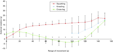
During squatting, the knees were gradually flexed from a mean of -2.8° (sd 1.3°) to a mean of 145.5° (sd 5.1°). From 0° to 40° of flexion, femurs displayed a sharp external rotation relative to the tibia, reaching a mean of 13.8° (sd 3.0°). From 40° of flexion, a gradual femoral external rotation was observed, reaching a total mean of 22.4° (sd 6.1°). The mean rotational from 100° to 150° of flexion was 7.0° (sd 5.5°),
During kneeling, the knees gradually flexed from a mean of 100.6° (sd 3.7°) to a mean of 155.7° (sd 3.0°), which was accompanied by a mean femoral external rotation, relative to the tibia, of 20.2° (sd 7.2°). The mean rotational range from 100° to 150° of flexion was 14.8° (sd 3.8°).
When sitting cross-legged, the knees gradually flexed from a mean of 4.9° (sd 4.4°) to a mean of 147.5° (sd 4.2°). During this activity, the femoral internal rotation relative to the tibia occurred from 10° to 100° of flexion, with a mean internal rotational angle of 11.2° (sd 6.9°). From 100° to 150° of flexion, femoral external rotation reached a mean of 22.4° (sd 7.0°) (Fig. 1). and 22.4° (sd 7.0°), respectively. The external rotation while sitting cross-legged was significantly more than during squatting (p = 0.041).
Fig. 1.
Graph showing the mean rotation angle when squatting, kneeling and sitting cross-legged (error bars indicate standard deviation). The markers indicate the femoral external rotation relative to the tibia (*Significant differences between squatting and sitting cross-legged; p < 0.05).
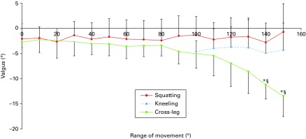
During squatting and kneeling, no significant difference in varus-valgus angle was seen for each knee flexion angle. However, when sitting cross-legged, a varus position was observed from 140° with knee flexion. The varus-valgus angle reached a mean of -13.5° (sd 3.7°) (Fig. 2).
Fig. 2.
Graph showing the mean varus-valgus angle when squatting, kneeling and sitting cross-legged legged (error bars indicate standard deviation). The markers indicate the femoral external rotation relative to the tibia (*Significant differences between squatting and sitting cross-legged p < 0.05)
AP translation: medial side
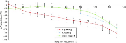
During squatting, the medial side moved a mean of 11.1% (sd 6.4%) anteriorly from 0° to 40° with knee flexion. From 40°, it moved a mean of 34.8% (sd 2.8%) posteriorly. During kneeling, no statistically significant movement was seen with knee flexion. During sitting cross-legged, it moved a mean of 13.8% (sd 4.3%) anteriorly from 0° to 30° with knee flexion. From 30° to 120°, it then moved a mean of 35.1% (sd 7.3%) posteriorly. Subsequently from 120° to 150° it translated a mean of 5.4% (sd 8.3%) anteriorly.
Among the three activities, the medial side when sitting cross-legged was located significantly posteriorly from 80° to 110° with knee flexion (Fig. 3).
Fig. 3.
Graph showing the mean anteroposterior (AP) translation of the femoral medial epicondylar sulcus when squatting, kneeling and sitting cross-legged (error bars indicate standard deviation). AP translation was calculated as a percentage relative to the AP length of tibia (*Significant differences between squatting and sitting cross-legged, p < 0.05).
AP translation: lateral side
During squatting, the lateral side moved a mean of 78.7% (sd 11.0%) posteriorly from 0° to 150° knee flexion. During kneeling, it moved a mean of 40.2% (sd 10.2%) posteriorly from 100° to 150°. While sitting cross-legged, no statistically significant movement was seen from 0° to 100°, but from 100° to 150°, the lateral side moved a mean of 51.0% (sd 12.3%) posteriorly.
Among three activities, the lateral side during squatting was located significantly posteriorly from 20° to 130° with knee flexion (Fig. 4).
Fig. 4.
Graph showing the mean anteroposterior (AP) translation of the femoral lateral epicondyle when squatting, kneeling and sitting cross-legged (error bars indicate standard deviation). AP translation was calculated as a percentage relative to the AP length of the tibia (*Significant differences between squatting and sitting cross-legged, p < 0.05).
Kinematic pathway
During squatting, the medial side displayed mild anterior movement from 0° to 40° with knee flexion. From 40° to 100° and from 100° to 150°, it moved posteriorly. At the same time, the lateral side showed -posterior movement with knee flexion. From 0° to 40°, the difference between the medial and lateral side represented a medial pivot pattern. From 40° to 100° and from 100° to 150°, bicondylar rollback was evident (supplementary -figure a). During kneeling, the medial side did not move -significantly with knee flexion. On the other hand, the lateral side moved posteriorly with knee flexion. The kinematic pathway showed a medial pivot pattern (supplementary -figure b). While sitting cross-legged, the medial side moved slightly anteriorly from 0° to 30° with knee flexion. From 30° to 120°, it moved posteriorly. Then it moved slightly anteriorly again from 120°. The lateral side did not show any significant movement from 0° to 120°. From 120°, it moved posteriorly. From 0° to 100°, the difference between the medial and lateral side represented a lateral pivot pattern. From 100°, a medial pivot pattern was seen (supplementary figure c).
Discussion
This study using CAD modelling of fluoroscopically--captured images in four male volunteers (eight knees) has examined in vivo kinematics while sitting cross-legged on the floor for the first time. Studies that have evaluated knees while sitting cross-legged using electromagnetic tracking systems have reported femoral external rotation movement in deep knee flexion.16,17 In our study, the femur displayed internal rotation relative to the tibia from 10° to 100° with knee flexion and external rotation beyond 100°. This fact suggests that the femoral rotatory motion while sitting cross-legged at the mid-flexion is variable, and may depend on the imagining modality. In addition, this suggests that the external rotation of the femur is responsible from mid- to high-flexion.
Previous static examinations have shown that knees display gradual femoral external rotation during squatting.1,2,6 However, in our study, normal knees displayed different kinematic patterns during squatting. Femurs displayed sharp external rotation relative to the tibia from 0° to 40° with knee flexion. From 40°, gradual femoral external rotation was observed. Sharp femoral external rotation in early knee flexion suggested a screw-home motion. The small amount of femoral external rotation from mid-flexion to deep flexion suggests femoral rollback. This suggests that despite the same squatting motions, kinematics differ between static and dynamic imaging, or weight-bearing and non-weight-bearing positions. Among the three activities, there were significant different rotational patterns with knee flexion. Additionally, in high flexion, rotational range while sitting cross-legged was significantly larger than that during squatting. This suggests that the rotational range of knees varies widely.
Regarding varus-valgus angle, varus position was observed while sitting cross-legged at angles of deep flexion. Previous studies evaluating cross-legged knees have reported varus movement in high flexion.16,17 Our results are similar to these studies, suggesting that the motions of sitting cross-legged tend to cause severe varus stress on the knees. Around the maximum flexion angle, no significant differences were seen among the three activities regarding the angle of rotation and AP translation. Hence, the lateral compartment of the knees in a cross-legged position might be more distracted than those of squatting and kneeling knees around maximum knee flexion.
AP translation of the medial side, sitting cross-legged, moved posteriorly in mid-flexion. However, laterally with squatting, posterior movement occurred up to 130° with flexion. However, from 130°, the differences among the three activities were small. This suggests that AP positions differ among the three activities up to mid-flexion, but in high flexion, there was no significant difference between each activity.
During squatting and kneeling, kinematic pathways were similar to the findings of Moro-oka et al.7 On the other hand, when sitting cross-legged, the kinematic pathway showed a lateral pivot pattern up to 100° with flexion. In particular, from 30° to 100°, the medial side moved posteriorly (supplementary figure c). This suggests that the medial compartment of normal knees is loose when sitting cross-legged. Several studies have reported that medial pivot-reproducing prostheses offer favourable clinical outcomes.25-29 The results of this study suggest that if the kinematics after TKA are targeted to replicate those of a normal knee, enabling some lateral pivoting for some activities must also be considered.
At high flexion, the kinematic pathway during squatting exhibited bicondylar rollback (supplementary figure a); on the other hand, kneeling and sitting cross-legged indicated a medial pivot pattern (supplementary figure b and c).
There are several limitations in our study. The local co-ordination system was not identical to that used in other studies. Therefore, the angles and distances reported are not directly comparable. Several authors have reported variability in the identification of the surgical epicondylar axis.30-32 Therefore, our procedure might have variability. However, some have reported that the accuracy using a CT model is better than the use of a cadaver or image-free navigation.33,34 In fact, the intra- and inter-observer error of our surgical epicondylar axis identification was less than 2.0°. Hence, we considered our procedure acceptable. We evaluated the pathway of the surgical epicondylar axis, known as the functional axis,33,35 therefore, this pathway might not recreate the pathway of contact points.
In addition, the transepicondylar axis is generally thought to be closer to the flexion axis than the geometric centre axis.36-38 Our study was restricted to four Japanese male volunteers. Women and individuals from other ethnic backgrounds might display different kinematics.
This study has demonstrated, using an in vivo modelling technique, that there is variability in the kinematics of normal knees, which were different depending on the high-flexion activity. In particular, when sitting cross-legged, femoral internal rotation was noted in mid-flexion, but the kinematic pattern changed from an effective lateral pivot to medial pivoting with increased knee flexion.
Take home message:
- The kinematics of normal knees during high flexion activities differ with each activity.
- Particularly when sitting cross-legged, the difference of the kinematics is remarkable.
- When sitting cross-legged, femurs show internal rotation in mid-flexion, and the kinematic pattern changes from lateral to medial pivot with knee flexion.
Author contributions:
K. Kono: Collecting and analysing the data, Producing the tables and figures, Literature review, Writing and approving the manuscript.
T. Tomita: Designing the study, Collecting and analysing the data, Editing and approving the manuscript.
K. Futai: Collecting and analysing the data, Producing the tables and figures, Approving the manuscript.
T. Yamazaki: Analysing the data, statistical analysis, Approving the manuscript.-
S. Tanaka: Literature review, Editing and approving the manuscript.
H. Yoshikawa: Editing and approving the manuscript.
K. Sugamoto: Literature review, Editing and approving the manuscript.
Funding statement:
No benefits in any form have been received or will be received from a -commercial party related directly or indirectly to the subject of this article.
This is an open-access article distributed under the terms of the Creative Commons Attributions license (CC-BY-NC), which permits unrestricted use, distribution, and reproduction in any medium, but not for commercial gain, provided the original author and source are credited.
This article was primary edited by S. Kutty and first proof edited by G. Scott.
Supplementary material. Figures showing kinematic pathways in various positions are available alongside this article at www.bjj.boneandjoint.org.uk
References
- 1.Nakagawa S, Kadoya Y, Todo S, et al. Tibiofemoral movement 3: full flexion in the living knee studied by MRI. J Bone Joint Surg [Br] 2000;82-B:1199–1200. [DOI] [PubMed] [Google Scholar]
- 2.Asano T, Akagi M, Tanaka K, Tamura J, Nakamura T. In vivo three-dimensional knee kinematics using a biplanar image-matching technique. Clin Orthop Relat Res 2001;388:157–166. [DOI] [PubMed] [Google Scholar]
- 3.Dennis D, Komistek R, Scuderi G, et al. In vivo three-dimensional determination of kinematics for subjects with a normal knee or a unicompartmental or total knee replacement. J Bone Joint Surg [Am] 2001;83-A(Suppl 2 Pt 2):104–115. [DOI] [PubMed] [Google Scholar]
- 4.You BM, Siy P, Anderst W, Tashman S. In vivo measurement of 3-D skeletal kinematics from sequences of biplane radiographs: application to knee kinematics. IEEE Trans Med Imaging 2001;20:514–525. [DOI] [PubMed] [Google Scholar]
- 5.Komistek RD, Dennis DA, Mahfouz M. In vivo fluoroscopic analysis of the normal human knee. Clin Orthop Relat Res 2003;410:69–81. [DOI] [PubMed] [Google Scholar]
- 6.Johal P, Williams A, Wragg P, Hunt D, Gedroyc W. Tibio-femoral movement in the living knee. A study of weight bearing and non-weight bearing knee kinematics using 'interventional' MRI. J Biomech 2005;38:269–276. [DOI] [PubMed] [Google Scholar]
- 7.Moro-oka TA, Hamai S, Miura H, et al. Dynamic activity dependence of in vivo normal knee kinematics. J Orthop Res 2008;26:428–434. [DOI] [PubMed] [Google Scholar]
- 8.Tsai TY, Lu TW, Chen CM, Kuo MY, Hsu HC. A volumetric model-based 2D to 3D registration method for measuring kinematics of natural knees with single-plane fluoroscopy. Med Phys 2010;37:1273–1284. [DOI] [PubMed] [Google Scholar]
- 9.Tanifuji O, Sato T, Kobayashi K, et al. Three-dimensional in vivo motion analysis of normal knees using single-plane fluoroscopy. J Orthop Sci 2011;16:710–718. [DOI] [PubMed] [Google Scholar]
- 10.Hamai S, Moro-oka TA, Dunbar NJ, et al. In vivo healthy knee kinematics during dynamic full flexion. Biomed Res Int 2013;2013:717546. [DOI] [PMC free article] [PubMed] [Google Scholar]
- 11.Murakami K, Hamai S, Okazaki K, et al. In vivo kinematics of healthy male knees during squat and golf swing using image-matching techniques. Knee 2016;23:221–226. [DOI] [PubMed] [Google Scholar]
- 12.DeFrate LE, Sun H, Gill TJ, Rubash HE, Li G. In vivo tibiofemoral contact analysis using 3D MRI-based knee models. J Biomech 2004;37:1499–1504. [DOI] [PubMed] [Google Scholar]
- 13.Fregly BJ, Rahman HA, Banks SA. Theoretical accuracy of model-based shape matching for measuring natural knee kinematics with single-plane fluoroscopy. J Biomech Eng 2005;127:692–699. [DOI] [PMC free article] [PubMed] [Google Scholar]
- 14.Iwaki H, Pinskerova V, Freeman MA. Tibiofemoral movement 1: the shapes and relative movements of the femur and tibia in the unloaded cadaver knee. J Bone Joint Surg [Br] 2000;82-B:1189–1195. [DOI] [PubMed] [Google Scholar]
- 15.Hill PF, Vedi V, Williams A, et al. Tibiofemoral movement 2: the loaded and unloaded living knee studied by MRI. J Bone Joint Surg [Br] 2000;82-B:1196–1198. [DOI] [PubMed] [Google Scholar]
- 16.Hemmerich A, Brown H, Smith S, Marthandam SS, Wyss UP. Hip, knee, and ankle kinematics of high range of motion activities of daily living. J Orthop Res 2006;24:770–781. [DOI] [PubMed] [Google Scholar]
- 17.Acker SM, Cockburn RA, Krevolin J, et al. Knee kinematics of high-flexion activities of daily living performed by male Muslims in the Middle East. J Arthroplasty 2011;26:319–327. [DOI] [PubMed] [Google Scholar]
- 18.Howell SM, Howell SJ, Kuznik KT, Cohen J, Hull ML. Does a kinematically aligned total knee arthroplasty restore function without failure regardless of alignment category? Clin Orthop Relat Res 2013;471:1000–1007. [DOI] [PMC free article] [PubMed] [Google Scholar]
- 19.Nishio Y, Onodera T, Kasahara Y, et al. Intraoperative medial pivot affects deep knee flexion angle and patient-reported outcomes after total knee arthroplasty. J Arthroplasty 2014;29:702–706. [DOI] [PubMed] [Google Scholar]
- 20.Yamazaki T, Watanabe T, Nakajima Y, et al. Visualization of femorotibial contact in total knee arthroplasty using X-ray fluoroscopy. Eur J Radiol 2005;53:84–89. [DOI] [PubMed] [Google Scholar]
- 21.Yamazaki T, Watanabe T, Nakajima Y, et al. Improvement of depth position in 2-D/3-D registration of knee implants using single-plane fluoroscopy. IEEE Trans Med Imaging 2004;23:602–612. [DOI] [PubMed] [Google Scholar]
- 22.Yamazaki T, Watanabe T, Tomita T, et al. Accuracy validation of 2D/3D registration of normal knee using X-ray fluoroscopic images and CT images. Japanese J Clin Biomech 2008;29:389–396. [Google Scholar]
- 23.Kawashima K, Tomita T, Tamaki M, et al. In vivo three-dimensional motion analysis of osteoarthritic knees. Mod Rheumatol 2013;23:646–652. [DOI] [PubMed] [Google Scholar]
- 24.Grood ES, Suntay WJ. A joint coordinate system for the clinical description of three-dimensional motions: application to the knee. J Biomech Eng 1983;105:136–144. [DOI] [PubMed] [Google Scholar]
- 25.Mannan K, Scott G. The Medial Rotation total knee replacement: a clinical and radiological review at a mean follow-up of six years. J Bone Joint Surg [Br] 2009;91-B:750–756. [DOI] [PubMed] [Google Scholar]
- 26.Fan CY, Hsieh JT, Hsieh MS, Shih YC, Lee CH. Primitive results after medial-pivot knee arthroplasties: a minimum 5-year follow-up study. J Arthroplasty 2010;25:492–496. [DOI] [PubMed] [Google Scholar]
- 27.Bae DK, Song SJ, Cho SD. Clinical outcome of total knee arthroplasty with medial pivot prosthesis a comparative study between the cruciate retaining and sacrificing. J Arthroplasty 2011;26:693–698. [DOI] [PubMed] [Google Scholar]
- 28.Shimmin A, Martinez-Martos S, Owens J, Iorgulescu AD, Banks S. Fluoroscopic motion study confirming the stability of a medial pivot design total knee arthroplasty. Knee 2015;22:522–526. [DOI] [PubMed] [Google Scholar]
- 29.Scott G, Imam MA, Eifert A, et al. Can a total knee arthroplasty be both rotationally unconstrained and anteroposteriorly stabilised? A pulsed fluoroscopic investigation. Bone Joint Res 2016;5:80–86. [DOI] [PMC free article] [PubMed] [Google Scholar]
- 30.Inui H, Taketomi S, Nakamura K, et al. An additional reference axis improves femoral rotation alignment in image-free computer navigation assisted total knee arthroplasty. J Arthroplasty 2013;28:766–771. [DOI] [PubMed] [Google Scholar]
- 31.Siston RA, Patel JJ, Goodman SB, Delp SL, Giori NJ. The variability of femoral rotational alignment in total knee arthroplasty. J Bone Joint Surg [Am] 2005;87-A:2276–2280. [DOI] [PubMed] [Google Scholar]
- 32.Kinzel V, Ledger M, Shakespeare D. Can the epicondylar axis be defined accurately in total knee arthroplasty? Knee 2005;12:293–296. [DOI] [PubMed] [Google Scholar]
- 33.van der Linden-van der Zwaag HM, Valstar ER, van der Molen AJ, Nelissen RG. Transepicondylar axis accuracy in computer assisted knee surgery: a comparison of the CT-based measured axis versus the CAS-determined axis. Comput Aided Surg 2008;13:200–206. [DOI] [PubMed] [Google Scholar]
- 34.Aunan E, Østergaard D, Meland A, Dalheim K, Sandvik L. A simple method for accurate rotational positioning of the femoral component in total knee arthroplasty. Acta Orthop 2017;1–7. [DOI] [PMC free article] [PubMed] [Google Scholar]
- 35.Laskin RS. Flexion space configuration in total knee arthroplasty. J Arthroplasty 1995;10:657–660. [DOI] [PubMed] [Google Scholar]
- 36.Asano T, Akagi M, Nakamura T. The functional flexion-extension axis of the knee corresponds to the surgical epicondylar axis: in vivo analysis using a biplanar image matching technique. J Arthroplasty 2005;20:1060–1067. [DOI] [PubMed] [Google Scholar]
- 37.Churchill DL, Incavo SJ, Johnson CC, Beynnon BD. The transepicondylar axis approximates the optimal flexion axis of the knee. Clin Orthop Relat Res 1998;356:111–118. [DOI] [PubMed] [Google Scholar]
- 38.Hollister AM, Jatana A, Singh AK, Sullivan WW, Lupichuk AG. The axes of rotation of the knee. Clin Orthop Relat Res 1993:290;259–268. [PubMed] [Google Scholar]


