
Extended Data Figure 5. Risperidone has distinct poses in solution and in complex with DRD2 and comparison of x-ray structure and model of DRD2.
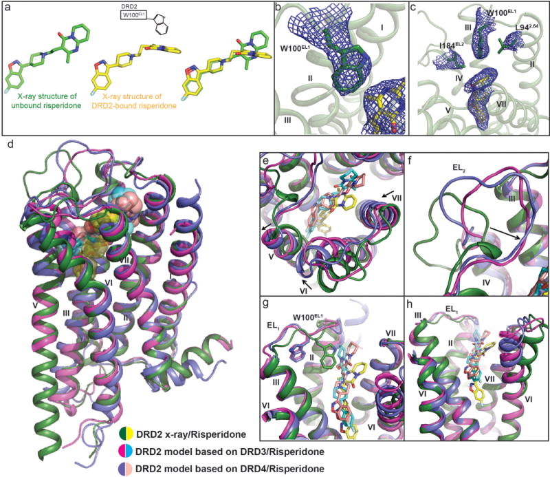
a, Trp100EL1 determines the configuration of the tetrahydropyridopyrimidinone moiety of risperidone. Structure of unbound risperidone colored in green and DRD2-bound risperidone in yellow. b, Electron density (2Fo-Fc maps, blue mesh) for W100EL1 in the DRD2/Risperidone complex (contoured at 1.0σ). c, 2Fo-Fc electron density map (blue mesh) of Leu942.64, Trp100EL1, Ile184EL2 and risperidone (yellow) contoured at 0.8σ. Ballesteros-Weinstein numbering is shown as superscript. d, Overall view of DRD2/Risperidone x-ray structure and model. e, f, g, h, Comparison of x-ray structure and model of DRD2. In d-h panels, DRD2 x-ray structure and model is shown as cartoons, with the x-ray structure colored in green and model in magenta or blue. Risperidone in x-ray structure is shown as yellow spheres or sticks and model as cyan or lightpink.