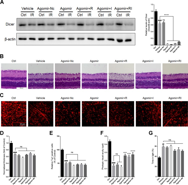
Figure 5. Overexpression of let-7d by agomir in mouse retina.
(A) Western blot for Dicer and β-actin in injured retinas administered with agomir-let-7d or agomir-Nc. (B) HE staining shows the thickness of retinas injured for 7 days after let-7d overexpression. (C) Immunofluorescent staining by Tuj1 on retinal flat mounting displays the survival of RGCs in IR-injured retinas after let-7d overexpression. (D) Analysis of retinal thickness of IR-injured retinas after let-7d overexpression. (E) Analysis of the survival of RGCs in IR-injured retinas by Tuj1 immunostaining on retinal flat mounting after let-7d overexpression. (F) Photopic visual acuity of RIR-injured mice after let-7 overexpression. Data were shown as mean ± SEM (G) Percent time spent in the light chamber by RIR-injured mice after intravitreal injection of let-7d Agomir. n = 8, ##p < 0.01, ####p < 0.0001, versus control; ***p < 0.001, ****p < 0.0001, versus vehicle; ns: no significance.

