Abstract
The purpose of this study was to identify which of the eye solutions is best for sodium fluorescein staining of the cornea to diagnose dry eye disease. The study included 173 eyes with suspected or known dry eye disease. The eyes were stained sequentially with sodium fluorescein and each of the following four conditions: balanced salt solution (BSS); BSS and cyclosporine 0.05% emulsion; BSS and lipids containing omega-3; and BSS, cyclosporine 0.05% emulsion, and lipids containing omega-3. Our results showed that compared to BSS alone, artificial tears with cyclosporine 0.05% emulsion and lipids containing omega-3 remain in the cornea for longer periods, thus allowing the clinician to evaluate tear break-up time and visualize corneal punctate erosions.
Key Words: Corneal Staining, Corneal Punctate Erosions, Dry Eye Disease, Sodium Fluorescein, Artificial Tear, Cyclosporine, Balanced Salt Solution, Lipids Containing Omega-3
INTRODUCTION
According to the National Eye Institute, it is estimated that 5 million Americans over the age of 50 have dry eye disease (DED) [1]. Many cases remain undiagnosed until they progress to a severe stage that is very difficult to treat. The International Dry Eye Workshop II (DEWS II) stated, “dry eye disease is a multifactorial disease of the ocular surface characterized by a loss of homeostasis of the tear film, and accompanied by ocular symptoms, in which tear film instability and hyperosmolarity, ocular surface inflammation and damage, and neurosensory abnormalities play etiological roles” [2]. There are two types of dry eye, aqueous-deficient and evaporative dry eye, with the evaporative type being the most common [3]. In some cases, both types occur simultaneously, which is referred to as mixed DED [2]. Tears comprise three layers: the outer lipid layer, the middle aqueous layer, and the inner mucin layer [4]. Aqueous-deficient DED is caused by the lack of lacrimal gland production of the aqueous layer. Evaporative DED is a result of the lack of quality of the lipid layer from the Meibomian glands [5]. Dry eye can be caused by a variety of factors, including medications, age, autoimmune disorders, allergies, corneal surgeries, and the environment [6]. Unfortunately, DED is under-diagnosed and under-treated. DED goes unnoticed because patients may not realize their symptoms are treatable, and thus do not discuss them with their doctors. Moreover, many physicians are not proactive in investigating DED symptoms and signs [7]. Beyond eye symptoms, DED can affect an individual’s overall health, impacting one’s quality of life. This includes aspects of “physical, social, psychological functioning daily activities and workplace productivity” [8]. There is even a significant correlation between DED and sleep and mood disorders [9]. Because DED is a chronic condition that can be debilitating, it is imperative to find ways to improve diagnosis of DED, so patients can seek appropriate management and care as soon as possible [10]. We conducted a prospective study to identify which of the eye solutions is best for sodium fluorescein staining of the cornea to evaluate tear break-up time (TBUT) and visualize punctate erosions in patients with suspicious or known DED. To our knowledge, this is the first study of its type with this methodology.
MATERIALS AND METHODS
All patients signed a consent form in order to be included in this study, and the study received ethical approval at the department level. This prospective controlled study compared the efficacy of sodium fluorescein in balanced salt solution (BSS) solution, lipid-based artificial tears containing omega-3, and/or cyclosporine 0.05% emulsion in evaluating TBUT, visualizing corneal punctate staining, and diagnosing DED. The ingredients of each solution are listed in Table 1. The inclusion criterion was eyes with suspected or known DED.
Table 1.
Ingredients of each Staining Solution
| BSS | Cyclosporine 0.05% emulsion | Lipid-based artificial tears containing omega-3 |
|---|---|---|
| Sodium chloride 0.64% | Cyclosporine 0.05% | Carboxymethylcellulose sodium 0.5% |
| Potassium chloride 0.075% | Glycerin | Glycerin 1% |
| Calcium chloride dihydrate 0.048% | Castor oil | Polysorbate 80 0.5% |
| Magnesium chloride hexahydrate 0.03% | Polysorbate 80 | Boric acid |
| Sodium acetate trihydrate 0.39% | Carbomer type A | Butylated hydroxyl toluene |
| Sodium citrate dihydrate 0.17% | Purified water | Castor oil |
| Sodium hydroxide | Sodium hydroxide | Erythritol |
| Hydrochloric acid | Flaxseed oil | |
| Water | Levocarnitine | |
| Permulen TR-2 | ||
| Polyoxyl 40 stearate | ||
| Purified water | ||
| Sodium hydroxide | ||
| Trehalose |
BSS: Balanced Salt Solution
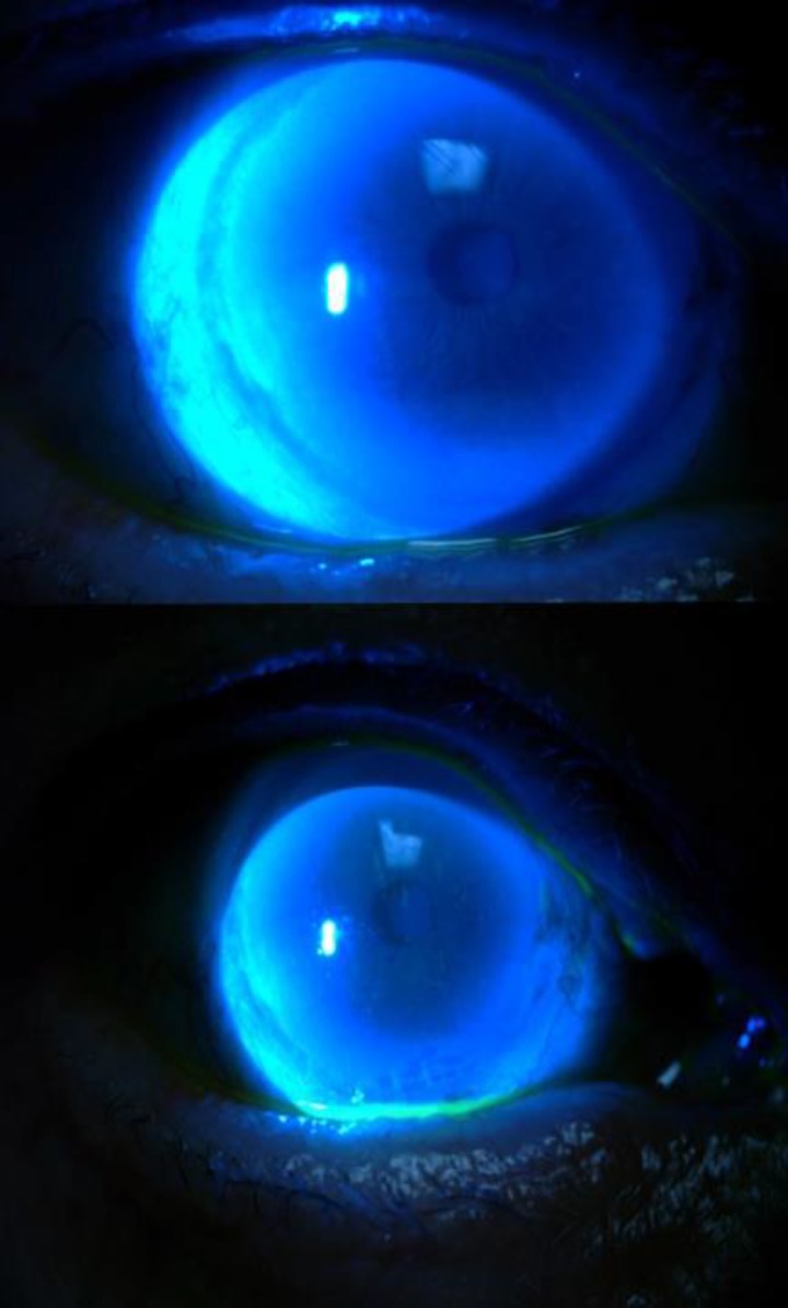
Differential diagnosis of aqueous-deficient or evaporative DED was not performed due to higher costs and longer times to testing. Therefore, the study population probably included both types. The exclusion criteria were eyes with severe eye disease, such as corneal ulcers, endophthalmitis, phthisis bulbi, acute conjunctivitis, and dacryocystitis. The solutions were tested sequentially for comparison. There were three solutions but four conditions: 1) BSS; 2) BSS and cyclosporine 0.05% emulsion; 3) BSS and lipids containing omega-3; and 4) BSS, cyclosporine 0.05% emulsion, and lipids containing omega-3. All eyes were stained with sodium fluorescein strips in BSS first. The eyes that showed no or faint sodium fluorescein strip staining were re-stained immediately with sodium fluorescein strips in either BSS + cyclosporine 0.05% emulsion or BSS + lipids containing omega-3. If the eyes stained with BSS + lipids containing omega-3 failed to show any corneal punctate staining, BSS + cyclosporine 0.05% emulsion was tested (Fig 1). All eyes were examined with a slit lamp under cobalt blue light by one of the authors (M.C.). Descriptive statistics were calculated to summarize patient demographics and medical information. The staining rate for each condition was calculated, together with its 95% confidence interval (Fig 2). The rate of either observing staining on the cornea with fluorescein or no staining observed.
Figure 1.
Flow Chart of Eyes Stained with Different Solutions. BSS: Balanced Salt Solution
Figure 2.
Sodium Fluorescein in Balanced Salt Solution (BSS) revealed no Discernible Punctate Staining on the Cornea (top); the Same Eye was Subsequently Stained with Sodium Fluorescein in BSS + Cyclosporine 0.05% Emulsion, and Obvious Punctate Staining was Observed (Bottom).
RESULTS
In this study, 173 right eyes from 173 patients met the inclusion criterion. The average age of the 173 patients was 68 years old, and 73% of them were women.
The difference between the BSS and the cyclosporine 0.05% emulsion is the main active ingredient, cyclosporine 0.05%, in a glycerin-in-castor oil emulsion (Table 1).
The difference between the cyclosporine 0.05% emulsion and the lipid-based artificial tears containing omega-3 is also cyclosporine 0.05% in a glycerin-in-castor oil emulsion (Table 1). More than 70% of the patients had symptoms of dry eye, 63% of the patients had a surgery history or other medical treatment, and 98% of the patients had an eye surgery history or other eye treatment (Table 2).
Table 2.
Demographic and Clinical Information of the 173 Patients
| Variable | Values |
|---|---|
| Age | 68.36 ± 11.98 |
| Gender (% of women) | 127 (73.41) |
| Dry eye symptoms | |
| Yes | 127 (73.41) |
| No | 46 (26.59) |
| Surgery history or other medical treatment | 109 (63.01) |
| Eye surgery history or other eye treatment | 170 (98.27) |
Data are presented as Mean ± SD or No. (%)
Of the 173 eyes stained with sodium fluorescein in BSS, 130 (75%) showed positive staining and 43 (25%) showed negative staining. Of the 43 eyes with negative staining, 28 were stained with sodium fluorescein in BSS + cyclosporine 0.05% emulsion, and 15 were stained with sodium fluorescein in BSS + lipids containing omega-3. Of the 28 eyes stained with sodium fluorescein in BSS + cyclosporine 0.05% emulsion, 25 (89%) showed positive staining. Of the 15 eyes stained with sodium fluorescein in BSS + lipids containing omega-3, 10 (67%) showed positive staining. Of the 5 eyes that showed negative staining with BSS + lipids containing omega-3, 4 (80%) showed positive staining with BSS + cyclosporine 0.05% emulsion (Table 3).
Table 3.
Staining Rate for each Condition
| Condition | n | Staining rate (95% CI) |
|---|---|---|
| BSS only | 173 | 0.75 (0.68–0.81) |
| BSS + Cyclosporine | 28 | 0.89 (0.73–0.96) |
| BSS + Lipids containing omega-3 | 15 | 0.67 (0.42–0.85) |
| BSS + Lipids containing omega-3 + Cyclosporine | 5 | 0.80 (0.38–0.99) |
CI: Confidence Interval
DISCUSSION
Our results showed that compared to BSS alone, artificial tears with cyclosporine 0.05% emulsion and lipids containing omega-3 remain in the cornea for longer periods.
Due to the poor aqueous solubility of cyclosporine itself, cyclosporine 0.05% solution is formulated as a glycerin-in-castor oil emulsion to allow easier administration of the drug to the eye [11]. The emulsion allows the lipids to remain in the cornea for longer times, thus allowing the clinician to detect small punctate corneal staining with sodium fluorescein. It also makes it more difficult to wash out the staining by the patient’s tears during examination under strong light. Commercially available Restasis® ; Allergan Inc, Irvine, CA comprises 0.05% cyclosporine in a homogenous emulsion of glycerin (2.2%), castor oil (1.25%), polysorbate 80 (1.00%), carbomer copolymer type A (0.05%), purified water (to 100%), and sodium hydroxide for pH adjustment [12]. Although all other ingredients besides cyclosporine can be found in other artificial tear products, they do not exist in emulsion form. Artificial tears contain lipids such as omega-3, which also allow for better visualization of punctate lesions compared to BSS alone. However, in this study, BSS + cyclosporine 0.05% emulsion was able to identify punctate lesions that were undetectable using BSS + lipids containing omega-3. Since both cyclosporine 0.05% emulsion and lipid-based artificial tears containing omega-3 contain water, just like BSS does, it would be expected that they would all allow sodium fluorescein staining of the cornea. Further comparative study in this regard is warranted to answer this question. There are many objective methods for DED diagnosis, namely the tear osmolarity test [13], tear matrix metalloproteinase 9 level [14], meibography [15, 16], lipid layer thickness [17], TBUT [16], confocal microscopy, and severity of punctate staining. In this study, we subjectively evaluated the corneal punctate staining and TBUT first before proceeding with objective methods, since this is a simple, fast, and economic method. In this study, we also tested if the eye drop caused stinging sensation. However, none of the patients complained of stinging sensation, probably due to the minimum amount of Restasis® ; Allergan Inc, Irvine, CA applied to each sodium fluorescein strip.
If excessive tear secretion occurs in a patient for various reasons, sodium fluorescein in BSS may be quickly washed out (wash-out effect) without sufficient staining for the examiner to visualize the punctuate lesions or determine the TBUT. The emulsion component of cyclosporine 0.05% solution remains longer in the cornea, thus providing more time for examination. However, in this study, sequential staining with different solutions may have contributed with the added effect of previous sodium fluorescein in the cornea. A future prospective controlled study to compare BSS with cyclosporine is warranted. In addition, it would be important to repeat this study in multiple centers, perhaps using additional artificial tear solutions. The results of this study can change the practice of initial diagnosis of DED before using expensive diagnostic equipment. It may also change the management associated with DED, such as in refractive surgery and glaucoma. In addition, lipid-containing emulsions should be investigated in “real-world” settings for diagnosing and treating DED as it may stabilize the tear film [18]. An efficient staining could assist in the differential diagnosis of non-DED ocular surface diseases such as anterior blepharitis, allergic conjunctivitis, corneal epithelial basement membrane dystrophy, contact lens intolerance, conjunctivochalasis, and keratoneuralgia [19]. The results of this study suggest that there are 25% of misdiagnosed DED cases by staining in BSS. Some DED cases may be diagnosed as neurotropic keratopathy while extensive diagnosis and treatment may be unnecessary [20]. As far as the authors know, numerous studies have described corneal staining with lissamine green, fluorescein, or rose Bengal dye, but none has discussed the best solution for efficient examination under slit lamp microscopy. We found that using lipid-free solutions such as BSS or artificial tears may lead to misdiagnosis of cornea punctate lesions in 14–25% of cases. This study has one important limitation. We did not diagnose the types of DED. However, it appeared that all patients had mixed DED (the most common type of DED). Differential diagnosis would require expensive testing and more time, and it was not pertinent to this study. In conclusion, if sodium fluorescein in BSS cannot confirm the punctate lesions in the cornea and determine TBUT, using cyclosporine 0.05% emulsion or lipid-based artificial tears could help retain sodium fluorescein for longer periods in the cornea, thus allowing the visualization of punctate lesions and evaluation of TBUT for DED diagnosis and follow-up.
DISCLOSURE
So Yung Choi was partially supported by grants (U54MD007584 and U54MD007601) from the National Institutes of Health (NIH). The content is solely the responsibility of the authors and does not necessarily represent the official views of the NIH. All named authors meet the International Committee of Medical Journal Editors (ICMJE) criteria for authorship for this manuscript, take responsibility for the integrity of the work as a whole, and have given final approval for the version to be published.
References
- 1.International Dry Eye Workshop. DEWS Report. Ocular Surface. 2007 doi: 10.1016/s1542-0124(12)70086-1. [DOI] [PubMed] [Google Scholar]
- 2.International Dry Eye Workshop. DEWS II: Redefining Dry Eye. Ocular Surface. 2017 [Google Scholar]
- 3.Schacter S. Schacter Factor: Evaporative dry eye vs aqueous tear deficiency. Optometry Times. 2015 [Google Scholar]
- 4.National Eye Institute. Facts about Dry Eye. National Eye Institute. 2017 [Google Scholar]
- 5.Lemp M, Geerling G. Distinguishing Evaporative from Aqueous Deficient Dry Eye. Cataract Refract Surg Today. 2011;9(2):140–59. [Google Scholar]
- 6.Boyd K. Causes of Dry Eye. USA: American Academy of Ophthalmology; 2017. [Google Scholar]
- 7.Phadatare SP, Momin M, Nighojkar P, Askarkar S, Singh KK. A Comprehensive Review on Dry Eye Disease: Diagnosis, Medical Management, Recent Developments, and Future Challenges. Adv Pharmac. 2015;2015:1–12. doi: 10.1155/2015/704946 . [Google Scholar]
- 8.Uchino M, Schaumberg DA. Dry Eye Disease: Impact on Quality of Life and Vision. Curr Ophthalmol Rep. 2013;1(2):51–7. doi: 10.1007/s40135-013-0009-1. doi: 10.1007/s40135-013-0009-1 pmid: 23710423. [DOI] [PMC free article] [PubMed] [Google Scholar]
- 9.Ayaki M, Kawashima M, Negishi K, Tsubota K. High prevalence of sleep and mood disorders in dry eye patients: survey of 1,000 eye clinic visitors. Neuropsychiatr Dis Treat. 2015;11:889–94. doi: 10.2147/NDT.S81515. doi: 10.2147/NDT.S81515 pmid: 25848288. [DOI] [PMC free article] [PubMed] [Google Scholar]
- 10.Javadi MA, Feizi S. Dry eye syndrome. J Ophthalmic Vis Res. 2011;6(3):192–8. pmid: 22454735. [PMC free article] [PubMed] [Google Scholar]
- 11.Ames P, Galor A. Cyclosporine ophthalmic emulsions for the treatment of dry eye: a review of the clinical evidence. Clin Investig (Lond) 2015;5(3):267–85. doi: 10.4155/cli.14.135. doi: 10.4155/cli.14.135 pmid: 25960865. [DOI] [PMC free article] [PubMed] [Google Scholar]
- 12.Gore A, Attar M, Pujara C, Neervannan S 1. Ocular emulsions and dry eye: a case study of a non-biological complex drug product delivered to a complex organ to treat a complex disease. Generic Biosimilar Initiative J. 2017;6(doi: 10.5639/gabij.2017.0601.004 ):13–23. [Google Scholar]
- 13.International Dry Eye Workshop. The definition and classification of dry eye disease: report of the definition and Classification Subcommittee of the international Dry Eye Workshop. 2007. [DOI] [PubMed] [Google Scholar]
- 14.Chotikavanich S, de Paiva CS, Li de Q, Chen JJ, Bian F, Farley WJ, et al. Production and activity of matrix metalloproteinase-9 on the ocular surface increase in dysfunctional tear syndrome. Invest Ophthalmol Vis Sci. 2009;50(7):3203–9. doi: 10.1167/iovs.08-2476. doi: 10.1167/iovs.08-2476 pmid: 19255163. [DOI] [PMC free article] [PubMed] [Google Scholar]
- 15.Tear Science, a streamlined method for evaluating the Meibomian glands. Tear Science Web site; 2017 [cited 2017 December 5]. Available from: https://tearscience.com.
- 16.Oculus Inc. The OCULUS keratography 5M: Oculus, Inc. 2017. [[cited 2017 December 5]]. Available from: https://www.oculus.de/us/products/topography/keratography-5m.
- 17.Finis D, Pischel N, Schrader S, Geerling G. Evaluation of lipid layer thickness measurement of the tear film as a diagnostic tool for Meibomian gland dysfunction. Cornea. 2013;32(12):1549–53. doi: 10.1097/ICO.0b013e3182a7f3e1. doi: 10.1097/ICO.0b013e3182a7f3e1 pmid: 24097185. [DOI] [PubMed] [Google Scholar]
- 18.Torkildsen G, Brujic M, Cooper MS, Karpecki P, Majmudar P, Trattler W, et al. Evaluation of a new artificial tear formulation for the management of tear film stability and visual function in patients with dry eye. Clin Ophthalmol. 2017;11:1883–9. doi: 10.2147/OPTH.S144369. doi: 10.2147/OPTH.S144369 pmid: 29089744. [DOI] [PMC free article] [PubMed] [Google Scholar]
- 19.Arnold WK, Savage CR, Brissette CA, Seshu J, Livny J, Stevenson B. RNA-Seq of Borrelia burgdorferi in Multiple Phases of Growth Reveals Insights into the Dynamics of Gene Expression, Transcriptome Architecture, and Noncoding RNAs. PLoS One. 2016;11(10):e0164165. doi: 10.1371/journal.pone.0164165. doi: 10.1371/journal.pone.0164165 pmid: 27706236. [DOI] [PMC free article] [PubMed] [Google Scholar]
- 20.Sacchetti M, Lambiase A. Diagnosis and management of neurotrophic keratitis. Clin Ophthalmol. 2014;8:571–9. doi: 10.2147/OPTH.S45921. doi: 10.2147/OPTH.S45921 pmid: 24672223. [DOI] [PMC free article] [PubMed] [Google Scholar]


