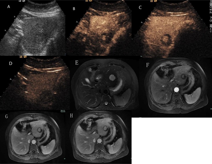
Figure 6. Hepatic hemangioma with centrifugal enhancement.
(A) Ultrasound revealed an isoechoic mass in the left liver. (B) and (C) CEUS showed a central enhancing foci in the arterial phase and followed by a centrifugal enhancement. (D) CEUS showed hypoechoic change in the delayed phase. (E) The lesion presented as markedly hyperintense on T2 weighted MR images. (F) Contrast enhanced MR images showed a central enhancing focus in the arterial phase. (G) and (H) The lesion showed centrifugal enhancement in the portal-venous phase and late phase.

