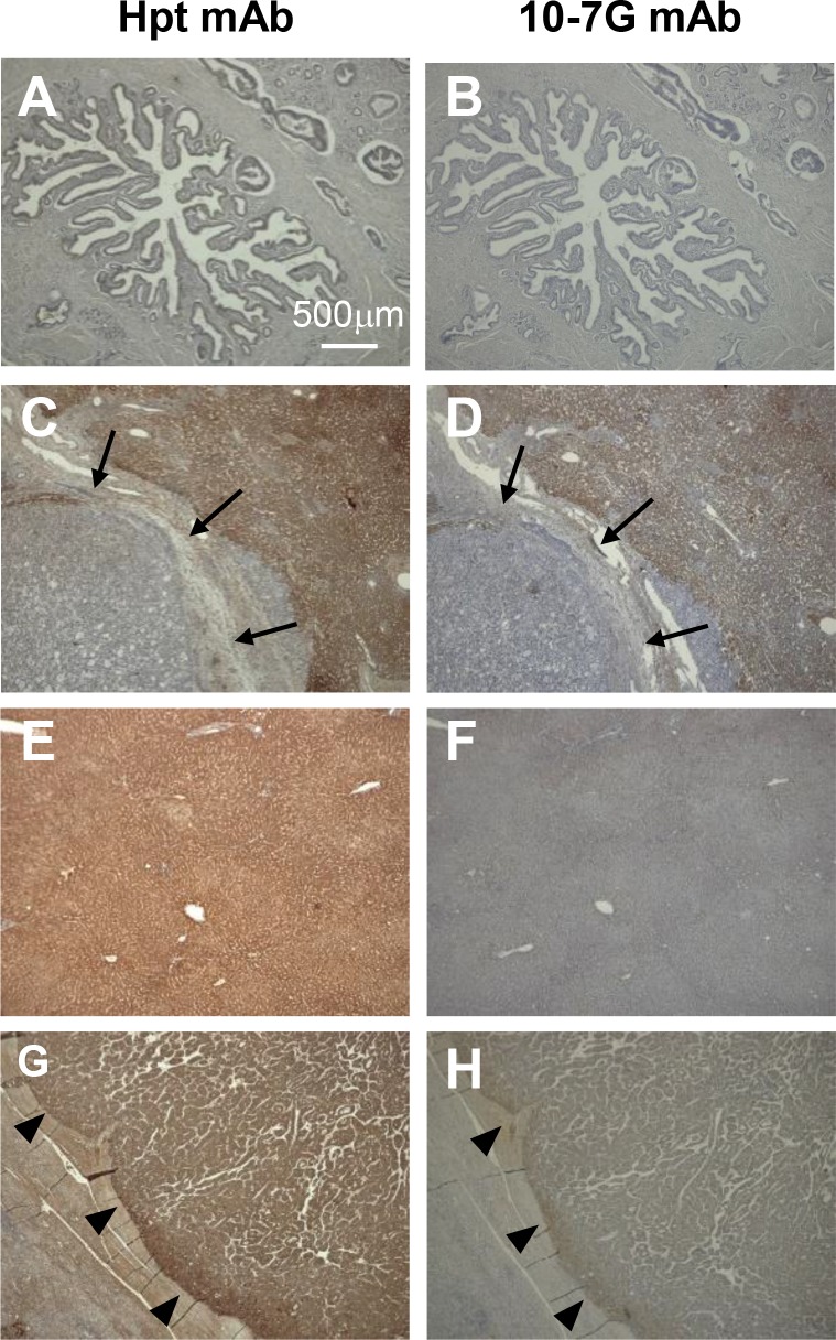
Figure 5. Immunohistochemical study of Fuc-Hpt in pancreatic cancer and liver tissue.
Immunohistochemical staining was performed with both the Hpt mAb and the 10-7G mAb as described in the Material and Methods section. (A) and (B), primary pancreatic cancer; (C) and (D), metastatic pancreatic cancer to the liver; (E) and (F), normal hepatic tissue; (G) and (H), hepatocellular carcinoma (HCC). Photographs were taken using a 4× objective. Scale bar, 500 μm. Arrow showed metastatic pancreatic cancer in the liver and arrowhead showed positive staining of each antibody.

