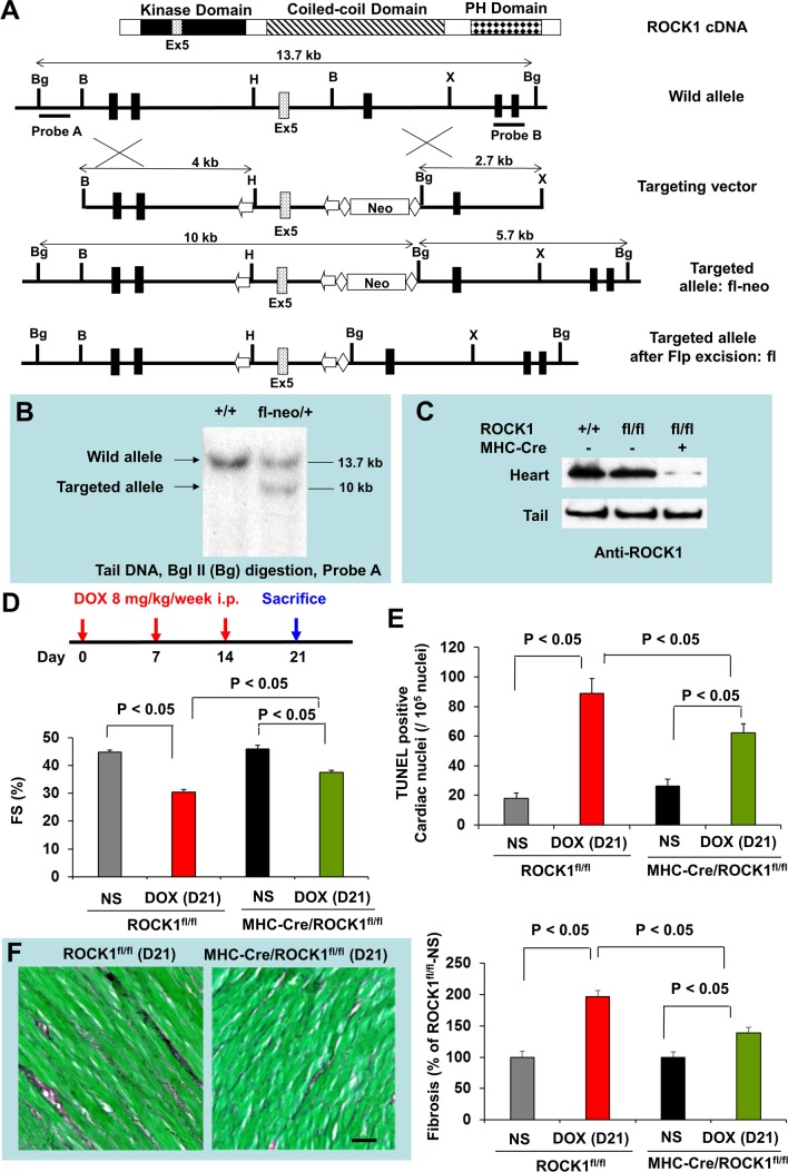
Figure 4. Cardiomyocyte-specific ROCK1 deletion also inhibited doxorubicin-induced cardiac dysfunction, apoptosis and fibrosis.
(A). Schematic illustration of the gene targeting strategy used for conditional disruption of the ROCK1 gene. Schematic representation of the domain structure of ROCK1 indicates the position of exon 5. A fragment of the ROCK1 allele that includes exons 3-6 is shown. A targeting vector was constructed containing loxP sites (arrows) flanking exon 5 of ROCK1 and Frt sites (diamonds) flanking the PGK-Neo cassette. The diagrams also indicate the positions of genomic probes used for distinguishing WT and the targeted alleles by Southern blot analysis and the positions of the restriction enzymes sites for Bam H I (B), Bgl II (Bg), Xba I (X), and Hind III (H). We first obtained germline transmission of the ROCK1fl-neo allele, which contains the loxP-flanked exon 5 and Neo cassette. The Neo cassette was removed via the Flp-Frt system by crossing ROCK1fl-neo/fl-neo mice with Flp mice, generating ROCK1fl/+ mice. (B). Southern blot analysis of genomic DNA obtained from the tail of WT and ROCK1fl-neo/+ mice. (C). Western blot analysis of ROCK1 levels in the heart and tail of WT, ROCK1fl/fl and MHC-Cre/ROCK1fl/fl mice, showing about 80% reduction of ROCK1 expression in the heart samples of the MHC-Cre/ROCK1fl/fl mice compared with WT or ROCK1fl/fl mice, but not in the tail samples of these mice. Residual ROCK1 expression in the heart is due to the presence of ROCK1 in other cell types in hearts (e.g., fibroblasts, vascular endothelial cells, and inflammatory cells). (D). Cardiomyocyte-specific ROCK1 knockout mice (MHC-Cre/ROCK1fl/fl) and ROCK1fl/fl mice 8 to 9 weeks old received three serial injections weekly of NS or DOX (8 mg/kg). Cardiac function was measured by echocardiography analysis on day 21 after the initial injection. (E-F). Quantitation of total TUNEL positive nuclei per 105 total nuclei (E) and the collagen deposition (F, scale bar, 50 μm) in ventricular myocardium from MHC-Cre/ROCK1fl/fl and ROCK1fl/fl mice hearts on day 21. N = 4-6 in each group.

