Abstract
This study was to report the comparison of outcomes between Ilizarov ring fixator (IRF) and Taylor Spatial Frame® (Smith & Nephew, Memphis, Tenn.; TSF) in terms of the effectiveness of ankle-foot deformities correction, follow-up results, and complications. Fourteen patients with ankle-foot deformities were corrected using circular external fixation (IRF group = 7 patients; TSF group = 7 patients) and related procedures. Baseline data and treatment variables were recorded. The patients’ mean age was 42.9 years. Mean follow-up time was 6.5 months. Most common cause of deformity/traumatic condition was posttraumatic equinus. There were successful results in 8 patients (57.1%), partial successful results in 5 patients (35.7%), and revision-needed in 1 patient (7.1%). TSF group demonstrated significantly higher rate of successful results than IRF group (P=0.033). A trend of lower complication rate was found in TSF group (P=0.286). Deformity corrections using TSF provided significantly better clinical scores and higher rate of successful outcome than conventional IRF.
Key words: Ilizarov, Taylor Spatial Frame, Deformity correction, Ankle, Foot
Introduction
Circular external fixation has been used for more than a century. During the 1950s, Gavriil Abramovich Ilizarov, developed the use of a modular circular fixator with transosseus tensioned wire fixation. Italian surgeon, Bianchi-Maiocchi, introduced Ilizarov’s methods to the western world in 1981.1 Today, this technique is widely used by a variety of surgeons.1,2 Regardless of the type of external fixation used, the surgeon’s understanding of external fixation principles and mechanics is required.1,2 It is typically preferred for static or dynamic gradual correction of hindfoot and ankle deformities because of the versatility of the components and strength for weight bearing. It is also chosen over acute correction when the deformity is complex, often involving an oblique plane, a rotational component, and/or limb shortening.1,3 The use of ring fixation, with or without gradual correction may allow the patient to be more functional during the healing period because a ring fixator will typically allow partial to full weight bearing during the recovery time.1,2
The Ilizarov Ring Fixator (IRF) is thought to have several advantages over other surgical options in the treatment of axial deformity.3-12 For the use in deformity correction, the surgeon uses hinges and translation mechanisms to build a custom made frame system for each distinct deformity. 3-6 During the treatment, correction of complex deformities may require changes of the frame construct, which may be very time consuming or even impossible.4,5,8 In this case, ring fixator may need modification occasionally throughout the correction of complex deformities.4-12 In 1994, J. Charles Taylor & Harold Taylor, developed a new type of external fixator called the Taylor Spatial Frame®(Smith & Nephew, Memphis, Tenn.).1,2 The TSF, which modified the Ilizarov system, is an apparatus that uses a Stewart platform with a computer program to allow for precise gradual bony or soft tissue correction in any plane.6,8,9 Thus, the deformity analysis and correction planning with this instrument were for the first time necessarily software dependent.3-12 It offers the possibility of simultaneous correction of multidirectional deformities without the need of additional apparatus to the system during correction.4,5,7 Thus, in comparison to the traditional IRF, the TSF uses one single frame construct, and no additional devices are needed for correction of translation or rotation deformities.3-12
However, there is still no consensus regarding the superiority between IRF versus TSF in terms of complex deformity correction. In addition, there was no report which published about the comparing results between the application of IRF and TSF in the corrections of ankle and foot deformities. The present study was to investigate the comparing outcomes following these circular external fixations in terms of the effectiveness in ankle and foot deformities correction, the follow-up results, and complications.
Materials and Methods
This study reviewed a total of 14 patients in whom ankle and foot deformity was corrected using circular external fixation and following procedure between May 2013 and June 2017.
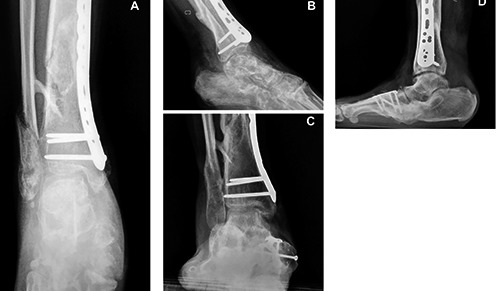
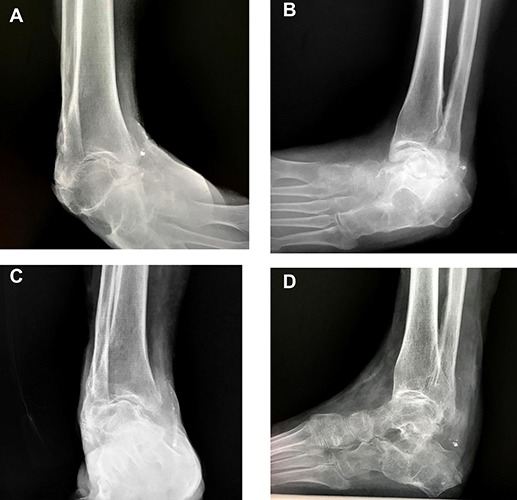
The inclusion criterion was a patient who accepted the use of either circular external fixator for any deformity correction in ankle and foot. The exclusion criterion was a patient with incomplete documentation included radiographs, medical record, and loss of follow up. With written informed consent from the patients before the procedure, the following parameters were assessed: gender, age at surgery, affected side, deformity/traumatic cause, surgical procedure, preoperative and postoperative deformity parameters (motion improvement or healing rate of fusion or fracture) in radiographs (Figures 1 and 2), and significant complications (soft tissue/bone-related, hardware-related). The pre- and postoperative pain, function and other complaints via clinical scores, including Visual Analog Scale Foot and Ankle (VAS-FA), and Health-related Quality of Life via Short- Form 36 (SF-36) scores were collected from the patients.13,14 Outcomes were also evaluated according to healing of ulcers and infection clearance. Final outcome was categorized as successful, partial successful (needed additional soft tissue procedure or delayed union), and revision-needed.
Figure 1.

Preoperative radiographs of a right ankle in the Ilizarov ring fixator group from the anteroposterior (A) and lateral (B) views. Follow-up radiographs of a right ankle in the Ilizarov ring fixator group following the fixator removal from the anteroposterior (C) and lateral (D) views.
Statistical Analyses
Statistical analysis was implemented using the IBM SPSS software version 22.0 (SPSS Inc., Chicago, IL, USA). ANOVA was used to analyze the statistical significance of the differences in numerical variables between the 2 groups. Categorical variables were analyzed using Chi-square or Fisher’s exact test. Pearson correlation analysis was used to calculate the correlations between numerical variables. The level of significance was identified as P<0.05.
Results
Of the 14 patients, there were 7 patients who were applied with Ilizarov ring fixator and 7 patients were carried out with TSF. There were 7 male patients and 7 female patients. The patients’ mean age was 42.9 years (range 18-63 years). There were 7 and 7 patients who had the deformities on right and left sides, respectively. Mean follow-up time for outcome scores and radiographic parameters was 6.5 months. Mean followup time for clinical scores was 9.4 months. The most common cause of deformity/traumatic condition was post-traumatic equinus (Table 1). Table 2 demonstrated the details of deformity regarding the deformity site(s) of patients in each treatment group. There was no significant difference regarding the number of patients in subgroups of deformity between the two treatment groups (P>0.05). The IRF application with minimally invasive fixation could be performed to treat ankle fracture with soft tissue compromise in a patient with diabetic mellitus.
Table 1.
Distribution of diagnoses.
| Diagnosis | Number (%) |
|---|---|
| Post-traumatic equinus | 10 (71.4) |
| Charcot hindfoot-ankle | 3 (21.4) |
| Ankle fracture with soft tissue compromise | 1 (7.1) |
Table 2.
Sites of deformity in each treatment group.
| Treatment | Ankle | Ankle and hindfoot | Ankle and midfoot | Total |
|---|---|---|---|---|
| Ilizarov ring fixator | ||||
| Number | 3 | 3 | 1 | 7 |
| % within treatment | 42.9 | 42.9 | 14.3 | 100.0 |
| % within sites of deformity | 42.9 | 50.0 | 100.0 | 50.0 |
| Taylor spatial frame | ||||
| Number | 4 | 3 | 0 | 7 |
| % within treatment | 57.1 | 42.9 | 0.0 | 100.0 |
| % within sites of deformity | 57.1 | 50.0 | 0 | 50 |
| Total | ||||
| Number | 7 | 6 | 1 | 14 |
| % within treatment | 50.0 | 42.9 | 7.1 | 100.0 |
| % within sites of deformity | 100.0 | 100.0 | 100.0 | 100.0 |
Regarding the results of treatment, successful result (outcome score = 2) was defined as no need of additional treatment. Partial successful result (outcome score = 1) was defined as the need of additional treatment for related complication following an initial treatment. Revision-needed (outcome score = 0) was defined as the unsatisfactory result which needed to revise the overall treatment. Of 14 patients, there were successful results in 8 patients (57.1%), partial successful results in 5 patients (35.7%), and revision-needed in 1 patient (7.1%). A patient with revision-needed was only found in the IRF group. In the IRF group, two patients had wound dehiscence or full thickness skin necrosis of wound edges which were treated with the reverse sural flap or rotational flap coverages/serial dressing, respectively. Two patients had delayed consolidation of fracture or arthrodesis site. One patient needed the revision due to infection, frame loosening, and delayed consolidation following the operation. In the TSF group, one patient had pin tract inflammation which was treated conservatively. One patient had pin tract infection which needed a pin removal and debridement.
Table 3 summarized the results of deformity correction in pre-and postoperative radiographs, outcome scores, and significant complications. The TSF group demonstrated the significantly higher rate of successful score than IRF group. This study also found a trend of lower complication rate in TSF group. Other result parameters were comparable between the two groups. Regarding the overall clinical scores, there were significant improvements between pre-and postoperative scores for both VAS-FA (from 55.5 to 68.0; P=0.009) and SF-36 scores (from 67.8 to 81.6; P=0.008). Table 4 summarized the clinical scores in both preand postoperative periods of each treatment groups. The TSF group showed the significantly better clinical scores than IRF group in both pre-and postoperative periods. The correlations between VAS-FA and SF-36 were significant in both preoperative (Pearson correlation coefficient (r) = 0.718, P=0.004) and postoperative (r=0.879, P<0.001) periods.
Table 3.
Outcomes according to operative procedure.
| Groups | Ilizarov ring fixator | Taylor spatial frame | P-value |
|---|---|---|---|
| Initial dorsiflexion (°) | −36.6 | −21.1 | 0.366 |
| Last follow-up dorsiflexion (°) | −4.4 | −7.1 | 0.451 |
| Outcome score | 1.14 | 1.86 | 0.033* |
| Complication rate | 71.4% | 28.6% | 0.286 |
*Significant difference.
Table 4.
Clinical scores in each treatment group.
| Treatment group | Preoperative VAS-FA | Postoperative VAS-FA | Preoperative SF-36 | Postoperative SF-36 |
|---|---|---|---|---|
| Ilizarov ring fixator | ||||
| Mean | 42.5 | 58.9 | 50.7 | 74.3 |
| N | 7 | 7 | 7 | 7 |
| S.D. | 9.9 | 7.4 | 12.8 | 8.0 |
| Taylor spatial frame | ||||
| Mean | 68.4 | 78.8 | 88.8 | 90.1 |
| Number | 7 | 6* | 7 | 6* |
| S.D. | 21.8 | 14.9 | 10.6 | 12.4 |
| P-value | 0.014** | 0.010** | <0.001** | 0.018** |
| Total | ||||
| Mean | 55.4 | 68.0 | 69.7 | 81.6 |
| Number | 14 | 13 | 14 | 13 |
| S.D. | 21.1 | 15.1 | 22.8 | 12.8 |
VAS-FA, Visual Analog Scale Foot and Ankle; SF-36, Short-Form 36; S.D., standard deviation.
*Patient number 12 had no availability of postoperative scores.
**Significant difference
Discussion
The current study highlighted the comparison of results using IRF or TSF in the ankle and foot deformity corrections. To our knowledge, a comparison between ankle and foot deformity corrections performed with the IRF and the TSF has not yet been reported. Nevertheless, the results of corrections performed with both devices, the IRF and the TSF, have been reported as favorable in many previous studies.
The previous study demonstrated the TSF comparing with the results on the use of an Ilizarov external fixator4. Manner et al. reported of the 129 cases that treated with the TSF device, the aim of the lower limb deformity correction was achieved in 117 cases (90.7%) and 79 cases treated with the IRF, the same aim was achieved in a total of 44 cases (55.7%).4 Floerkeimer et al. reported good results in 88.9% of all patients but one foot has complication after 2 years follow-up time.12 Eidelman et al. reported of 10 feet with various arthrogrypotic foot deformities.15 All patients achieved the preoperative correction goal, while one patient had recurrent equinus and another had the partial recurrence of forefoot supination.15 Ganger et al. demonstrated the using of TSF allows accurate results in the correction of complex post-traumatic lower limb deformities with minimal morbidity. 10 The present study demonstrated the more favorable results in the TSF group than the IRF group in terms of better clinical scores and higher rate of successful outcome and lower rate of complication. These results were in consistent with previous studies. However, multidimensional deformity corrections performed with the IRF deserve an expert’s skill, but even then complex deformities may limit the exact use of the IRF and its additional apparatus system. Accordingly the IRF needs steep learning curve in the surgical planning and application especially on the beginning of surgeon’s familiarity, while the TSF may have allowed for the favorable results because of its user-friendly system via the internet-based software program which guides the surgeon to perform preoperative planning and demonstrate the expected final result in accordance to his/her initial plan. Moreover, the satisfactory results of the TSF in multidimensional deformities could be produced via its possibility of simultaneous multidimensional deformity corrections, which were also enabled by the mentioned program for the surgical planning in combination with a rigid hexapod construction.
There were some limitations in the present study. The numbers of patient were quite limited as 7 IRF versus 7 TSF patients. This needs further study with larger number of patients and longer follow-up time to validate the proposed issue of the present study. However, the present study might be a potential report which proposed the basic information for further study.
Conclusions
The present study was one of earlier study which demonstrated the treatment outcomes of TSF comparing with conventional IRF in the ankle and foot deformity corrections. The deformity corrections using the TSF provided significantly better clinical scores and higher rate of successful outcome than the conventional IRF. There was a trend of lower complication rate in the TSF than in the IRF.
Figure 2.

Preoperative radiographs of a right ankle in the Taylor spatial frame group from the anteroposterior (A) and lateral (B) views. Follow-up radiographs of a right ankle in the Taylor spatial frame group following the frame removal from the anteroposterior (C) and lateral (D) views.
References
- 1.Southerland JT, Boberg JS, Downey MS, et al. Comprehensive textbook of foot and ankle surgery. Lippincott Williams & Wilkins; 2013. [Google Scholar]
- 2.Coughlin ML, Saltzman CL, Anderson RB. Mann’s surgery of the foot and ankle. Amsterdam: Elsevier; 2014. [Google Scholar]
- 3.Reitenbach E, Rödl R, Gosheger G, et al. Deformity correction and extremity lengthening in the lower leg: comparison of clinical outcomes with two external surgical procedures. Springerplus 2016;5:2003. [DOI] [PMC free article] [PubMed] [Google Scholar]
- 4.Manner HM, Huebl M, Radler C, et al. Accuracy of complex lower-limb deformity correction with external fixation: a comparison of the Taylor Spatial Frame with the Ilizarov ring fixator. J Child Orthop 2007;1:55-61. [DOI] [PMC free article] [PubMed] [Google Scholar]
- 5.Küçükkaya M, Karakoyun O, Armağan R, Kuzgun U. Correction of complex lower extremity deformities with the use of the Ilizarov-Taylor spatial frame. Acta Orthop Traumatol Turc 2009;43:1-6. [DOI] [PubMed] [Google Scholar]
- 6.Fadel M, Hosny G. The Taylor spatial frame for deformity correction in the lower limbs. Int Orthop 2005;29:125-9. [DOI] [PMC free article] [PubMed] [Google Scholar]
- 7.Sluga M, Pfeiffer M, Kotz R, Nehrer S. Lower limb deformities in children: two-stage correction using the Taylor spatial frame. J Pediatr Orthop B 2003;12:123-8. [DOI] [PubMed] [Google Scholar]
- 8.Thiryayi WA, Naqui Z, Khan SA. Use of the Taylor spatial frame in compression arthrodesis of the ankle: a study of 10 cases. J Foot Ankle Surg 2010; 49:182-7. [DOI] [PubMed] [Google Scholar]
- 9.Tellisi N, Fragomen AT, Ilizarov S, Rozbruch SR. Limb salvage reconstruction of the ankle with fusion and simultaneous tibial lengthening using the Ilizarov/Taylor spatial frame. HSS J 2008;4:32-42. [DOI] [PMC free article] [PubMed] [Google Scholar]
- 10.Ganger R, Radler C, Speigner B, Grill F. Correction of post-traumatic lower limb deformities using the Taylor spatial frame. Int Orthop 2010;34:723-30. [DOI] [PMC free article] [PubMed] [Google Scholar]
- 11.Matsubara H, Tsuchiya H, Takato K, Tomita K. Correction of ankle ankylosis with deformity using the Taylor spatial frame: a report of three cases. Foot Ankle Int 2007;28:1290-4. [DOI] [PubMed] [Google Scholar]
- 12.Floerkemeier T, Stukenborg-Colsman C, Windhagen H, Waizy H. Correction of severe foot deformities using the Taylor spatial frame. Foot Ankle Int 2011;32:176-82. [DOI] [PubMed] [Google Scholar]
- 13.Angthong C, Chernchujit B, Suntharapa T, Harnroongroj T. Visual analogue scale foot and ankle: validity and reliability of Thai version of the new outcome score in subjective form. J Med Assoc Thai 2011;94:952-7. [PubMed] [Google Scholar]
- 14.Jirarattanaphochai K, Jung S, Sumananont C, Saengnipanthkul S. Reliability of the medical outcomes study short-form survey version 2.0 (Thai version) for the evaluation of low back pain patients. J Med Assoc Thai 2005;88:1355-61. [PubMed] [Google Scholar]
- 15.Eidelman M, Katzman A. Treatment of arthrogrypotic foot deformities with the Taylor Spatial Frame. J Pediatr Orthop 2011;31:429-34. [DOI] [PubMed] [Google Scholar]


