Abstract
This pilot project was preliminary and essential to a larger effort to define the ability of certain human-subject groups across the infection spectrum to serve as reservoirs of Leishmania donovani infection to sand flies in areas of anthroponotic transmission such as in Bihar state, India. This is possible only via xenodiagnosis of well-defined subject groups using live vector sand flies. The objective was to establish at the Kala Azar Medical Research Center (KAMRC), Muzaffarpur, Bihar, India, a self-sustaining colony of Phlebotomus argentipes (Annandale & Brunneti), closed to infusion with wild-caught material and certified safe for human xenodiagnosis. Prior to this endeavor, no laboratory colony of this vector existed in India meeting the stringent biosafety requirements of this human-use study. From March through mid-December, 2015, over 68,000 sand flies were collected in human dwellings and cattle sheds using CDC-type light traps over 254 nights. Blood-fed and gravid P. argentipes females were selected and placed individually in isoline-rearing vials for oviposition, and >2,500 egg clutches were harvested. Progeny were reared according to standard methods, providing a continuous critical mass of F1 males and females to stimulate social feeding behavior. With construction of a large feeding cage and use of a custom-made rabbit restrainer, the desired level of blood-feeding on restrained rabbits was achieved to make the colony self-sustaining and expand it to working level. Once self-sustaining, the colony was closed to infusion with wild-caught material and certified free of specific human pathogens.
Keywords: sand fly, colonization, Phlebotomus argentipes, xenodiagnosis
Visceral leishmaniasis (VL), also known as kala-azar, is a neglected tropical disease caused by infection with Leishmania donovani, a flagellated protozoan parasite transmitted to humans by bites of phlebotomine sand flies. If left untreated, the disease is almost always fatal. Annually, an estimated 200,000 to 400,000 cases of VL occur worldwide (WHO 2016). Kala azar is highly endemic in the Indian subcontinent and in East Africa. In India, 90% of kala azar cases occur in Bihar state, where this work was conducted (Singh et al. 2016). The Kala Azar Medical Research Center (KAMRC) is a field research station of the Infectious Disease Research Laboratory, Department of Medicine, Banaras Hindu University (BHU), Varanasi, Uttar Pradesh, India. Its mission is to conduct research on the epidemiology and transmission dynamics of VL, directed toward the diagnosis, treatment, and elimination of this devastating disease. The pilot project described herein was preliminary and essential to a larger follow-on effort aimed at defining the ability of specific human-subject groups across the infection spectrum to serve as reservoirs of L. donovani infection to sand flies in areas of anthroponotic transmission, such as Bihar state, India. This can be accomplished only via direct xenodiagnosis of well-defined subject groups using live vector sand flies, Phlebotomus argentipes (Annandale & Brunneti). For such studies, an on-site, robust, self-sustaining sand fly colony, closed to infusion with wild-caught material and certified for human xenodiagnostic use, is essential. Prior to this endeavor, no laboratory colony existed in India that met the stringent requirements of this human-use study.
Materials and Methods
Infrastructure and Training
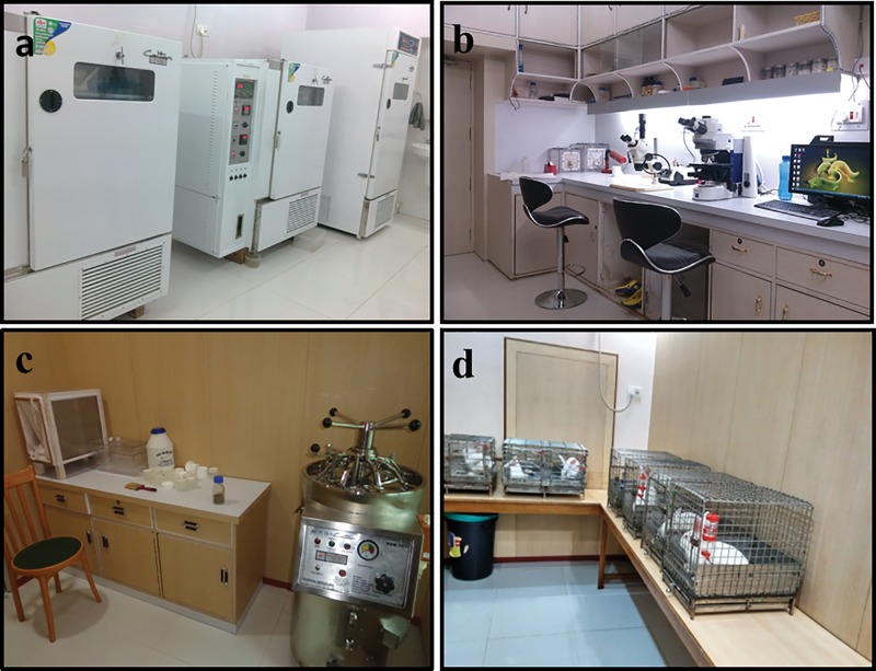
Requisite for initiating and establishing a permanent sand fly colony were construction of a proper insectary facility, procurement of equipment and supplies, as well as enlistment and training of personnel to perform the complex and labor-intensive procedures required to support the colony. All this was accomplished within a 7-mo period from March 2014 to September of 2014. At the KAMRC, a state-of-the-art insectary was built and equipped according the requirements set forth in the Arthropod Containment Levels published by the American Committee of Medical Entomology, American Society of Tropical Medicine and Hygiene (ACME 2003). The facility includes an insectary room, preparation and processing room, autoclave room, donning room, and animal housing (Fig. 1a–d). It is equipped with three locally purchased environmental chambers (Narang Scientific Works PVT, LTD, New Delhi), a refrigerator, freezer, autoclave, vacuum pump, dissection and compound microscopes, computer, as well as custom-made containers, cages and apparatus necessary for rearing and handling live sand flies. In March and April of 2014, one of the authors (P.T.) from BHU was trained for 6 wk in sand fly biology and mass rearing at the Laboratory of Parasitic Diseases, NIAID, NIH, Bethesda, MD, and at the Department of Entomology, Walter Reed Army Institute of Research (WRAIR), Silver Spring, MD. Local field collectors were trained on site in study villages on methods for collecting sand flies alive for colony stock.
Fig. 1.
Sand fly insectary facility, KAMRC, Muzaffarpur, Bihar, India: (a) colony room showing incubators and environmental cabinets; (b) preparation and processing room; (c) larva food preparation and autoclave room; (d) animal housing.
Collecting Sand Flies From the Field for Colony Stock
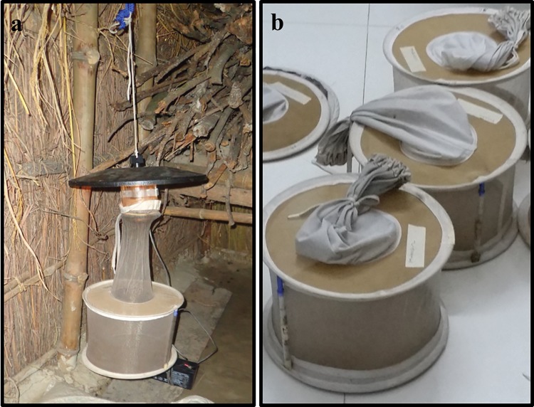
From September through mid-December of 2014, sand flies were collected from several VL-endemic rural villages in Muzaffarpur district, Bihar (26.07° N, 85.45° E) that had been selected for previous studies by the KAMRC based on reported VL cases. Then, in March of 2015, when sand fly populations were increasing (Picado et al. 2010) and after coordinating with Ministry of Health IRS (indoor residual spraying) teams, one village was identified in which residual spraying had not been done recently and in which the sand fly density was consistently high enough to enable trapping of large numbers of sand flies with which to build the colony. Sand flies were collected with CDC-type miniature light traps (John W. Hock Company, Inc., Gainesville, FL) equipped with double-ring, fine-mesh collection bags (Fig. 2a). Plastic struts, made from disposable plastic pipettes, were inserted vertically between the rings to expand the nets and prevent them from collapsing and crushing or injuring the sand flies (Fig. 2b). During the 2014 collection period (September–early December), 15 light traps were run five nights per week and in 2015 (March-early December), 15 traps were run seven nights per week. Traps were set inside bedrooms in human dwellings and in nearby cattle sheds and operated through the night from 5:00 pm until 6:00 am. Each morning, the collection bags were carefully removed from the light traps, sleeves tied shut and with expansion struts remaining in place, then enveloped in plastic bags to maintain humidity during transport to the KAMRC insectary facility.
Fig. 2.
Collecting sand flies and keeping them alive: (a) CDC-type light trap installed in a corner of a cattle shed; (b) double-ring fine-mesh collection nets expanded with plastic struts to prevent the nets from collapsing and crushing or injuring the sand flies.
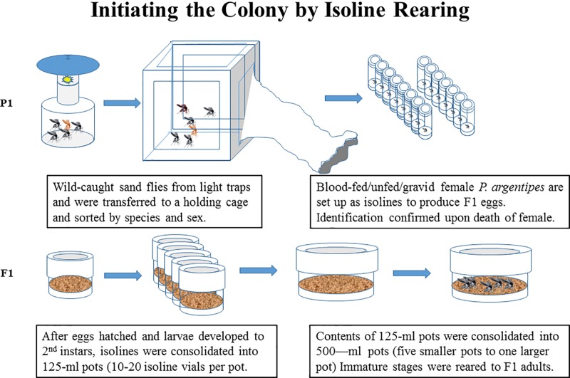
Initiating the Colony Through Isoline Rearing (Fig. 3)
Fig. 3.
Initiating the colony through isoline rearing.
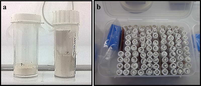
In the insectary, the captured, live flies were transferred from the collection bags via mouth aspirators to a 30- by 30- by 30-cm (1-cubic-ft.) polycarbonate holding cage. When sand flies are collected live from the field, the collected material is often a mixture of two or more species and it is necessary to separate the species to insure colonization of pure strains. This was accomplished through isoline rearing, where each female sand fly was allowed to lay eggs in a separate oviposition vial and her progeny then reared in the same vial, separate from the progeny of any other female. Because P. argentipes is a darkly pigmented species and larger than most Sergentomyia species, the larger, darkly pigmented female sand flies, blood-fed or gravid, were captured into 10–15-ml isoline-rearing vials for egg laying (Fig. 4a). All other flies were discarded. The bottom of each vial was lined with a 2-cm layer of dental plaster, which was moistened to provide a suitable oviposition surface and to maintain humidity in the vial. The center of the snap-on cap was cut out, forming a ring with which to secure a fabric-screen cover over the mouth of the vial. A small piece of cotton soaked in 30% sucrose solution was placed on the screen top of each vial as an energy source and the vials were stored in vented plastic boxes at 27 °C and 80% RH (Fig. 4b). Each vial was labeled with collection site, collection date, and set-up date. Because most sand fly species cannot be identified with certainty based on size, pigmentation or external morphology, and because P. argentipes may be confused with some of the larger, dark-pigmented Sergentomyia species, no assumptions were made as to species identity. When an isolined female laid eggs and died, she was removed immediately from the vial, dissected under a microscope and identified, based on internal morphological characters, using appropriate dichotomous keys (Lewis 1978, Kalra and Bang 1988), thus identifying also the female’s progeny. If a specimen could not be identified or in case of doubt, it was discarded along with the associated progeny. When the eggs hatched, usually after about 5 or 6 d, that date was marked on the vial and a small amount of finely ground larva food was sprinkled onto the plaster surface in the bottom of the vial and the immature stages were reared to adults following the general procedures of Lawyer et al (1991) and Modi and Rowton (1996) with some modifications. After developing to second instars, progeny of isolined females were pooled into 125-ml oviposition and rearing pots and reared together. Subsequently, four or five 125-ml pots could be consolidated into a single 500-ml pot and reared to adults (Fig. 3).
Fig. 4.
Isoline rearing: (a) Female sand flies were captured into 10–15-ml isoline-rearing vials for egg laying; (b) isoline rearing vials were provided with a small piece of cotton soaked in 30% sucrose solution placed on the screen top of each vial as an energy source and the vials were stored in plastic boxes at 28 °C and 80% humidity.
Establishing and Expanding the Colony (Fig. 5)
Fig. 5.
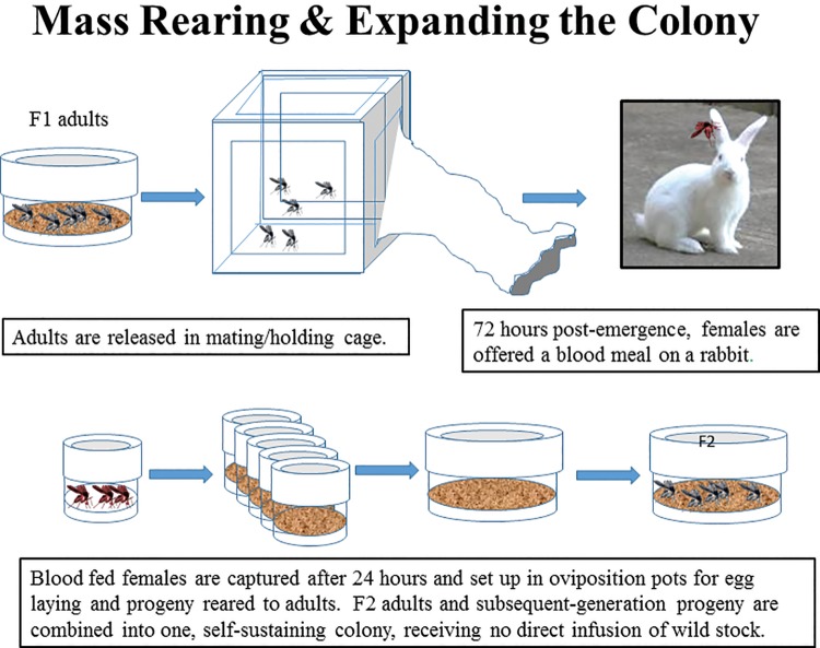
Establishing and expanding the colony.
Blood Feeding the Adults
When the F1 adults emerged in the rearing pots, they were released into 30- by 30- by 30-cm holding or mating cages or, depending on the number of flies, transferred to 1,000-ml holding pots and held for up to 72 h to mate and to develop blood hunger. Because it was uncertain at first which animal host the flies would feed on readily, the F1 females were offered bloodmeals on anesthetized mice, hamsters, chickens, and restrained rabbits until it was determined which host was preferred. All host animals were purchased from the Central Drug Research Institute (CDRI), Lucknow, Uttar Pradesh, and were certified pathogen free. A large feeding cage (55 by 48 by 40 cm) was constructed to accommodate the restrained rabbit. Three- to five-day-old female P. argentipes were released into the cages and allowed to feed on host animals for 1 to 3 h. After blood feeding, the host animals were removed and blood-engorged females were left in the cage overnight to permit more mating and allow for the peritrophic membranes surrounding the bloodmeals to harden. After 24 h, engorged flies were transferred to either 125-ml- or 500-ml oviposition and rearing pots, depending on the number of flies that fed, and the immature rearing cycle started over. Females that did not blood-feed at the first offering were transferred to holding containers to be fed the next time around. All feeds on live animals were conducted in compliance with the following animal-use protocol: Use of Rabbits, New Zealand Strain (Oryctolagus cuniculus), Hamsters (Mesocricetus auratus) and Mice (Mus musculus) for Blood Feeding Colonies of Phlebotomine Sand Flies (Protocol No. BHU13LM); approved 27/04/2015 by the Central Animal Ethical Committee, Banaras Hindu University.
Feeding the Larvae
Initially, larva food, consisting of a composted 1:1 mixture of commercial rabbit chow and rabbit feces (Young et. al., 1981) was provided by the Division of Entomology, Walter Reed Army Institute of Research (WRAIR, Silver Spring, MD), until suitable food could be prepared at the KAMRC from locally available ingredients. A variety of natural ingredients such as rabbit feces, cow manure and goat feces, all readily available at the collection sites, were composted 1:1 with locally purchased rabbit chow to make larva food. Before using the larva food to feed the colony, it was sterilized by autoclaving. Thereafter it was stored in a refrigerator at 4 °C until used.
Expanding the Colony to Working Level
F1-generation adults and their F2-immature progeny were kept in a separate incubator apart from the main colony to provide some physical and temporal separation between the wild-caught parents (P1) and the laboratory colony. F2- and subsequent-generation adults and progeny were combined in a single colony for expansion to a targeted working-level of 1,000–1,500 blood-fed females per week, of which one third could be used for research without jeopardizing the health and vigor of the colony. To accelerate expansion of the colony, emergent females were fed three to five times per week (Fig. 5).
Routine Colony Maintenance
To ensure that all colony maintenance tasks are accomplished in a timely and accurate manner each day, a weekly colony data log sheet was developed on which to record performance of routine tasks, as well as daily temperature and humidity readings, the number of blood-fed females captured after each feeding, and the number of flies (males and females) removed from the colony for research purposes. Such data provide researchers a clear picture of colony size, rate of growth, the impact of research demands, and the overall health of the colony.
Life Table Study
The progeny of 45 fifth-generation females were reared as isolines through adult emergence and all life events such as oviposition, hatching, transition between instars, pupation, and eclosion were chronicled to generate a stage-specific life table for future reference.
Closing and Certifying the Colony
By early December of 2015, field collections were terminated because the sand fly populations in the field had dwindled to near zero. At that time the laboratory P. argentipes colony was closed to further infusions of wild-caught material. Because the colonized sand flies are to be used for xenodiagnosis on humans, an important consideration was to ensure that the sand flies in the closed colony were free of sand fly-borne human pathogens. All handling equipment, holding containers, and working surfaces had to be sanitized regularly and larva food sterilized by autoclaving. The closed colony was then subjected to PCR assays to confirm the species identification as P. argentipes and to certify that the colony was free of specific sand fly-borne pathogens of actual or potential concern to humans and known to have caused disease in India.
Confirmation of Sand Fly Species
Molecular confirmation of morphologically identified flies was performed on a random sampling of 25 flies from the colony. Each sand fly was processed individually for DNA isolation using a Fast Tissue-to-PCR Kit (Thermo Fisher Scientific, Waltham, MA) according to the manufacturer’s instructions. For PCR, the target gene was the 18s rRNA coding region with forward primer 5′-TAGTGAAACCGCAAAAGGCTCAG-3′ and reverse primer 5′-CTCGGATGTGAGTCCTGTATTGT-3′ (Tiwary et al. 2012). The reaction was carried out in a volume of 25 µl using the pair of primers (10 pM each), 1 U normal Taq DNA polymerase (New England Biolabs, Ipswich, MA) supplemented with MgCl2, 10 X buffer, 1 X BSA, and 0.2 mM of dNTPs. For a template, 50 ng of extracted DNA were used, while nuclease-free water (Qiagen, Hilden, Germany) was used as negative control. PCR conditions were initial denaturation at 95°C for 5 min, followed by 35 cycles of denaturation at 95°C for 30 s, annealing at 58°C for 40 s, extension at 72°C for 30 s, and final extension at 72°C for 10 min. Amplified PCR products were used directly for sequencing. The amplicons were observed as a single band on the agarose gel, leftover primers and dNTPs from PCR products having been removed using a QIAquick PCR Purification Kit (Qiagen, Hilden, Germany). Further, the eluted PCR products were sent to an independent facility (SciGenom Lab, Hyderabad, India) for sequencing using the same set of primers. The PCR products were sequenced using a BigDye Terminator 3.1 cycle sequencing kit (Applied Biosystems, Foster City, CA) and run through an ABI 3133 sequencer. This process will be repeated quarterly for the first year to reconfirm the purity of the colony.
Screening for Specific Pathogens
All sand fly-transmitted arboviruses worldwide were considered for detection in our colony-reared sand flies. However, based on which sand fly-borne viruses are known to occur in India, the list for viral screening in sand flies was limited to Chandipura virus and Sand Fly Fever viruses (Sicilian, Naples) (Menghani et al. 2012, Alkan et al. 2013). Control RNA samples were prepared from viral material generously provided by Dr. Robert Tesh, Center for Biodefense and Emerging Infectious Diseases, The University of Texas Medical Branch, Galveston, TX.
From the closed colony, 100 sand flies were randomly selected and divided into 10 pools, each containing 10 sand flies. Pools were stored in RNAlater (Qiagen USA, Germantown, MD) at −80°C until RNA extraction. RNA was isolated using RNeasy mini kit (Qiagen USA, Germantown, MD) as per the manufacturer’s protocol and eluted in 30 µl RNase-free water (Qiagen USA, Germantown, MD). RNA of each pool was quantified using Nano Drop 2000c and quality checked based on the ratio of A260/A280. One µg RNA isolated from each pool was reverse transcribed in a 20-μl reaction using a High Capacity cDNA Reverse Transcription Kit (Invitorgen, Thermo-Fisher Scientific, Waltham, MA) and diluted 10 times with RNase-free water to a final concentration of 5 ng/μl.Pre-described primers and probes specific for each virus were used for PCR (Kumar et al. 2008, Weidmann et al. 2008). Q-PCR reactions were set up with a 20-µl reaction mixture containing 10 µl of TaqMan master mix (Applied Biosystems, Thermo Fisher Scientific, Waltham, MA), 10 pM each of forward primer, reverse primer and probe, and 25 ng of each sample. Reactions were analyzed in MicroAmp optical 96-well reaction plates (Applied Biosystems, Thermo Fisher Scientific, Waltham, MA) using a 7500 Real-Time PCR System (Applied Biosystems, Thermo Fisher Scientific, Waltham, MA).
Pathogen screening is repeated quarterly to ensure that the colony remains pathogen free for the purposes of the xenodiagnosis study.
Blood-Feeding Experiments on Human Volunteers
In preparation for actual xenodiagnostic studies, feeding experiments were conducted on healthy (uninfected) human volunteers to optimize the number of flies to be fed on human subjects, as well as the exposure site and the exposure time. Small, 5 cm- (2-inch-) diameter polycarbonate feeding cups (Fig. 6), specifically designed for this purpose (Precision Plastics Inc., Beltsville, MD), were loaded with 20 to 32 four-day-old female P. argentipes, then strapped to the inside of the arm or leg of a volunteer. The flies were allowed to feed for 30 min, after which they were removed and the number fed and unfed were counted. All sand fly feeds on humans were conducted in compliance with the following human-use protocol: Xenodiagnosis of Visceral Leishmaniasis, Post Kala-azar Dermal Leishmaniasis and Asymptomatic Subjects Using Phlebotomine Sand Flies (Revised Protocol Version 4.0); approved 26/05/2016 by the Institute Ethical Committee, Banaras Hindu University.
Fig. 6.
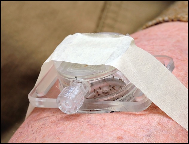
Custom-made feeding cup used for xenodiagnostic feeds (Precision Plastics, Beltsville, MD).
Infection Experiments
To assess the limits and sensitivity of parasite detection by microscopic examination and by RT-PCR, 4-d-old fifth-generation female sand flies were fed through chick-skin membranes on heat-inactivated human blood laced with concentrations of 5 × 10³, 5 × 10⁴, and 5 × 10⁵ of cultured L. donovani parasites per ½ ml of blood estimated to provide 2, 20, and 200 parasites per 0.2 µl bloodmeal, respectively. Serial dissections and microscopic examinations were performed in sterile 1x-PBS on the blood-fed flies 24, 48, and 72 h postprandial to assess how soon after feeding parasites could be detected in the sand fly midgut via light microscopy.
Detection and Identification of Leishmania donovani in Experimentally Infected Flies
Material recovered from flies that were dissected and microscopically examined was analyzed by molecular methods to confirm microscopy results. Leishmania-specific minicircle kDNA-targeting primers were used to quantify parasite load in the experimentally infected flies. Determination of parasite load was done using a standard curve made with 10-fold serial dilutions spiked with Leishmania DNA and a fixed amount of sand fly DNA. Parasite load in the infected sand flies was calculated by comparing Ct values with the standard plot. Q-PCR reactions were set up with a 20-µl reaction mixture containing 10 µl of SYBR Green reagent (Applied Biosystems, Thermo Fisher Scientific, Waltham, MA), 10 pM each of forward and reverse primers, and 100 ng of each DNA sample. Reactions were analyzed in MicroAmp optical 96-well reaction plates (Applied Biosystems, Thermo Fisher Scientific, Waltham, MA) using a 7500 Real-Time PCR System (Applied Biosystems, Thermo Fisher Scientific, Waltham, MA).
Results
Collecting Sand Flies From the Field for Colony Stock
In September, 2014, collections began at a time when sand fly density was on the down side of the seasonal population peak and numbers were declining (Picado et al. 2010). From September through early-December of 2014, 3,644 sand flies (1,642 males, 2,022 females) were collected in 50 nights of trapping (15 traps per night) or 750 trap nights. In spite of low density, numbers were sufficient to enable harvesting of 412 egg clutches from blood-fed and gravid females set up as isolines. Attempts to feed wild-caught female flies on anesthetized mice or hamsters placed inside the holding cage were unsuccessful but some feeding was observed when a small, restrained rabbit was placed inside the cage. It was found that feeding success could be increased by placing wild-caught males in the cage with the females. When F1 adults began to emerge from the isolined broods, numbers of males and females increased sufficiently to stimulate marginal feeding activity. A small colony was initiated with regular infusions of females from the field to produce F1 progeny. However, when sand fly population density in the field decreased to near zero during the first week in December, regular infusions with wild stock ceased. Adult emergence from the isolines was only sufficient to sustain the colony through the F3 generation. Once the number of emerging F1 adults declined below a critical threshold, blood-feeding success declined and the colony did not expand. Ultimately, due to a “perfect storm” of issues including cessation of infusions of wild stock to produce F1 adults, marginal blood-feeding success, poor survival of blood-fed females resulting in few viable egg clutches, poor quality plaster in the isoline vials that caused unacceptable mortality of eggs and first instars, changes in insectary staff and lack of training and experience, temporary absence of key personnel and a severe mite infestation, the colony dwindled and died.
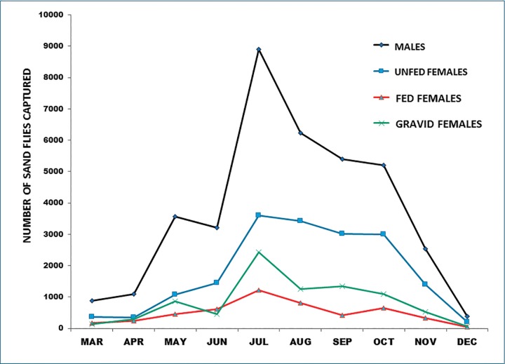
Building on the experiences and lessons learned from the previous year’s effort, and taking advantage of an anticipated seasonal increase in sand fly populations (Picado et al., 2010), collections resumed in March of 2015 and continued through mid-December 2015, using 15 traps for a total of 254 nights or 7,620 trap nights. As shown in Fig. 7 and in Table 1, sand fly populations in the field increased steadily, peaking in July with a monthly total of 16,144, then declined gradually through October, and precipitously to zero in mid-December. A total of 68,601 flies was collected, from which 13,348 blood-fed or presumptively gravid P. argentipes females were visually selected and set up as isolines (Table 1).
Fig. 7.
Graph showing seasonal profile of sand fly collections using CDC light traps, 1 March to mid-December, 2015.
Table 1.
Results of light-trap collections, 1 March to mid-December 2015; 15 traps operating seven nights per week for 254 d (7,620 trap nights)
| Females |
||||||
|---|---|---|---|---|---|---|
| Month | Males | Unfed | Fed | Gravid | Total Fem | Total SF |
| March | 880 | 358 | 166 | 134 | 658 | 1,538 |
| April | 1,101 | 339 | 235 | 301 | 875 | 1,976 |
| May | 3,569 | 1081 | 455 | 854 | 2,390 | 5,959 |
| June | 3,208 | 1,446 | 608 | 456 | 2,510 | 5,718 |
| July | 8,906 | 3,604 | 1,212 | 2,422 | 7,238 | 16,144 |
| August | 6,231 | 3,419 | 804 | 1,249 | 5,472 | 11,703 |
| September | 5,388 | 3,018 | 423 | 1,339 | 4,780 | 10,168 |
| October | 5,198 | 2,990 | 645 | 1,093 | 4,728 | 9,926 |
| November | 2,533 | 1,401 | 325 | 518 | 2,244 | 4,777 |
| December | 383 | 200 | 39 | 70 | 309 | 692 |
| Seasonal totals | 37,397 | 17,856 | 4,912 | 8,436 | 31,204 | 68,601 |
Initiating the Colony Through Isoline Rearing
From the isolined females mentioned above, 2,598 clutches averaging 28 eggs each (range 1–98; SD = 17.0098) were harvested, ∼90% of which hatched. All but two of the isolined females that oviposited were identified postmortem as P. argentipes, validating the accuracy of visual selection of P. argentipes females from wild-caught material for colony stock. The two exceptions were Sergentomyia sp. and their eggs were discarded. Although feeding success of F1 females was meager at first because of low numbers, as collections steadily increased with the season, a continuous critical mass of F1 males and females was available to stimulate feeding.
Establishing and Expanding the Colony
Blood Feeding the Adults
Attempts to feed P. argentipes on anesthetized mice and hamsters were largely unsuccessful owing possibly to the small number of flies at first. However, when a retrained rabbit was placed in the holding cage, blood feeding improved, indicating that the larger animal was more attractive to the flies. Use of the larger feeding cage and a larger and a more comfortable restrainer to accommodate an adult rabbit further enhanced blood-feeding success to around 90%.
Feeding the Larvae
Larva food made with locally collected rabbit feces and locally purchased rabbit chow did not work well, owing most likely to excess moisture and urine content. Surprisingly, composted cow manure, ever present at the collection sites, was not well accepted by the larvae. Finally, dried goat feces, aerobically composted 1:1 with locally purchased rabbit chow, made excellent larva food, and is now used to maintain the colony. Autoclaving the larva food inevitably destroyed some nutrient content but the only noticeable effect was extension of the developmental cycle by 2 or 3 d. Otherwise, the immature and adult stages were healthy and robust and very suitable for the xenodiagnosis study.
Expanding the Colony to Working Level
With blood-feeding success consistently at ∼90% and larvae thriving on locally made larva food, the colony quickly expanded with each new generation and was soon self-sustaining. By the time field populations of sand flies dwindled to near zero around the second week of December, 2015, the colony was self-sustaining and was closed to further infusion with wild stock. Since closure, the self-sustaining colony has expanded through 15 generations as much as eight fold per generation. It is healthy and robust, and produces 1,500 to 2,500 blood-fed females per week for egg production, exceeding the target of 1,000–1,500 per week.
Life-Table Study
Table 2 shows the results of isoline-rearing at 27 °C and 80% RH of the progeny of 45 F5-females from the colony. The mean number of eggs laid per female was 49.51 and the mean generation time from bloodmeal to adult emergence was only 38.6 d, reflecting near optimal rearing conditions and a healthy and robust colony.
Table 2.
Stage-specific life table attributes of the F5 progeny of 45 P. argentipes females reared at 27 °C and 80% RH
| Life table attributes | ||
|---|---|---|
|
P. argentipes, 27 °C and 80% RH | ||
| Development time in days | Mean | SD |
| Bloodmeal to oviposition | 4.21 | 1.93 |
| Oviposition to egg hatch | 5.93 | 0.53 |
| Egg hatch to 2nd instar (1st stadium) | 5.86 | 0.53 |
| 2nd to 3rd instar (2nd stadium) | 4.79 | 1.25 |
| 3rd to 4th instar (3rd stadium) | 3.86 | 1.51 |
| 4th instar to pupa (4th stadium) | 5.36 | 1.5 |
| Pupa to 1st adult (pupa stage) | 6.14 | 1.23 |
| Oviposition to 1st adult | 33.14 | 2.21 |
| Oviposition to 1st male | 33.21 | 2.29 |
| Oviposition to 1st female | 33.24 | 2.13 |
| Mean generation time | 36.15 | 2.06 |
| Fecundity | ||
| Mean number eggs/female (n = 45) | 49.51 | 12.69 |
| Range | 4–70 | |
| Total no. of eggs from 45 clutches | 2,284 | |
Closing and Certifying the Colony
Confirmation of Sand Fly Species
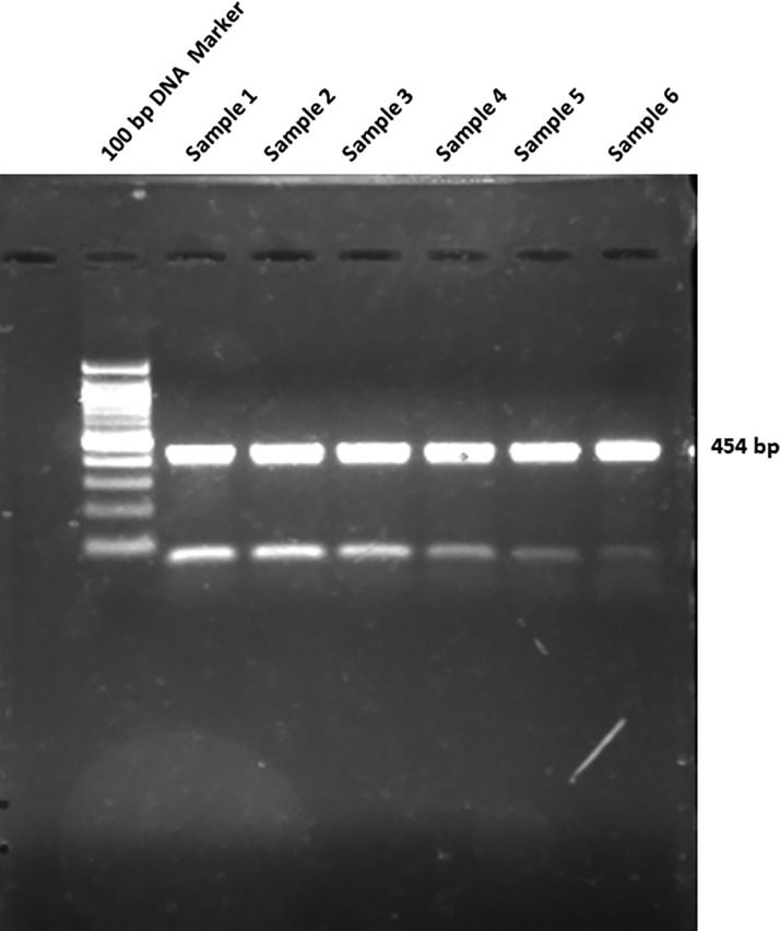
Twenty-five randomly selected sand flies from the laboratory colony were processed individually for DNA extraction, 18s rRNA-coding region-specific PCR, and sequencing. All of the samples were P. argentipes, confirming colonization of a pure species strain (n = 25; Fig. 8).
Fig. 8.
Image showing amplified PCR products derived from DNA of colony sand flies on agarose gel, confirming their identity as P. argentipes.
Screening for Specific Pathogens
Ten pools of colony-reared sand flies, each with 10 flies, were processed for RNA extraction, cDNA preparation, and real-time RT-PCR. None of the control virus-segment probes detected viral RNA in colony-reared flies (n = 100).
Blood-Feeding Experiments on Humans
A series of uninfected feeds on human volunteers was conducted to assess feeding success and to determine the optimum number of sand flies to be fed on patients in the xenodiagnostic study. Feeding success ranged from 5% to 65% depending on the human blood source and number of flies in the feeding cup. The more flies per cup, the higher the blood-feeding success. Near optimum feeding (>60%) was accomplished with 32–50 females per feeding cup (plus 10 males). Based on these results, it was decided to limit the number of females per feed to 30.
Infection Experiments
In serial dissections of flies artificially infected through chick-skin membranes, at 24 h, the bloodmeal was still intact, red at the center and black at the periphery and a few procyclic promastigotes could be seen at the highest concentration (2 × 10⁵) by microscopic examination. By 48 h the peritrophic membrane had broken down and many procyclic promastigotes and nectomonads were readily visible. By 72 h, most of the bloodmeal had been digested and the bulk of the parasites had passed out with it, but numerous nectomonads could be seen attached to the wall of the anterior abdominal midgut and haptomonads were already attached to the stomodeal valve. No parasites were seen in these experiments via light microscopy at concentrations of <50,000 parasites/ml while Q/RT-PCR detected parasites with infection concentration of 10,000 parasites/ml at 48–72 h post infectious feed.
Discussion
In 1924, under the directorship of Christophers, a Kala-azar commission was set up in India at the School of Tropical Medicine, Calcutta, to “resolve the important problem of kala-azar transmission.” One of the first accomplishments of the Commission was to establish a colony of the suspected vector, Phlebotomus argentipes, from wild-caught material, “…using a new technique for the breeding and feeding of sandflies… “ Female P. argentipes produced from that colony were fed directly on kala-azar patients in a series of 11 experiments. In 10 of the 11 experiments, upon subsequent dissection and microscopic examination of blood-fed flies, “typical herpetomonad forms” similar to L. donovani cultured from patients’ blood were observed, thus confirming the suspicion that P. argentipes was indeed the vector of kala-azar (Knowles et al. 1924). These early experiments also demonstrated the value of direct xenodiagnosis in the study of the transmission cycle of kala-azar. Now, nearly a century later, direct xenodiagnosis on canine and human subjects once again is being used to resolve important aspects of kala-azar epidemiology. With the advent of modern molecular techniques, direct xenodiagnosis on human subjects is not considered an option for determining if a person is infected with the parasite. However, for assessing the potential of specific human-subject groups to serve as reservoirs of Leishmania donovani infection to sand flies, direct xenodiagnosis using live vector sand flies is the only viable option.
Key to the success of any xenodiagnostic study is the availability of a robust, self-sustaining colony of the vector, closed to infusions with wild-caught material and certified free of specific sand fly-borne human pathogens. Direct xenodiagnosis relative to VL epidemiological studies has been conducted in areas where canine leishmaniasis due to Leishmania infantum is endemic and where coinfections of HIV and L. infantum have occurred, using laboratory colonies of Lutzomyia longipalpis and Phlebotomus perniciosus (Molina et al. 1992, 1999; Magalhães-Junior et al. 2016). Although colonies of P. argentipes have been established for various purposes at other institutions in India (Ghosh et al. 1991), to our knowledge, none have been maintained as closed colonies nor do any exist that meet the stringent biosafety requirements of this human-use study.
When collecting sand flies from the field, knowledge of local seasonal changes is crucial to insure appropriate timing to take advantage of peak population densities or, in some instances, to avoid collecting flies that are already programmed for an obligatory or facultative winter diapause (Lawyer et al. 1991). Because our initial opportunity to collect wild stock from the field began in September, 2014, when sand fly density was on the downside of the seasonal population peak (Dinesh et al. 2001, Picado et al. 2010), it was impossible to collect sufficient numbers of flies to sustain a constant supply of F1 adults to stimulate good social feeding behavior and grow the colony. Having learned from the 2014 collection experience, we commenced the 2015 collection effort in May when field populations of P. argentipes were approaching highest densities (Dinesh et al. 2001, Picado et al. 2010), enabling the production of a steady supply of F1 adults throughout the sand fly season to grow the colony to a self-sustaining level.
The use of isoline rearing was another key factor contributing to the successful establishment of the colony. Our experience has revealed some advantages of isoline rearing: 1) females set up in isoline vials, as opposed to larger oviposition containers, are more sedentary and expend less energy flying around, resulting in better survival and higher egg production; 2) after the parent female dies, she can be removed and accurately identified to species, thus also identifying her progeny and ensuring colonization of a pure strain; 3) the progeny of each parent female can be reared separately, facilitating life-table studies to chronical immature development and determine fecundity, productivity, and overall generation time.
Successful blood feeding of emergent F1 females is essential to the survival and expansion of an incipient colony and, with some species, may be the biggest challenge. Many sand fly colonies are reared successfully on anesthetized mice, hamsters or Guinea pigs, but finding an acceptable host for some species may be problematic. Attraction of the female sand fly to a host is necessary for good feeding success. With this P. argentipes colony, very poor feeding success was achieved on anesthetized rodents. This may have been due in part to the small numbers of flies available to feed. However, a few flies were attracted to a restrained rabbit. In the natural mating system of P. argentipes, males aggregate in large numbers on or in the vicinity of the host, a behavior called “lekking.” Host-seeking females are then attracted to the lek and when in proximity to the host, they feed and simultaneously the males couple and mate with them (Yuval 2006, Lane et al. 1990). Although it has not been shown in P. argentipes, there is substantial evidence in some sand fly species that pheromone-mediated communication may play an important role in mating and feeding behaviors (Jones and Hamilton 1998, Yuval 2006). Because the natural social mate- and host- seeking behavior of P. argentipes requires male lekking to attract the females to the host and stimulate mating and feeding, initial efforts to feed female P. argentipes in small numbers in the absence of males were largely unsuccessful and insufficient to sustain the colony without regular infusions of wild-stock. However, in the subsequent effort, with the sustained production of large numbers of F1 adults, both male and female, optimum social feeding behavior was achieved and the colony became self-sustaining. Expansion of the colony was accelerated by feeding the flies at least three times per week.
Equally important to the survival and growth of the colony was the successful feeding of the larvae. Killick-Kendrick (1978) stated that “Among the major problems in rearing phlebotomine sand flies is excessive larval mortality caused by fungal growth, improper diet or moisture, disease and other factors. These problems are especially acute during the first stadium when the first instar larvae become easily entrapped in fungal mycelia or in moisture condensation.” Because the aerobic composting process (Young et al. 1981, Lawyer et al. 1991, Modi and Rowton 1996, Volf and Volfova 2011) takes 2 wk to 1 mo, the food had to be prepared well in advance of initiating the colony so as to be available to feed the developing larvae. Fortunately, a supply of excellent larva food was available from the WRAIR until such time as suitable larvae food could be produced locally from goat feces and rabbit chow.
Not until the colony was self-sustaining could it be closed to infusion with wild-caught flies, nor could the process of screening for specific pathogens begin, both of which were absolutely necessary before the colony-bred flies could be used for xenodiagnosis on humans. “Specific-pathogen-free (SPF)” is a term used for laboratory animals that are guaranteed free of particular pathogens. Use of SPF animals ensures that specified diseases do not interfere with an experiment. In this study, the objects of specific-pathogen screening were the sand flies as well as the host animals (rabbits) used as a bloodmeal source.
This sand fly colonization effort is especially noteworthy in that the elapsed time from start to finish was just under 18 mo (June 2014 to May 2016), which included building the infrastructure, training personnel, collecting wild stock, initiating and establishing a self-sustaining colony, expanding it to working level, closing it to infusion with wild-caught material, and certifying it specific-pathogen free. Having accomplished all of the milestones of this pilot project, a one-of-a-kind, healthy, robust, self-sustaining and closed colony, certified free of specific sand fly-borne human pathogens known to occur in India, is now available and is being used for direct xenodiagnosis to define the ability of specific human-subject groups across the infection spectrum to serve as reservoirs of Leishmania donovani infection to sand flies in Bihar, India, directed toward the eventual elimination of this important infectious disease.
Acknowledgments
We thank Mr. Anil Sharma and his staff at the KAMRC for administrative and logistical support, without which this project could not have been accomplished. Thanks also to the excellent and hard work of the many technicians who collected sand flies from the field, and especially Ratan Kumar, Roshan Kumar, and Vinod Kumar who processed the collections in the laboratory and performed the detailed and time-consuming labor of maintaining the growing colony. Dr. Mukesh Mishra provided valuable field expertise in the initial phase of sand fly collection for colony stock. We also thank the friendly residents of the study villages, without whose willing and outstanding cooperation this effort would have been impossible. This work was funded by the Foundation for the National Institutes of Health under grants #BHU13LM and #KAMRC13LM through grant #OPP1096046 from the Bill & Melinda Gates Foundation, and in part by the Intramural Research Program of the National Institute of Allergy and Infectious Diseases, National Institutes of Health.
References Cited
- American Committee of Medical Entomology. 2003. Arthropod Containment Guidelines. Vector-Borne and Zoonotic Diseases, Mary Anne Liebert, Inc. 32: 75–90. (http://online.liebertpub.com/doi/pdf/10.1089/153036603322163475) (accessed 2 May 2017). [Google Scholar]
- Alkan C., Bichaud L., de Lamballerie X., Alten B., Gould E. A., Charrel R. N.. 2013. Sandfly-borne phleboviruses of Eurasia and Africa: Epidemiology, genetic diversity, geographic range, control measures. Antiviral Res. 100: 54–74. http://dx.doi.org/10.1016/j.antiviral.2013.07.005 (accessed 2 May 2017). [DOI] [PubMed] [Google Scholar]
- Dinesh D. S., Ranjan A., Palit A., Kishore K., Kar S. K.. 2001. Seasonal and nocturnal landing/biting behaviour of Phlebotomus argentipes (Diptera: Psychodidae). Ann. Trop. Med. Parasitol. 95: 197–202. [DOI] [PubMed] [Google Scholar]
- Ghosh K. N., Ghosh D. K., De A., Battacharya A.. 1991. Biology of Phlebotomus argentipes Annandale and Brunetti and P. papatasi (Scopoli) in the laboratory. Ann. Parasitol. Hum. Comp. 67: 55–61. [Google Scholar]
- Jones T. M., Hamilton J.G.C.. 1998. A role of pheromones in mate choice in a lekking sandfly. Anim. Behav. 56: 891–898. [DOI] [PubMed] [Google Scholar]
- Kalra N. L., Bang Y. H.. 1988. Manual on Entomology in Visceral Leishmaniasis. WHO, Regional Office for South-East Asia, New Delhi. SEA/VBC/35. [Google Scholar]
- Killick-Kendrick R. 1978. Recent advances and outstanding problems in the biology of phlebotomine sandflies. Acta Trop. 35: 297–313. http://www.e-periodica.ch/digbib/view?var=true&pid=act-001:1978:35::460#315 (accessed 2 May 2017). [PubMed] [Google Scholar]
- Knowles R., Napier L. E., Smith R.O.A.. 1924. On a Herpetomonas found in the gut of the sand fly, Phlebotomus argentipes, fed on kala-azar patients. A Preliminary Note. Ind. Med. Gaz. 59: 593–597. [PMC free article] [PubMed] [Google Scholar]
- Kumar S., Jadi R. S., Anakkathil S. B., Tandale B. V., Mishra A. C., Arankalle V. A.. 2008. Development and evaluation of a real-time one step Reverse-Transcriptase PCR for quantitation of Chandipura Virus. BMC Infect. Dis. 8: 168.. [DOI] [PMC free article] [PubMed] [Google Scholar]
- Lane R. P., Pile M. M., Amerasinghe F. P.. 1990. Anthropophagy and aggregation behaviour of the sandfly Phlebotomus argentipes in Sri Lanka. Med. Vet. Entomol. 4: 79–88. [DOI] [PubMed] [Google Scholar]
- Lawyer P. G., Rowton E. D., Perkins P. V., Johnson R. N., Young D. G.. 1991. Recent advances in laboratory mass rearing of phlebotomine sand flies. Parassitologia 33: 361–364. [PubMed] [Google Scholar]
- Lewis D. 1978. The phlebotomine sandflies (Diptera:Psychodidae) of the Oriental Region. Department of Entomology, British Museum (Natural History; ), Cromwell Road, London: SW7 5BD. [Google Scholar]
- Magalhães-Junior J. T., Molta T. F., Porfirion-Passos G., Larangeira D. F., Franke C. R., Barrouin-Melo S. M.. 2016. Xenodiagnosis on dogs with visceral leishmaniasis: Canine and sand fly aspects related to the parasite transmission. Vet. Parasitol. 15: 120–126. [DOI] [PubMed] [Google Scholar]
- Menghani S., Rupesh C., Raval A., Wadibhasme P., Khedekar P.. 2012. Chandipura virus: An emerging tropical pathogen. Acta Trop. 124: 1–14. [DOI] [PubMed] [Google Scholar]
- Modi G. B., Rowton E. D.. 1996. Laboratory maintenance of phlebotomine sand flies, pp. 109–121. InMaramorsch K., Mahmood F. (eds.), Maintenance of human, animal and plant pathogen vectors. Oxford & IBH Publishing Co. Pvt. Ltd, New Delhi, India. [Google Scholar]
- Molina R., López-Vélez R., Jiménez M.. 1992. Isolation of Leishmania infantum from the blood of a patient with AIDS using sandflies. Trans. R. Soc. Trop. Med. Hyg. 86: 516. DOI: 10.1016/0035-9203(92)90092-Q· 86, 516. [DOI] [PubMed] [Google Scholar]
- Molina R., Lohes J. M., Pulido F., Laguna F., Lopez-Velez R., Alvar J.. 1999. Infection of sandflies by humans coinfected with Leishmania infantum and human immunodeficiency virus. Am. J. Trop. Med. Hyg. 60: 51–53. [DOI] [PubMed] [Google Scholar]
- Picado A., Das M. L., Kumar V., Dinesh D. S., Rijal S., Singh S. P., Das P., Coosemans M., Boelaert M., Davies C.. 2010. Phlebotomus argentipes seasonal patterns in India and Nepal. J. Med. Entomol. 47: 283–286. DOI: 10.1603/ME09175 [DOI] [PubMed] [Google Scholar]
- Singh O. P., Hasker E., Boelaert M., Sundar S.. 2016. Elimination of visceral leishmaniasis on the Indian subcontinent: Critical knowledge gaps to be answered to get us there. Lancet Infect. Dis. Published online September 28, 2016 http://www.thelancet.com/journals/laninf/article/PIIS1473-3099(16)30140-2/fulltext (accessed 2 May 2017). [DOI] [PMC free article] [PubMed] [Google Scholar]
- Tiwary P., Kumar D., Rai M., Sundar S.. 2012. PCR-RFLP based method for molecular differentiation of sand fly species Phlebotomus argentipes, Phlebotomus papatasi, and Sergentomyia babu found in India. J. Med. Entomol. 49: 1515.. [DOI] [PMC free article] [PubMed] [Google Scholar]
- Volf P., Volfova V.. 2011. Establishment and maintenance of sand fly colonies. J. Vector Ecol. 36 (Suppl 1): S1–S9. doi: 10.1111/j.1948-7134.2011.00106.x. [DOI] [PubMed] [Google Scholar]
- Weidmann M., Sanchez-Seco M. P., Sall A. A., Ly P. O., Thiongane Y., Lô M., Schley H., Hufert F. T.. 2008. Rapid detection of important human pathogenic Phleboviruses. J. Clin. Virol. 41: 138–142. [DOI] [PubMed] [Google Scholar]
- (WHO) World Health Organization. 2016. WHO Fact Sheet, March, 2016 (http://www.who.int/mediacentre/factsheets/fs375/en/) (accessed 3 May 2017).
- Young D. G., Perkins P. V., Endris R. G.. 1981. A larval diet for rearing phlebotomine sand flies (Diptera: Psychodidae). J. Med. Entomol. 18: 446. [Google Scholar]
- Yuval B. 2006. Mating system of blood-feeding flies. Annu. Rev. Entomol. 51: 413–440. doi: 10.1146/annurev.ento.51.110104.151058. [DOI] [PubMed] [Google Scholar]


