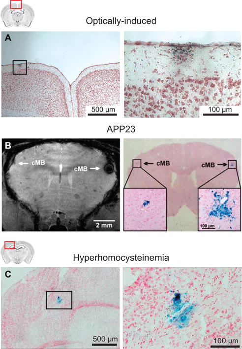
Figure 4. Microhemorrhages in mouse models.

(A) A cortical microhemorrhage observed in Prussian blue stained mouse brain sections after optically-induced rupture of a single cortical penetrating arteriole. From Rosidi et al.34 (B) Microbleeds detected by T2*-weighted MRI (left) and corresponding microhemorrhages detected by Prussian blue (right) in an APP23 mouse. From Reuter et al.35 (C) Microhemorrhages detected by Prussian blue staining in a mouse that received a specialized diet to induce hyperhomocysteinemia. From Sudduth et al.36
