Abstract
Rationale:
Prostatic cyst is a rare disease of the prostate especially in general practice. As it is often asymptomatic, how to manage it is still unfamiliar with, general practitioners (GPs).
Patient concerns:
The 24-year-old man presented with left back discomfort for 1 week without severe pain, dysuria, or fever.
Diagnoses:
Ultrasonography revealed the presence of a 14×14 mm cystic lesion.
Interventions:
The patient was given the medicine and regular follow-up.
Outcomes:
Several days later, he recovered without lower back discomfort.
Lessons:
Patients with prostatic cyst of small size and no symptom should be follow-up regularly. Although prostatic cyst of progressive symptoms, large size (2.5 cm or larger), or high serum prostate-specific antigen (PSA) should be timely referred to urological specialists.
Keywords: general practice, prostate, prostatic cyst
1. Introduction
Prostatic cyst is a rare disease of the prostate with 0.5% to 7.9% prevalence.[1] It is often asymptomatic and found accidentally with abdominal ultrasound, computerized tomography (CT), or magnetic resonance imaging (MRI). As some large prostatic cysts would have high level of serum prostate-specific antigen (PSA),[2,3] it should be differentiated from other disorders such as prostatic neoplasm. Although it has been reported by some urological specialists, to our knowledge, this is the first case report and literature review that conclude how to do when a general practitioner (GP) encountered patients with prostatic cyst.
2. Case presentation
Institutional Review Board approval was obtained from Research Ethics Committee of the First Affiliated Hospital College of Medicine, Zhejiang University. Oral informed consent was obtained from the patient for publication of this report.
A 24-year-old man came to general clinic presented with left lower back discomfort for 1 week. Apart from the discomfort, there were no other symptoms like perineal pain, dysuria, nocturia or urgency, frequent urination, weak of slow urinary steam, and postvoiding incontinence, hematospermia, or fever. His past medical and surgical history was unremarkable.
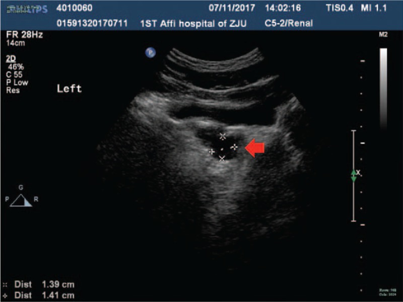
All his general physical examinations were normal. After the physical examination, abdominal ultrasonography was performed. As shown in Fig. 1, a 14 × 14 mm cystic lesion was seen within the prostate and prostatic cyst was diagnosed. Then the patient was given medicine and regular follow-up. Several days later, he recovered without the lower back discomfort and no other symptoms were observed.
Figure 1.

Ultrasonography of the prostate showing a prostatic cyst (red arrow).
3. Discussion
Existing literatures of prostatic cysts are mainly reported by specialists and few, highlighting the infrequent occurrence.[4] According to Mou et al,[5] the etiological factors of prostatic cysts include inflammatory disease, benign prostatic hyperplasia, ejaculatory duct obstruction, atrophy of the prostate gland, and tumor. And the clinical manifestations depend on the size of the cyst. Symptoms varied from asymptomatic to recurrent urinary tract infections, epididymitis, hematuria, pyuria, urinary incontinence, oligospermia, lower abdominal discomfort/heaviness, urinary retention or constipation, bladder outlet obstruction, perineal pain, hematospermia, painful ejaculation,[6] irritative and/or obstructive lower urinary tracts, decreased ejaculate volume, infertility,[7] pelvic pain, or dysuria.[8] In our case, the young man just had lower back discomfort. The manifestation maybe related to the small size of cyst. Gualco et al[9] reported a case of primary clear cell adenocarcinoma of the prostatic utricle, suggesting that prostate cyst adenocarcinoma may be secondary to prostatic cyst. As our patient is unmarried and has no symptom of hematospermia, genital examination, semen analysis, and culture was not conducted.
Apart from the clinical manifestations, imaging technology such as ultrasound, CT, and MRI could assist the diagnoses. It had been reported that MRI could demonstrate the liquid content of prostatic,[10] further MRI scan could show clear image of prostatic cyst and the result was consistent with CT scan.[11] Based on transrectal ultrasound and pathological features, prostatic cysts could be classified into 6 distinct cyst types, including midline cyst, cyst of the erectile dysfunction, cyst of the parenchyma, multiple cysts, complicated cyst, cystic tumor, and cyst secondary to other diseases.[1] A total of 3% to 7.5% of asymptomatic patients have medial cysts. As the biopsy in our case was not performed, the types here could not be clarified.
Although the ultrasound result suggested a benign disease, GPs should clearly know its differential diagnosis. In addition, some large benign prostatic cysts presented with PSA elevation, needing the differential diagnosis.[2,3] The differential diagnoses include Mullerian duct cysts, bladder diverticulum, teratoma, seminal vesicle cyst, epididymal cyst, and Wolffian duct cyst.[12] Once GPs have difficulty for the differentiation for patients with progressive symptoms, timely referral should be performed.
How to manage prostatic cysts by GPs when they encounter the disease again? Based on the reported cases, a prostatic cyst with about 2.5 cm size would make patients feel discomfort and have no abnormal laboratory test results, only regular follow-up is needed. At the present time, therapeutic options consist of transurethral resection, endoscopic or transurethral marsupialization, endoscopic urethrotomy and incision, transrectal ultrasound-guided aspiration with or without sclerotherapy, and open surgery.[8,13] For GPs with special interests with qualification of urology, treatment could be conducted directly. Although most GPs have no qualification of urology, patients with progressive symptoms, large size (2.5 cm or larger), or higher serum PSA should be referred to the specialists.
4. Conclusions
Although prostatic cyst is rare, it is easily found with the help of ultrasound. However, most GPs are unfamiliar with the management. Through the case report and literature review, patients with prostatic cyst of small size and no symptom should have routine follow-up. Although prostatic cyst of progressive symptoms, large size (2.5 cm or larger) or high serum PSA should be timely referred to urological specialists.
Footnotes
Abbreviations: CT = computerized tomography, GP = general practitioner, MRI = magnetic resonance imaging, PSA = prostate-specific antigens.
Consent for publication: The patient gave consent for publication of this case report and images.
Funding/support: The case report was funded by the National Scientific and Technological Major Project of China (2014ZX10004008, 2017ZX10105001).
The authors have no conflicts of interest to disclose.
References
- [1].Galosi AB, Montironi R, Fabiani A, et al. Cystic lesions of the prostate gland: an ultrasound classification with pathological correlation. J Urol 2009;181:647–57. [DOI] [PubMed] [Google Scholar]
- [2].Yu J, Wang X, Luo F, et al. Benign or malignant? Two case reports of gigantic prostatic cyst. Urol Case Rep 2016;8:40e43. [DOI] [PMC free article] [PubMed] [Google Scholar]
- [3].Chen HK, Pemberton R. A large, benign prostatic cyst presented with an extremely high serum prostate-specific antigen level. BMJ Case Rep 2016;pii: bcr2015213381. [DOI] [PMC free article] [PubMed] [Google Scholar]
- [4].Saha B, Sinha RK, Mukherjee S, et al. Midline prostatic cyst in a young man with lower urinary tract symptoms. BMJ Case Rep 2014;pii: bcr2014207816. [DOI] [PMC free article] [PubMed] [Google Scholar]
- [5].Mou P, Zijun W, Mao D, et al. Giant multilocular prostatic cysts treated by laparoscopic prostatectomy: a rare case report in China mainland. Int J Clin Exp Med 2016;9:13227–30. [Google Scholar]
- [6].Gong C, Bianjiang L, Zhen S, et al. A novel surgical management for male infertility secondary to midline prostatic cyst. BMC Urol 2015;15:18. [DOI] [PMC free article] [PubMed] [Google Scholar]
- [7].Issa MM, Kalish J, Petros JA. Clinical features and management of anterior intra urethral prostatic cyst. Urology 1999;54:923. [DOI] [PubMed] [Google Scholar]
- [8].F. Hernández-Galván R, Jaime-Dávila LS, Gómez-Guerra A, et al. Prostatic cyst: an unusual cause of hemospermia. Med Univ 2015;17:162–4. [Google Scholar]
- [9].Gualco G, Ortega V, Ardao G, et al. Clear cell adenocarcinoma of the prostatic utricle in an adolescent. Ann Diagn Pathol 2005;9:153–6. [DOI] [PubMed] [Google Scholar]
- [10].Gevenois PA, Van Sinov ML, Sintzoff SA, et al. Cysts of the prostate and seminal vesicles: MR imageing findings in 11 cases. AJR Am J Roentgenol 1990;155:1021–4. [DOI] [PubMed] [Google Scholar]
- [11].Jingjin Y, Xingkai L, Yong Z. A case report of an uncommon large size of prostatic cyst. Int J Case Rep Images 2015;6:407–10. [Google Scholar]
- [12].Wasim AM, Muhammad SK, Abdus S. Enlarged prostatic utricle cyst with incidental finding of renal agenesis: a case report and literature review. Pak J Radiol 2016;26:345–7. [Google Scholar]
- [13].Jarow JP. Diagnosis and management of ejaculatory duct obstruction. Tech Urol 1996;2:79–85. [PubMed] [Google Scholar]


