Abstract
Congenital cataract is a clinically and genetically heterogeneous disease. The present study was undertaken to find the genetic cause of congenital cataract families. DNA samples of a large consanguineous Pakistani family were genotyped with a high resolution single nucleotide polymorphism Illumina microarray. Homozygosity mapping identified a homozygous region of 4.4 Mb encompassing the gene GJA3. Sanger sequence analysis of the GJA3 gene revealed a novel homozygous variant c.950dup p.(His318ProfsX8) segregating in an autosomal recessive (AR) manner. The previously known mode of inheritance for GJA3 gene mutations in cataract was autosomal dominant (AD) only. The screening of additional probands (n = 41) of cataract families revealed a previously known mutation c.56C>T p.(Thr19Met) in GJA3 gene. In addition, sequencing of the exon-intron boundaries of the GJA8 gene in 41 cataract probands revealed two additional mutations: a novel c.53C>T p.(Ser18Phe) and a known c.175C>G p.(Pro59Ala) mutation, both co-segregating with the disease phenotype in an AD manner. All these mutations are predicted to be pathogenic by in silico analysis and were absent in the control databases. In conclusion, results of the current study enhance our understanding of the genetic basis of cataract, and identified the involvement of the GJA3 in the disease etiology in both AR and AD manners.
Keywords: congenital cataract, homozygosity mapping, sanger sequencing, proband, GJA3 gene, GJA8, mutation, segregation
1. Introduction
Congenital cataract is the leading cause of visual impairment and blindness worldwide during infancy and early childhood. The disease is characterized by opacities of the lens and decreased visual acuity. The prevalence of congenital cataract was estimated to be 1–6 cases per 10,000 live births in developed countries, and 5–15 cases per 10,000 in the underdeveloped countries [1]. Worldwide estimates show that approximately 200,000 children every year are affected by lifelong vision impairment due to cataract [2].
Multiple factors are known to be involved in the pathogenesis of the disease. Among them, genetic factors are the most common, and explain approximately 50% of the cases. The disease segregates in a typical Mendelian manner, i.e., autosomal dominant (AD), autosomal recessive (AR) and X-linked. However, autosomal dominant transmission with high penetrance seems to be the most frequent [3]. In congenital cataract patients, early diagnosis is very important to treat the disease by surgically removing the visually significant cataracts and to achieve good vision. Nowadays, the most frequent surgical method used for the treatment and management of childhood cataracts is micro-incision cataract aspiration together with primary intraocular lens (IOL) implantation [4].
Currently, over 48 genes have been identified underlying congenital cataract. About 50% of the families have pathogenic mutations in crystalline genes; almost 25% have changes in the connexin genes [5]; and the remainder have causative mutations in genes such as aquaporin (MIP) [6], beaded filament structural protein 2 (BFSP2) [7], paired-like homeodomain 3 (PITX3) [8], avian musculoaponeurotic fibrosarcoma (MAF) [9], heat shock transcription factor 4 gene (HSF4) [10], lens intrinsic membrane protein (LIM2) [11], glucosaminyl (N-acetyl) transferase 2 (GCNT2) [12] as well as many others, as delineated in the Cat-Map database [13].
In this study, we report for the first time a homozygous novel mutation c.950dup p.(His318ProfsX8) in the homozygous region encompassing gap junction alpha 3 (GJA3) gene in a Pakistani family with a recessively inherited congenital cataract. In addition, sequencing of GJA3 and GJA8 genes in 41 congenital cataract probands revealed one known mutation in GJA3 gene inherited dominantly and two mutations in GJA8 gene (one novel and one known) segregating in an autosomal dominant fashion in cataract families.
2. Materials and Methods
The participants were recruited at the pediatric ophthalmology department of Al-Shifa Eye Trust Hospital, Rawalpindi, Pakistan. The study was approved by the Institutional Review Board of the Al-Shifa Eye Trust Hospital (Rawalpindi, Pakistan), and adhered to the tenets of the Declaration of Helsinki, with the approval code PK2014:102. Written informed consent was obtained for study participation from the participants and/or their parents, as appropriate. Comprehensive, ocular, medical, and family histories were obtained from the parents/available family member. Detailed ophthalmic examination was performed for both affected and unaffected individuals of families. Blood samples were collected from affected and unaffected siblings, and from the parents. Genomic DNA was extracted using QIAGEN DNA Blood Midi Kit (QIAGEN, Germantown, Maryland, USA).
Genomic DNA samples of two affected and three unaffected family members of a large consanguineous cataract family were genotyped with Affymetrix 250K single nucleotide polymorphism (SNP) microarray (Affymetrix: Santa Clara, CA, USA), and genotype data were analyzed for homozygous regions using the online mapping tool HomozygosityMapper (http://www.homozygositymapper.org/). Haplotypes of the affected and unaffected members of the family were compared to find the identical homozygous regions among the affected individuals of the family.
The coding exons and intronic boundaries of GJA3 (NM_021954) gene were amplified in a family with the homozygous region encompassing the GJA3 gene. Both GJA3 and GJA8 (NM_005267) genes were sequenced in probands of 41 additional cataract families. Primers were designed using Primer 3 (http://bioinfo.ut.ee/primer3-0.4.0/) (primer sequences and polymerase chain reaction (PCR) conditions are available on request). PCR products were analyzed on 2% agarose gels followed by Sanger sequencing using ABI BigDye chemistry (Applied Biosystems Inc., Foster City, CA, USA), and were processed through an automated ABI 3730 Sequencer (Applied Biosystems, Inc.). Sequences obtained were aligned with the reference sequence using CodonCode Aligner (version 6.1) (CodonCode Co., Centerville, MA, USA). Intra-familial segregation analysis was performed upon the identification of variants among the probands in the GAJ3 and GJA8 genes.
Pathogenicity of missense variants was evaluated by publically available tools including PhyloP, Grantham, polymorphism phenotyping v-2 (PolyPhen-2) (version 2.1.0 r367) (http://genetics.bwh.harvard.edu/pph2/), MutationTaster (http://www.mutationtaster.org/), and sorting intolerant from tolerant (SIFT, http://sift.bii.a-star.edu.sg/) to predict the functional impact of the sequence variants on the encoded protein. The amino acid sequences were obtained from protein sequence database UniProt (http://www.rcsb.org/pdb/protein/Q6P2Q9) from different species to check the amino acid conservation. Kalign (2.0) was used for the multiple nucleotide sequence alignment.
3. Results
3.1. Clinical Findings
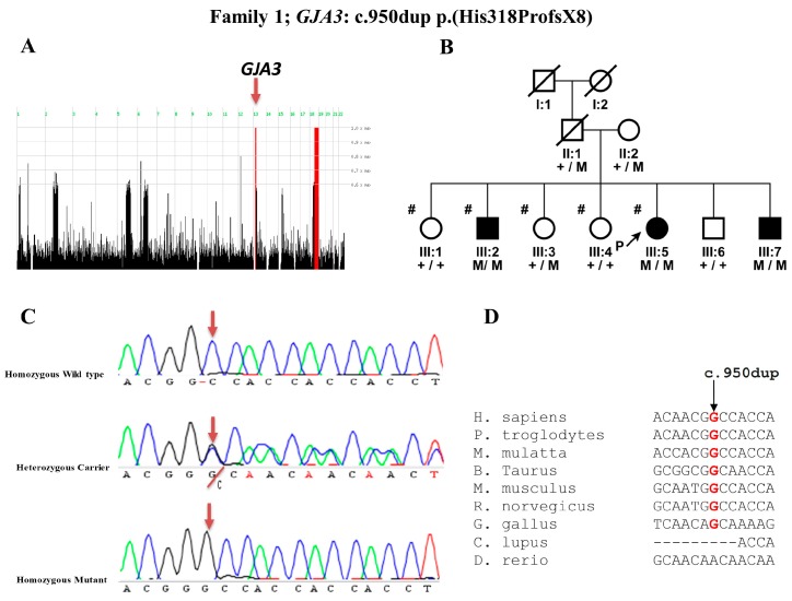
In Family 1, Proband (III:5) was a two-year-old boy presented with squinting eyes (Figure 1B). His corneal diameter was 11 mm horizontally and 10.5 mm vertically in both eyes measured with caliper under general anesthesia before surgery. Nuclear cataract was noted in both the eyes. All the affected individuals of the family after cataract surgery developed secondary glaucoma phenotype with intraocular pressure (IOP) > 22 mmHg and cup-to-disc ratio (CDR) > 0.7 which resulted in severe visual impairment. The heterozygous carrier individuals of the family which includes parents (II:1 and II:2) and unaffected siblings (III:1, III:3, III:4 and III:6) had normal crystalline lenses on clinical examination.
Figure 1.
Homozygosity mapping and mutation analysis of Family 1: (A) homozygosity mapping results indicating homozygous region (red line) on chromosome 13 encompassing a Gap junction alpha 3 (GJA3) gene; (B) pedigree of a family with congenital cataract, and segregation of a novel mutation c.950dup p.(His318ProfsX8) in the GJA3 gene; (C) DNA sequence chromatogram of GJA3 for the normal (+/+), heterozygous carrier (+/M), and affected individuals (M/M); and (D) nucleotide conservation among the orthologues.
In Family 2, proband (II:1) was a three-year-old boy with bilateral congenital nuclear cataracts with the central, dense nuclear lens opacity. The proband’s mother (I:2) also suffered from bilateral lens opacities.
The pedigree of Family 3 comprised three generations with four affected and three unaffected individuals. The proband (III:4) was a three-month-old boy presented with leucocoria. He had bilateral central cataract, nystagmus and amblyopia. Intraocular pressure was 8 mm Hg in both eyes. Lensectomy was performed in both eyes and visual rehabilitation was done using aphakic spectacle correction of +20 diopter sphere (DS) in the right and +21DS in the left eye. His corrected vision on last follow-up was 6/30 in the right and 6/19 in the left eye. His mother, aunt and grandmother had a history of bilateral cataract. His elder sister also had congenital cataract shortly after birth, and had undergone cataract extraction at around three years of age.
The large Family 4 also consisted of three generations with four affected and one unaffected individual. The proband (III:1), his mother, aunt, and grandfather all presented with bilateral nuclear cataract. They all had bilateral surgery for cataract extraction.
3.2. Mutation Detection in GJA3 Gene
In Family 1, we found two homozygous chromosomal regions among the two affected individuals of the family. One 4.4 Mb region was located on chromosome 13 containing the previously identified gene (GJA3) implicated in cataract (Figure 1A). Sanger sequencing of the coding exons of GJA3 gene revealed a novel homozygous variant c.950dup p.(His318ProfsX8) co-segregating with the disease phenotype within the family in AR manner (Figure 1B, C). This duplication shifts the conceptual reading frame of the protein leading to a premature chain termination. Nucleotide sequence alignment shows that there was no gap between the nucleotides among different orthologous species (Figure 1D). This indicates that the duplication of the “G” nucleotide at the c.950dup position is not tolerated.
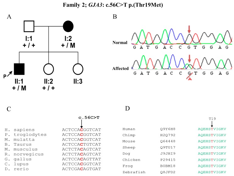
In Family 2, Sanger sequencing of the proband revealed a previously described mutation in the GJA3 gene c.56C>T p.(Thr19Met), segregating in our family with the disease phenotype in an AD manner (Figure 2A, B). This particular variant was located in the connexin domain of the protein and was predicted to be disease causing, deleterious, damaging by MutationTaster, SIFT, and Polyphen-2, respectively, with a PhyloP score of 6.18 (indicates nucleotide conservation) (Figure 2C) and Grantham score of 81, as threonine is a highly conserved amino acid among different species (Figure 2D).
Figure 2.
Pedigree of a Family 2 with dominant GJA3 mutation: (A) Family with congenital cataract, showing segregation of a mutation c.56C>T p.(Thr19Met) in the GJA3 gene. Normal individuals are represented with +/+ and affected individuals with the +/M symbol for the mutation. (B) Sanger sequencing DNA chromatogram of GJA3 for the normal and affected individuals. (C, D) Nucleotide and amino acid conservation in orthologous species for the c.56C>T p.(Thr19Met). The wild type nucleotide (C) and amino acid (T) are represented with arrow and in red color.
3.3. Mutation Detection in GJA8 Gene
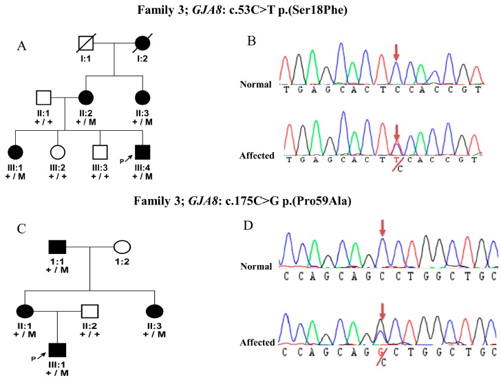
In Family 3, direct sequencing of the probands of cataract families identified a novel missense variant c.53C>T p.(Ser18Phe) in the GJA8 gene. This variant segregate with the disease phenotype in the family in AD manner (Figure 3A, B). The Ser18Phe variant is located in the connexin domain of the protein. The amino acid residue Ser18 is highly conserved among different species. It is predicted to be disease causing, deleterious, and damaging by MutationTaster, SIFT, and Polyphen-2, respectively, with a Grantham score of 155 and a PhyloP score of 5.86, which indicates both amino acid and nucleotide conservation among different species.
Figure 3.
Pedigrees of two families with GJA8 mutations: (A) pedigree of Family 3 with congenital cataract, and segregation of a novel mutation c.53C>T p.(Ser18Phe) in the GJA8 gene; (B) DNA sequence chromatogram of c.53C>T p.(Ser18Phe) mutation; (C) Family 4 segregation of a mutation c.175C>G p.(Pro59Ala) in the GJA8 gene; and (D) DNA sequence chromatogram of c.175C>G p.(Pro59Ala) mutation in GJA8.
In Family 4, a missense variant c.175C>G p.(Pro59Ala) was identified in GJA8, and was found to be segregating with the disease phenotype dominantly (Figure 3C, D). This variant was previously known for its association with cataract and is localized in the connexin domains. This change in amino acid was also predicted to be disease causing by in-silico predictions with a Grantham score of 27 and a PhyloP score of 5.94.
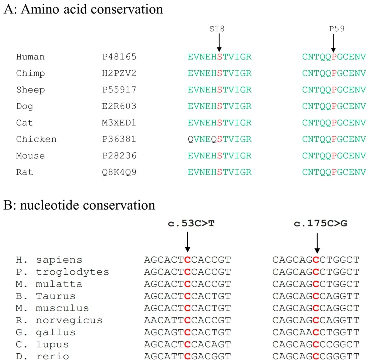
Both the wild type nucleotides at positions c.53C, and c.175C as well as amino acids at positions Ser18 and Pro59 are highly conserved among the orthologous species (Figure 4).
Figure 4.
Multiple sequence alignment of GJA8 orthologues: (A) mutated amino acids p.(Ser18Phe) (S), and p.(Pro59Ala) (P) are indicated with an arrow and in red color; and (B) nucleotide conservation among the orthologous species is indicated with arrow and the wild type nucleotide is represented in red color.
All variations identified in the GJA3 and GJA8 genes have been excluded from the population matched 100 controls by performing Sanger Sequencing of the particular exons. All these variants are absent in the publically available Gnomad database for GJA3 (http://gnomad.broadinstitute.org/gene/ENSG00000121743) and GJA8 (http://gnomad.broadinstitute.org/gene/ENSG00000121634) genes.
4. Discussion
Previously, multiple genes have been implicated in cataract [13]. In this study, we have focused on GJA3 and GJA8 underlying human cataract frequent mutations in these genes explaining about 25% of the patients [5]. We sequenced both genes in our current panel of probands of Pakistani families. In this study, we report a novel homozygous mutation c.950dup p.(His318ProfsX8) in the GJA3 gene causing autosomal recessive cataract in a large Pakistani family. Previously, mutations in GJA3 in animal models were associated with recessively inherited form of cataract [14], but, in humans, only autosomal dominant GJA3 mutations have, so far, been implicated in the disease [15,16].
In case of our Family 1, the proband was initially recruited with a (secondary) glaucoma phenotype. Taking into account the recessive inheritance pattern of family, homozygosity analysis was performed. However, later, clinical re-examination of all the affected members of the family revealed autosomal recessive cataract as a primary phenotype. Since the homozygous region identified also encompassed the GJA3 gene, we subsequently focused on Sanger sequencing of this cataract candidate gene to discover the novel homozygous mutations, segregating in the family with the disease. Previously, pathogenic mutations have been found all over the GJA3 protein [17]. Notably, our p.(His318ProfsX8) homozygous variant is present in the middle of the carboxy-terminal domain (CT) of the GJA3 where previously no other dominant change has been identified. This CT domain is specific for the connexin isotypes and is important for the post-translational modifications and for interactions with other protein partners [18]. This duplication creates a frame shift starting at codon His318. The new reading frame ends in a stop codon 7 positions downstream. Thus, the protein will be smaller in length with mistranslation of few amino acids. Due to the presence of early stop, the abnormal mRNA might be subjected to nonsense-mediated decay. The recessive mutations are mostly responsible for causing severe disease phenotypes. In the current study, individual III:2 of Family 1, with the recessive mutation in the GJA3 gene indeed suffered from severe secondary glaucoma after cataract surgery with high cup-to-disc ratio of 0.8 in both eyes, and intraocular pressure was 28 mmHg and 34 mmHg for the right and left eye respectively. Upon the follow-up of all the affected individuals of the family, secondary glaucoma was observed with IOP > 22 mmHg and CDR > 0.7. The severe secondary glaucoma phenotype in all the affected individuals could be one of the outcomes of the recessive mutation in GJA3 in this particular family. Similarly, in the Indian family, a recessive mutation c.670insA p.(Thr203AsnfsX47) was reported in the GJA8 gene with an additional secondary phenotype of nystagmus and amblyopia due to severe visual impairment [19]. However, the majority of mutations reported in the GJA8 were inherited in an autosomal dominant manner. In the current family with GJA3 mutation and previous families with GJA8 mutations recessive inheritance was associated with the severe phenotype together with the additional secondary phenotypes.
Approximately 38–40 GJA3 mutations were previously implicated in dominantly inherited cataract. In one of our families, a previously known mutation c.56C>T p.(Thr19Met) was identified, which is also inherited in an autosomal dominant manner. In the current study, the proband (II:1) having this mutation (Family 2) was presented with nuclear cataract at the age of three years. Interestingly, this particular mutation has also been reported in an Indian family with posterior polar cataract. The proband of the Indian family was presented also at the age of three years [20]. In addition, in a rat model suffering from autosomal recessive congenital nuclear cataract, a missense mutation in gja3, p.Glu42Lys, has been reported [14]. A homozygous deletion of the gja3 in a knockout mouse resulted in congenital cataract [21]. These findings suggest that at least one cataract mutation may result in a pleiotropic phenotype, which contributes to the understanding of the complexity of cataract genetics.
In addition to GJA3 variants, we also identified pathogenic changes in GJA8 gene which include a previously known c.175C>G p.(Pro59Ala) and a novel c.53C>T p.(Ser18Phe) mutation. The p.(pro59Ala) mutation was previously reported in a Chinese family presented with the congenital nuclear cataract [22]. Interestingly, the phenotype and age of onset of the cataract in the Pakistani family was similar to a Chinese family. In a previous study, the functional effect of the p.(Pro59Ala) change was characterized by stably transfecting recombinant DNA constructs into Hek293 cells. In contrast to the wild type protein, the GJA8 protein with the pathogenic variant failed to form gap junction plaques at appositional membrane. The mutation had also an effect on the cell viability, growth and proliferation [22].
The novel p.(Ser18Phe) mutation is also predicted to be a pathogenic change and is located close to previously known mutations p.(Leu7Pro) and p.(Arg23Thr) at the N-terminal domain of GJA8 [23,24]. In addition, the p.(Arg23Thr) change was previously functionally characterized in vitro. HeLa cells were stably transfected with the mutant and wild types. Decreased intercellular communication was reported in the presence of mutated amino acid [24]. In p.(Arg23Thr) a basic arginine has been replaced with the polar uncharged threonine amino acid. Since the p.(Ser18Phe) novel mutation identified in a Pakistani family is located in proximity to the previous mutation and polar uncharged serine has been replaced with the hydrophobic phenylalanine amino acid, it could be that, due to the p.(Ser18Phe) mutation, intercellular communication is affected. This mutation may affect the secondary/tertiary structure of the protein or the interaction of the GJA8 with the other proteins.
In previous studies in mice, it has been observed that the complete knockout of either of the two connexins; GJA3 or GJA8, resulted in cataract formation and decreased lens growth. These mutated connexins affect the formation of primary and secondary lens fiber cells which resulted in the cataract phenotype [25]. The lens is an avascular organ and due to the lack of vasculature it is dependent on the formation of a network of gap junctions. Lens tissues express three types of connexins: CX43 (GJA1), CX46 (GJA3) and CX50 (GJA8). GJA3 (CX46) and GJA8 (CX50) are connexins involved in the formation of these networks. Gap junctions are important in the intracellular communication essential for the cell survival, function and maintenance of the lens homoeostasis and transparency [25]. Both connexins are part of a large multigene family comprising of 21 members. All connexins, consists of four transmembrane domains (M1–M4), two extracellular loops, a cytoplasmic loop, NH2-terminal, and COOH-terminal domains. They all form connexin hemi-channels important for the permeation of ions, as well as small metabolites such as adenosine triphosphate (ATP), cyclic adenosine monophosphate (cAMP), inositol triphosphate (IP3) and glutamate.
The expression pattern of CX43, CX46 and CX50 connexins in the lens is highly dynamic. GJA3 (CX46) is significantly upregulated during the differentiation of the lens epithelial-to-fiber cell. GJA3 plays a vital role in the regulation of several cellular processes such as cell growth, proliferation, differentiation, and apoptosis [26,27]. GJA8 (CX50) is also highly expressed in lens fiber cells and epithelial cells. It is involved in the growth and differentiation of the lens tissues. The higher expression was observed during the lens epithelial fiber cell differentiation [28]. In addition to gap junction formation, these connexins are also involved in the regulation of the cell cycle, especially GJA8 (CX50) which is involved in the cell cycle arrest [29].
Apparently, the lens has a controlled mechanism for the opening and closing of the connexin hemi-channels, but, in the case of mutated connexins, it may be they are inefficient to prevent the openings of the hemi-channels and to decrease the intake by them. Thus, the presence of c.56C>T p.(Thr19Met), c.427G>A p.(Gly143Arg) in GJA3 [25,26] and c.137G>T p.(Gly46Val) in GJA8 [30] and the novel mutations identified in current study may result in reduced gap junction channel formation and aberrant/increased hemi-channel activity and, eventually, cell death [31,32]. The in-silico analysis of rare variants identified in the current study (GJA3 and GJA8 genes) and reported in the discussion section predicted all these variants as deleterious and disease causing (Table 1). GJA3 (CX46), GJA8 (CX50) and CX43 (GJA1) have been reported vital for interconnecting the lens fiber and epithelial cells in humans [33].
Table 1.
In-silico analysis of rare variants in GJA3 and GJA8 genes.
| Gene ID | cDNA Position | Amino Acid Position | Study | phyloP | Grantham Score | SIFT | Mutation Taster | Poly Phen-2 |
|---|---|---|---|---|---|---|---|---|
| GJA3 | c.950dup | p.(His318ProfsX8) | Current | N/A | N/A | D | Disease causing | Damaging |
| c.56C>T | p.(Thr19Met) | Current | 6.18 | 81 | D | Disease causing | damaging | |
| c.427G>A | p.(Gly143Arg) | Previous | 5.86 | 125 | D | Disease causing | damaging | |
| c.137G>T | p.(Gly46Val) | Previous | 5.94 | 109 | D | Disease causing | damaging | |
| GJA8 | c.53C>T | p.(Ser18Phe) | Current | 5.86 | 155 | T | Disease causing | damaging |
| c.175C>G | p.(Pro59Ala) | Current | 5.96 | 27 | D | Disease causing | damaging | |
| c.20T>C | p.(Leu7Pro) | Previous | 3.35 | 98 | T | Disease causing | damaging | |
| c.68G>C | p.(Arg23Thr) | Previous | 4.16 | 71 | T | Disease causing | damaging |
Foot Note: D; deleterious, T; Tolerated.
Interestingly, mutations in yet another connexin member, GJA1, are associated both dominantly and recessively with oculodentodigital dysplasia (ODDD) including the ocular phenotypes of microphtalmia, microcornea, cataract and glaucoma [34]. In few patients with ODDD, iris anomalies and secondary glaucoma have also been reported [34,35]. Previous studies have reported both dominant and recessive modes of inheritance of cataract associated with both the GJA1 in ODDD and GJA8 in congenital cataract. This study is the first one reporting the recessive mode of inheritance for the GJA3 gene mutations in cataract.
In summary, the results of the current study demonstrate a new inheritance pattern of GJA3 gene mutations with cataract. We report a novel homozygous duplication p.(His318ProfsX8) in GJA3 associated with nuclear cataract in a Pakistani family. In addition, we identified one novel mutation p.(Ser18Phe) in the GJA8 gene inherited dominantly in a Pakistani family. The results of the current study help broaden the spectrum of mutations identified in GJA3 and GJA8 genes, and are of utmost importance for genetic counselling and family planning of parents at risk. Additional cataract families with the recessive inheritance should be screened for the mutations in GJA3 gene.
Author Contributions
S.M. and A.A.B.B. conceived and designed the experiments; S.M. and I.T.G.N. performed the experiments; S.N.S., S.N.Z., M.I.K. recruited patients and collected samples; S.N.S., S.N.Z., M.I.K. and A.A.B.B. contributed reagents/materials/analysis tools; and S.M. wrote the manuscript. All authors have read and approved the final manuscript.
Conflicts of interest
The authors declare no conflict of interest.
References
- 1.Apple D.J., Ram J., Foster A., Peng Q. Elimination of cataract blindness: A global perspective entering the new millenium. Surv. Ophthalmol. 2000;45(Suppl. 1):S1–S196. [PubMed] [Google Scholar]
- 2.Foster A., Gilbert C., Rahi J. Epidemiology of cataract in childhood: A global perspective. J. Cataract Refract. Surg. 1997;23(Suppl. 1):601–604. doi: 10.1016/S0886-3350(97)80040-5. [DOI] [PubMed] [Google Scholar]
- 3.Pichi F., Lembo A., Serafino M., Nucci P. Genetics of congenital cataract. Dev. Ophthalmol. 2016;57:1–14. doi: 10.1159/000442495. [DOI] [PubMed] [Google Scholar]
- 4.Wang M., Xiao W. Congenital cataract: Progress in surgical treatment and postoperative recovery of visual function. Eye Sci. 2015;30:38–47. [PubMed] [Google Scholar]
- 5.Hejtmancik J.F. Congenital cataracts and their molecular genetics. Semin. Cell Dev. Biol. 2008;19:134–149. doi: 10.1016/j.semcdb.2007.10.003. [DOI] [PMC free article] [PubMed] [Google Scholar]
- 6.Berry V., Francis P., Kaushal S., Moore A., Bhattacharya S. Missense mutations in MIP underlie autosomal dominant ‘polymorphic’ and lamellar cataracts linked to 12q. Nat. Genet. 2000;25:15–17. doi: 10.1038/75538. [DOI] [PubMed] [Google Scholar]
- 7.Jakobs P.M., Hess J.F., FitzGerald P.G., Kramer P., Weleber R.G., Litt M. Autosomal-dominant congenital cataract associated with a deletion mutation in the human beaded filament protein gene BFSP2. Am. J. Hum. Genet. 2000;66:1432–1436. doi: 10.1086/302872. [DOI] [PMC free article] [PubMed] [Google Scholar]
- 8.Semina E.V., Ferrell R.E., Mintz-Hittner H.A., Bitoun P., Alward W.L., Reiter R.S., Funkhauser C., Daack-Hirsch S., Murray J.C. A novel homeobox gene PITX3 is mutated in families with autosomal-dominant cataracts and ASMD. Nat. Genet. 1998;19:167–170. doi: 10.1038/527. [DOI] [PubMed] [Google Scholar]
- 9.Vanita V., Singh D., Robinson P.N., Sperling K., Singh J.R. A novel mutation in the DNA-binding domain of MAF at 16q23.1 associated with autosomal dominant “cerulean cataract” in an Indian family. Am. J. Med. Genet. A. 2006;140:558–566. doi: 10.1002/ajmg.a.31126. [DOI] [PubMed] [Google Scholar]
- 10.Behnam M., Imagawa E., Chaleshtori A.R., Ronasian F., Salehi M., Miyake N., Matsumoto N. A novel homozygous mutation in HSF4 causing autosomal recessive congenital cataract. J. Hum. Genet. 2016;61:177–179. doi: 10.1038/jhg.2015.127. [DOI] [PubMed] [Google Scholar]
- 11.Irum B., Khan S.Y., Ali M., Kaul H., Kabir F., Rauf B., Fatima F., Nadeem R. Mutation in LIM2 is responsible for autosomal recessive congenital cataracts. PLoS ONE. 2016;11:e0162620. doi: 10.1371/journal.pone.0162620. [DOI] [PMC free article] [PubMed] [Google Scholar]
- 12.Pras E., Raz J., Yahalom V., Frydman M., Garzozi H.J., Pras E., Hejtmancik J.F. A nonsense mutation in the glucosaminyl (N-acetyl) transferase 2 gene (GCNT2): Association with autosomal recessive congenital cataracts. Investig. Ophthalmol. Vis. Sci. 2004;45:1940–1945. doi: 10.1167/iovs.03-1117. [DOI] [PubMed] [Google Scholar]
- 13.Shiels A., Bennett T.M., Hejtmancik J.F. Cat-Map: Putting cataract on the map. Mol. Vis. 2010;16:2007–2015. [PMC free article] [PubMed] [Google Scholar]
- 14.Yoshida M., Harada Y., Kaidzu S., Ohira A., Masuda J., Nabika T. New genetic model rat for congenital cataracts due to a connexin 46 (GJA3) mutation. Pathol. Int. 2005;55:732–737. doi: 10.1111/j.1440-1827.2005.01896.x. [DOI] [PubMed] [Google Scholar]
- 15.Zhou D., Ji H., Wei Z., Guo L., Li Y., Wang T., Zhu Y., Dong X., Wang Y., Xing Q., et al. A novel insertional mutation in the connexin 46 (gap junction alpha 3) gene associated with autosomal dominant congenital cataract in a Chinese family. Mol. Vis. 2013;19:789–795. [PMC free article] [PubMed] [Google Scholar]
- 16.Guleria K., Sperling K., Singh D., Varon R., Singh J.R., Vanita V. A novel mutation in the connexin 46 (GJA3) gene associated with autosomal dominant congenital cataract in an Indian family. Mol. Vis. 2007;13:1657–1665. [PubMed] [Google Scholar]
- 17.Pfenniger A., Wohlwend A., Kwak B.R. Mutations in connexin genes and disease. Eur. J. Clin. Investig. 2011;41:103–116. doi: 10.1111/j.1365-2362.2010.02378.x. [DOI] [PubMed] [Google Scholar]
- 18.Lampe P.D., Lau A.F. The effects of connexin phosphorylation on gap junctional communication. Int. J. Biochem. Cell Biol. 2004;36:1171–1186. doi: 10.1016/S1357-2725(03)00264-4. [DOI] [PMC free article] [PubMed] [Google Scholar]
- 19.Ponnam S.P., Ramesha K., Tejwani S., Ramamurthy B., Kannabiran C. Mutation of the gap junction protein alpha 8 (GJA8) gene causes autosomal recessive cataract. J. Med. Genet. 2007;44:e85. doi: 10.1136/jmg.2007.050138. [DOI] [PMC free article] [PubMed] [Google Scholar]
- 20.Santhiya S.T., Kumar G.S., Sudhakar P., Gupta N., Klopp N., Illig T., Söker T., Groth M., Platzer M., Gopinath P.M., et al. Molecular analysis of cataract families in India: New mutations in the CRYBB2 and GJA3 genes and rare polymorphisms. Mol. Vis. 2010;16:1837–1847. [PMC free article] [PubMed] [Google Scholar]
- 21.Gong X., Li E., Klier G., Huang Q., Wu Y., Lei H., Kumar N.M., Horwitz J., Gilula N.B. Disruption of alpha3 connexin gene leads to proteolysis and cataractogenesis in mice. Cell. 1997;91:833–843. doi: 10.1016/S0092-8674(00)80471-7. [DOI] [PubMed] [Google Scholar]
- 22.Yu Y., Wu M., Chen X., Zhu Y., Gong X., Yao K. Identification and functional analysis of two novel connexin 50 mutations associated with autosome dominant congenital cataracts. Sci. Rep. 2016;6:26551. doi: 10.1038/srep26551. [DOI] [PMC free article] [PubMed] [Google Scholar]
- 23.Mackay D.S., Bennett T.M., Culican S.M., Shiels A. Exome sequencing identifies novel and recurrent mutations in GJA8 and CRYGD associated with inherited cataract. Hum. Genomics. 2014;8:19. doi: 10.1186/s40246-014-0019-6. [DOI] [PMC free article] [PubMed] [Google Scholar]
- 24.Thomas B.C., Minogue P.J., Valiunas V., Kanaporis G., Brink P.R., Berthoud V.M., Beyer E.C. Cataracts are caused by alterations of a critical N-terminal positive charge in connexin50. Investig. Ophthalmol. Vis. Sci. 2008;49:2549–2556. doi: 10.1167/iovs.07-1658. [DOI] [PMC free article] [PubMed] [Google Scholar]
- 25.White T.W. Unique and redundant connexin contributions to lens development. Science. 2002;295:319–320. doi: 10.1126/science.1067582. [DOI] [PubMed] [Google Scholar]
- 26.Banerjee D., Das S., Molina S.A., Madgwick D., Katz M.R., Jena S., Bossmann L.K., Pal D., Takemoto D.J. Investigation of the reciprocal relationship between the expression of two gap junction connexin proteins, connexin46 and connexin43. J. Biol. Chem. 2011;286:24519–24533. doi: 10.1074/jbc.M110.217208. [DOI] [PMC free article] [PubMed] [Google Scholar]
- 27.Oyamada M., Oyamada Y., Takamatsu T. Regulation of connexin expression. Biochim. Biophys. Acta. 2005;1719:6–23. doi: 10.1016/j.bbamem.2005.11.002. [DOI] [PubMed] [Google Scholar]
- 28.Gu S., Yu X.S., Yin X., Jiang J.X. Stimulation of lens cell differentiation by gap junction protein connexin 45.6. Investig. Ophthalmol. Vis. Sci. 2003;44:2103–2111. doi: 10.1167/iovs.02-1045. [DOI] [PubMed] [Google Scholar]
- 29.Shi Q., Jiang J.X. Connexin arrests the cell cycle through cytosolic retention of an E3 ligase. Mol. Cell. Oncol. 2016;3:e1132119. doi: 10.1080/23723556.2015.1132119. [DOI] [PMC free article] [PubMed] [Google Scholar]
- 30.Minogue P.J., Tong J.J., Arora A., Russell-Eggitt I., Hunt D.M., Moore A.T., Ebihara L., Beyer E.C., Berthoud V.M. A mutant connexin50 with enhanced hemichannel function leads to cell death. Investig. Ophthalmol. Vis. Sci. 2009;50:5837–5845. doi: 10.1167/iovs.09-3759. [DOI] [PMC free article] [PubMed] [Google Scholar]
- 31.Tong J.J., Minogue P.J., Kobeszko M., Beyer E.C., Berthoud V.M., Ebihara L. The connexin46 mutant, Cx46T19M, causes loss of gap junction function and alters hemi-channel gating. J. Membr. Biol. 2015;248:145–155. doi: 10.1007/s00232-014-9752-y. [DOI] [PMC free article] [PubMed] [Google Scholar]
- 32.Ren Q., Riquelme M.A., Xu J., Yan X., Nicholson B.J., Gu S., Jiang J. Cataract-causing mutation of human connexin 46 impairs gap junction, but increases hemichannel function and cell death. PLoS ONE. 2013;8:e74732. doi: 10.1371/journal.pone.0074732. [DOI] [PMC free article] [PubMed] [Google Scholar]
- 33.Beyer E.C., Berthoud V.M. Connexin hemichannels in the lens. Front. Physiol. 2014;5:20. doi: 10.3389/fphys.2014.00020. [DOI] [PMC free article] [PubMed] [Google Scholar]
- 34.Paznekas W.A., Boyadjiev S.A., Shapiro R.E., Daniels O., Wollnik B., Keegan C.E., Innis J.W., Dinulos M.B., Christian C., Hannibal M.C., et al. Connexin 43 (GJA1) mutations cause the pleiotropic phenotype of oculodentodigital dysplasia. Am. J. Hum. Genet. 2003;72:408–418. doi: 10.1086/346090. [DOI] [PMC free article] [PubMed] [Google Scholar]
- 35.Frasson M., Calixto N., Cronemberger S., de Aguiar R.A., Leao L.L., de Aguiar M.J. Oculodentodigital dysplasia: Study of ophthalmological and clinical manifestations in three boys with probably autosomal recessive inheritance. Ophthalmic Genet. 2004;25:227–236. doi: 10.1080/13816810490513424. [DOI] [PubMed] [Google Scholar]


