Abstract
To evaluate the antigenic relationship between bat mumps virus (BMV) and the JL5 vaccine strain of mumps virus (MuVJL5), we rescued a chimeric virus bearing the F and HN glycoproteins of BMV in the background of a recombinant JL5 genome (rMuVJL5). Cross-reactivity and cross-neutralization between this chimeric recombinant MuV bearing the F and HN glycoproteins of BMV (rMuVJL5-F/HNBMV) virus and rMuVJL5 were demonstrated using hyperimmune mouse serum samples and a curated panel of human serum. All mouse and human serum samples that were able to neutralize rMuVJL5 infection had cross-neutralizing activity against rMuVJL5-F/HNBMV. Our data suggest that persons who have neutralizing antibodies against MuV might be protected from infection by BMV.
Keywords: neutralizing antibodies, mumps virus, viral envelope proteins, zoonoses, cross-reactive antibodies
Mumps virus (MuV) is a contagious virus of the Rubulavirus genus that typically causes painful swelling of the parotid and other salivary glands [1]. Live attenuated mumps vaccines were introduced in the 1960s and 1970s, with vaccine effectiveness ranging from 91% to 94.6% after 2 doses [2]. Consequently, the incidence of mumps morbidity has been greatly reduced in countries where high vaccine coverage has been achieved [3]. Recently however, the epidemiology of mumps has changed, and mumps is reemerging in the developed world, possibly as a result of waning immunity and/or a lack of broadly protective immunity.
MuV was thought to be an exclusive human pathogen with no animal reservoir until recently, when the complete genomic sequence of a conspecific mumpslike virus was obtained from an African bat (bat mumps virus [BMV]) [4]. It is not currently known whether BMV is capable of infecting humans, but the discovery of a possible animal reservoir for MuV raises concerns that elimination of circulating virus and subsequent cessation of vaccination might leave humans susceptible to disease from such a reservoir-borne virus. The genetic proximity of these 2 viruses suggests that antibodies against MuV might provide protection from zoonotic spillover events; thus, it is critical for risk assessment to evaluate the BMV-neutralizing activity of antibodies from MuV-seropositive individuals.
Evidence of functional and antigenic relatedness between BMV and MuV has been demonstrated by syncytia formation resulting from heterotypic expression of the fusion (F) and attachment (HN) envelope glycoproteins in tissue culture and the presence of MuV-reactive antibodies in serum from African bats [4, 5]. In this study, we used hyperimmune mouse serum samples and a panel of human serum samples to characterize the cross-reactive and cross-neutralizing activity of antibodies against the F and HN glycoproteins of MuV and BMV.
METHODS
Cloning and Rescue of Recombinant Viruses
Each of the chimeric viruses and the recombinant parental virus (rMuV) were rescued in 1 well of a 6-well plate of BSRT7 cells by cotransfection of 5 µg of the antigenomic plasmid, 2 µg of a plasmid encoding a codon-optimized T7 polymerase, and 0.3, 0.1, and 0.2 µg of T7-driven support plasmids encoding the N, P, and L proteins, respectively. For efficient rescue, we used the maximal T7 promoter to drive our antigenomic transcript and a hammerhead ribozyme sequence in the transcribed 5′-end of antigenome [6] (Supplementary Figure 1). Transfection was performed using Lipofectamine LTX reagent (Invitrogen) according to the manufacturer's instructions. At 7 days after transfection, supernatants were collected from the rescue wells and amplified by 2 sequential passages on DF-1 cells (American Type Culture Collection CRL 12203), and clarified supernatants were stored at −80°C.
Generation of Mouse Hyperimmune Serum
Purified stocks of rMuV and recombinant MuV bearing the F and HN glycoproteins of BMV (rMuVJL5-F/HNBMV) were inactivated by treatment with 0.03% formalin at 4°C for 48 hours and delivered intramuscularly into the hind limb of 6–8 week-old female BALB/c mice, adjuvanted with polyinosinic-polycytidylic acid (5 µg per injection; Invivogen). Blood samples were collected at 3 weeks after vaccination, and serum samples were prepared by centrifugation at 12 000 rpm for 5 minutes.
Quantification of Seroreactivity by Flow Cytometry
Vero cells were infected with rMuV or rMuVJL5-F/HNBMV at a multiplicity of infection of 0.75 and incubated at 37°C for 24 hours. Infected cells were incubated in phosphate-buffered saline (PBS) with 10 mmol/L ethylenediaminetetraacetic acid at 4°C for 20 minutes before being scraped and collected. Cells were pelleted by centrifugation at 1250 rpm for 5 minutes, washed with PBS, and resuspended in PBS plus 2% fetal bovine serum (FBS). Cells were resuspended in hyperimmune mouse serum diluted 1:200 in PBS plus 2% FBS and incubated at 4°C for 1 hour, washed twice with PBS plus 2% FBS and incubated with a fluorescent secondary antibody (goat anti-mouse AlexaFluor647; 1:1000 dilution) at 4°C for 1 hour. Cells were washed twice with PBS plus 2% FBS and resuspended in PBS plus 2% paraformaldehyde, and fluorescence intensity was measured by flow cytometry.
Study Subjects and Serum Samples
Serum samples were purchased from Innovative Research as deidentified research reagents. Serum was collected between August and November 2014 in Michigan, from donors aged 18–64 years. The serum was heat inactivated by incubation at 56°C for 30 minutes and stored at −20°C.
Generation of Purified F and HN Glycoproteins
Recombinant baculoviruses were used to produce the various soluble F and HN glycoproteins. as described elsewhere [7], and protein preps were purified by metal-resin affinity chromatography, as described by Margine et al [8].
Mumps Immunoglobulin G Enzyme-Linked Immunosorbent Assay
Purified soluble F and HN glycoproteins were used to coat 96-well plates (ThermoScientific 3855) and reactivity was quantified by means of enzyme-linked immunosorbent assay (ELISA), as described elsewhere [9]. For comparison, serum samples were also tested using a commercially available mumps immunoglobulin G ELISA kit (Sigma-Aldrich SE120093) according to manufacturer's instructions.
Seroneutralization Assay
For each serum, serial dilutions were made in 200 µL of plain Dulbecco's modified Eagle medium and preincubated with 1000 infectious units of rMuV, rMuVJL5-F/HNBMV, or recombinant Newcastle disease virus (rNDV) for 1 hour at 37°C before the inoculum was transferred to 4 × 10⁴ Vero cells that had been seeded the day before infection. Infected cells were incubated at 37°C for 24 hours and fixed with formalin, and cells expressing enhanced green fluorescent protein (EGFP) were quantified using a plate reader. For each serum tested, the dilution series for neutralization was performed in triplicate.
RESULTS
Phylogenetic Analysis of F and HN Glycoproteins From BMV and Diverse Strains of MuV
The F and HN glycoproteins of MuV play critical roles in determining the viral host range and tissue tropism, and they contain the primary antigenic determinants involved in virus neutralization. Thus, to assess the risk for zoonotic transmission of BMV, we first performed phylogenetic analysis of the F and HN amino acid sequences using sequences from BMV and representative MuV strains covering all 10 currently recognized genotypes. For both the F and HN glycoproteins, BMV is more distantly related to all MuV strains than any of the MuV genotypes are to each other (Supplementary Figures 2A and 2B and 3). Previous studies have shown that cross-neutralizing activity can vary across the distinct MuV genotypes [10], so cross-neutralization of the more distantly related BMV would not necessarily be expected a priori.
Syncytia Formation and Growth Kinetics of Chimeric rMuVJL5-BMV Viruses
We generated a series of isogenic chimeric viruses that would allow for testing the cross-neutralizing and cross-reactive relationship between MuV and the distantly related BMV. We cloned the F and HN genes of BMV into the homologous open reading frames of a recombinant MuV (rMuV) encoding the Jeryl Lyn 5 (JL5) vaccine strain containing an enhanced green fluorescent protein reporter transgene. Three chimeric viruses were generated by substituting either the F gene only (rMuVJL5-FBMV), the HN gene only (rMuVJL5-HNBMV), or both F and HN genes (rMuVJL5-F/HNBMV) (Supplementary Figure 4A). The parental rMuV and all 3 chimeras were rescued successfully and formed syncytia in the rescue well, demonstrating a functional relationship between heterotypic envelope glycoproteins for the rMuVJL5-FBMV and the rMuVJL5-HNBMV viruses. Whereas the heterotypic complementation previously observed by Kruger et al [5] illustrates functional compatibility of the F and HN glycoproteins in the absence of other viral proteins, the viral replication and virus-induced syncytia observed in our study further show that fusion-competent heterotypic F-HN complexes are incorporated into viral particles and that heterotypic complementation can occur in the context of multicycle viral replication.
To characterize the fusion activity and replication kinetics of the chimeric rMuVJL5-BMV viruses, we infected Vero cells with the MuV-BMV chimeras or rMuV. Very large syncytia were observed in cells infected with rMuV; however, fusion was dramatically reduced in cells infected with rMuVJL5-F/HNBMV or rMuVJL5-FBMV, and an intermediate fusion phenotype was observed in cells infected with rMuVJL5-HNBMV (Supplementary Figure 4B), consistent with previous findings [5]. Reduced syncytia formation in cells infected with rMuVJL5-FBMV and rMuVJL5-HNBMV might reflect some degree of inefficiency in cell-cell fusion driven by heterotypic F-HN complexes. The hypofusogenic phenotype of the rMuVJL5-FBMV virus compared with rMuVJL5-HNBMV suggests that the BMV F glycoprotein might be responsible for a reduction in syncytia formation.
We sought to compare the replication kinetics of the chimeric MuV-BMV viruses with that of rMuV by multicycle growth curves. Although rMuVJL5-HNBMV replication was delayed relative to rMuV, it reached a similar peak titer (Supplementary Figure 4C). In contrast, both chimeras containing the F glycoprotein from BMV (rMuVJL5-FBMV and rMuVJL5-F/HNBMV) grew to significantly higher titers than rMuV. Thus, it seems that substitution of the MuV F glycoprotein with the F glycoprotein of BMV confers an increase in viral replication, regardless of which HN glycoprotein is encoded in the viral genome.
Cross-Reactivity in Mouse and Human Serum Samples
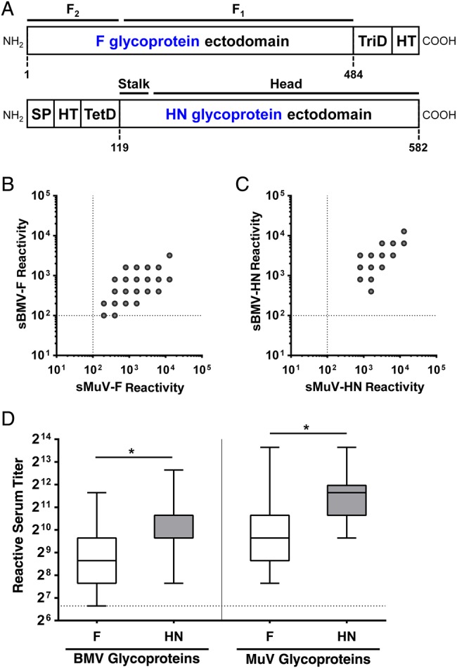
To evaluate the serological relationship between rMuV and rMuVJL5-F/HNBMV, we tested serum samples from mice immunized with each virus for cross-reactivity. Vero cells infected with either virus were stained with mouse hyperimmune serum, and seroreactivity was quantified by flow cytometry. Although naive mouse serum samples were nonreactive to rMuV-infected cells, serum samples from all immunized mice were highly reactive to cells infected with either rMuV or rMuVJL5-F/HNBMV (Supplementary Figure 5A and 5B). These results suggest that standard MuV vaccination in humans might elicit a cross-reactive response that could provide protection from BMV. Reactivity and cross-reactivity were also evaluated in a panel of 54 serum samples from human donors. For each serum, direct binding of reactive antibodies to viral envelope glycoproteins was quantified by an enzyme-linked immunosorbent assay (ELISA) using purified soluble versions of the envelope glycoproteins (sBMV-F, sBMV-HN, sMuV-F, and sMuV-HN), as depicted in Figure 1A. Human serum samples were reactive to the F and HN glycoproteins from both viruses, and we found a highly significant cross-reactive relationship between homologous glycoproteins (Figure 1B and 1C). The HN-specific reactivity was significantly greater than F-specific reactivity for both BMV and MuV, suggesting that mumps vaccination or natural infection might elicit a stronger antibody response against the HN glycoprotein than the F glycoprotein (Figure 1D).
Figure 1.

Reactivity of human serum to purified F and HN glycoproteins. A, Schematic of soluble F and HN glycoproteins that were produced in a baculovirus system. To mimic the oligomeric structure of the envelope glycoproteins, a T4 fibritin trimerization domain and a hexahistidine tag was appended to the C-terminus of the F ectodomain (amino acids 1–484), whereas a baculovirus gp64 signal peptide, a hexhistidine tag, and a human vasodilator-stimulated phosphoprotein tetramerization domain was appended to the N-terminus of the HN ectodomain (amino acids 119–582) [11]. Thrombin cleavage sites were place between the glycoprotein reading frames and the oligomerization domains and tags in both constructs. Numbers below each glycoprotein diagram indicate the amino acid residues that correspond to the full-length glycoprotein. The soluble version of the bat mumps virus (BMV) F glycoprotein (sBMV-F) could not be recovered in significant quantities, so the native proteolytic cleavage site (RRRKR) was mutated to a linker sequence (GSGSG), which allowed for the expression and recovery of higher quantities of sBMV-F. B, C, Reactivity represents the end-point dilution of serum for reactivity against purified F or HN glycoproteins from mumps virus (MuV) or BMV. sBMV-HN, sMuV-F, and sMuV-HN represent soluble versions of BMV HN, MuV F, and MuV HN glycoproteins, respectively; dashed lines represent the threshold limit of detection. B, F glycoprotein: Pearson r = 0.6160; P < .001. C, HN glycoprotein: Pearson r = 0.8001, P < .001. D, Box-and-whisker plots indicate human serum reactivity to purified F and HN glycoproteins, as measured by enzyme-linked immunosorbent assay enzyme-linked immunosorbent assay end-point serum dilution. The difference in reactivity to F and HN glycoproteins was evaluated using a 2-tailed paired t test. *P < .001. Dashed lines represent the threshold limit of detection. Abbreviations: COOH, carboxy terminus; HT, hexahistidine tag; NH2, amino terminus; SP, signal peptide; TetD, tetramerization domain; TriD, trimerization domain.
Cross-Neutralization in Human Serum Samples
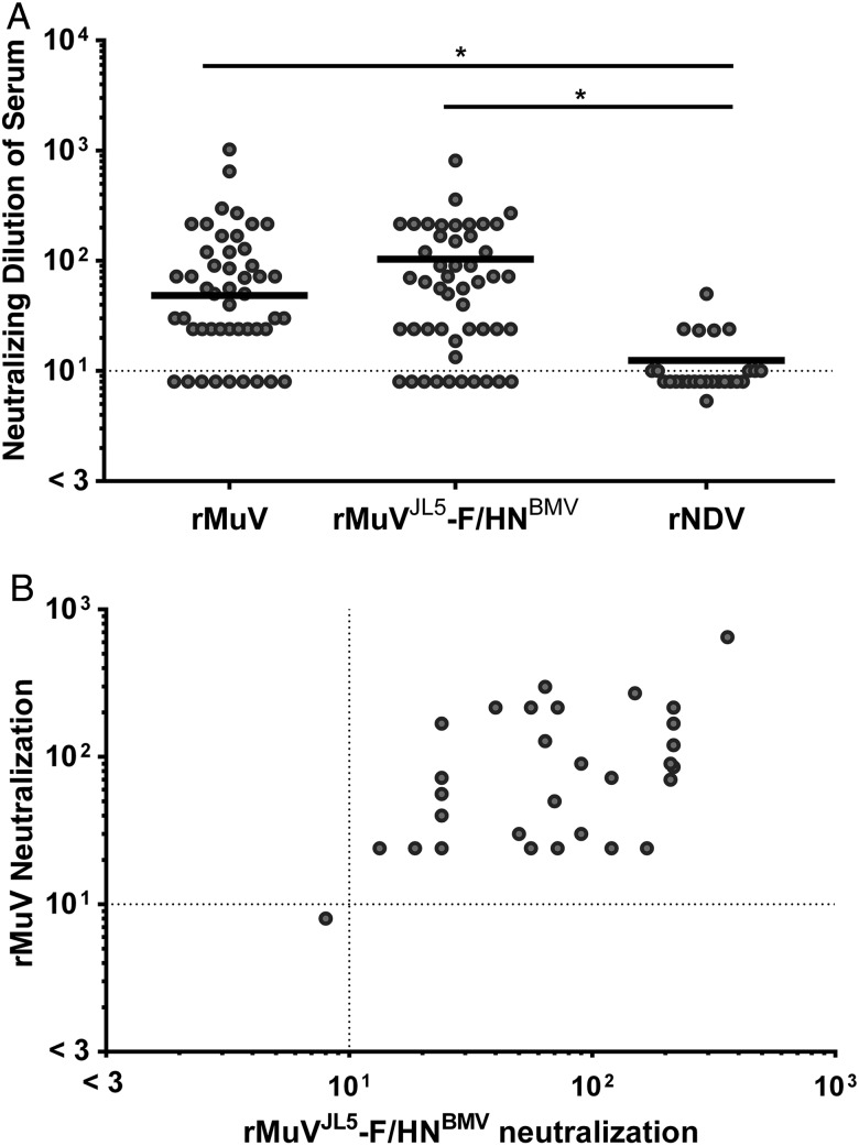
Next, we sought to determine whether the cross-reactive human serum samples also exhibited cross-neutralizing activity between MuV and BMV. Thus, we performed a neutralization assay on the same panel of human serum samples (Figure 2A). Of the 54 serum samples that were tested, 39 samples (72.2%) were neutralizing against rMuV. The mean neutralizing dilutions against rMuV, rMuVJL5-F/HNBMV, and rNDV were 85.6, 75.3, and 4.5, respectively (threshold for seropositivity, 10). All of the rMuV-positive serum samples were cross-neutralizing against rMuVJL5-F/HNBMV, and the positive correlation between the neutralizing activity against rMuV and rMuVJL5-F/HNBMV was highly significant (Figure 2B). These results demonstrate a strong antigenic relationship between BMV and MuV and underscore the potential for cross-protective immunity by MMR vaccination.
Figure 2.
Neutralization activity in a panel of human serum samples. Each data point represents the average neutralizing dilution of serum from 3 experimental replicates (error bars not shown for the purpose of clarity in the figure). A, Bold horizontal lines indicate the geometric mean neutralization values of each data set; differences between mean neutralization values were evaluated by a 1-way analysis of variance. * P < .001. The inhibitory dilution was determined as the lowest dilution in each series with <50% infection. Dashed lines represent the threshold of seropositivity. A threshold inhibitory dilution of 1:10 was used as a cutoff for seropositivity, based on several factors: (1) the limit of detection of our assay was 1:8, (2) apparent nonspecific inhibition was observed at serum concentrations >1:10, and (3) the vast majority of serum samples had an inhibitory dilution <1:10 for the negative control group recombinant Newcastle disease virus (rNDV). B, Pearson's correlation was used to determine the significance of the cross-neutralizing activity (Pearson r = 0.5793; P < .001). Dashed lines represent the threshold of seropositivity as defined above. Serum samples that were able to neutralize rNDV in addition to recombinant parental virus (rMuV) and recombinant MuV bearing the F and HN glycoproteins of BMV (rMuVJL5-F/HNBMV) were considered cytotoxic or nonspecific (5 of 59 serum samples), and as such were removed from further analyses.
DISCUSSION
Taken together, cross-complementation of the envelope glycoproteins, cross-reactivity, and cross-neutralization between the 2 mumps viruses implies significant structural and functional overlap in the F and HN glycoproteins of MuV and BMV. Because the F and HN glycoproteins are the primary determinants of viral entry and antigenicity, their high conservation provides a structural basis for the antigenic relationship that allows for cross-neutralization of the 2 viruses. Strikingly, mapping of the sequence conservation between the envelope glycoproteins of MuV and BMV onto the structure of MuV F or HN glycoproteins shows very few surface-exposed areas that are not identical or similar and absolute conservation of all 6 residues known to be involved in sialic acid binding (Supplementary Figure 6A and 6B) [12]. Furthermore, whereas previously identified neutralizing epitopes on helices α3 (N329-F340) and α4 (G352-R360) of the HN glycoprotein have only 58% and 66% sequence identity between MuV and BMV, respectively, the neutralizing epitope on helix α2 (T265-D266) is completely conserved, which could indicate an important role in cross-neutralization (Supplementary Figure 6C and 6D) [13].
The results of this study demonstrate a strong antigenic relationship between MuV and BMV. All mouse and human serum samples used in this study that were found to have rMuV-reactive or rMuV-neutralizing antibodies also have some degree of activity against rMuVJL5-F/HNBMV, which supports the hypothesis that the single serotype of MuV might extend to include BMV. During our preparation of this article, Katoh et al [14] published similar findings showing evidence of cross-neutralizing activity between MuV and BMV. Our results confirm and extend theirs by showing cross-reactivity and direct binding of human serum to MuV and BMV envelope glycoproteins. Although cross-protective immunity is difficult to determine, the cross-reactive antibody response against rMuVJL5-F/HNBMV in all rMuV-immunized mice suggests that standard mumps vaccination may confer some degree of protection against BMV.
Supplementary Material
Notes
Acknowledgments. We thank all of the serum donors, whose contribution made this study possible.
Financial support. This work was supported by the National Institutes of Health (grants AI115226, AI125536, and AI123449 to B. L.), the Medical Research Council UK (grant MR/L009528/1 to T. A. B.), The University of California, Los Angeles (UCLA Molecular Pathogenesis training grant T32-AI07323 to S. M. B.), and the Icahn School of Medicine at Mount Sinai (Host-Pathogens Interactions training grant T32 AI007647-15 to S. M. B.).
Potential conflicts of interest. All authors: No reported conflicts. All authors have submitted the ICMJE Form for Disclosure of Potential Conflicts of Interest. Conflicts that the editors consider relevant to the content of the manuscript have been disclosed.
References
- 1. Hviid A, Rubin S, Mühlemann K. Mumps. Lancet 2008; 371:932–44. [DOI] [PubMed] [Google Scholar]
- 2. Dayan GH, Rubin S. Mumps outbreaks in vaccinated populations: are available mumps vaccines effective enough to prevent outbreaks? Clin Infect Dis 2008; 47:1458–67. [DOI] [PubMed] [Google Scholar]
- 3. Galazka AM, Robertson SE, Kraigher A. Mumps and mumps vaccine: a global review. Bull World Health Organ 1999; 66:509. [PMC free article] [PubMed] [Google Scholar]
- 4. Drexler JF, Corman VM, Müller MA et al. Bats host major mammalian paramyxoviruses. Nat Commun 2012; 3:796. [DOI] [PMC free article] [PubMed] [Google Scholar]
- 5. Kruger N, Hoffmann M, Drexler JF et al. Functional properties and genetic relatedness of the fusion and hemagglutinin-neuraminidase proteins of a mumps virus-like bat virus. J Virol 2015; 89:4539–48. [DOI] [PMC free article] [PubMed] [Google Scholar]
- 6. Yun T, Park A, Hill TE et al. Efficient reverse genetics reveals genetic determinants of budding and fusogenic differences between Nipah and Hendra viruses and enables real-time monitoring of viral spread in small animal models of henipavirus infection. J Virol 2015; 89:1242–53. [DOI] [PMC free article] [PubMed] [Google Scholar]
- 7. Krammer F, Margine I, Tan GS, Pica N, Krause JC, Palese P. A carboxy-terminal trimerization domain stabilizes conformational epitopes on the stalk domain of soluble recombinant Hemagglutinin substrates. PLoS One 2012; 7:e43603. [DOI] [PMC free article] [PubMed] [Google Scholar]
- 8. Margine I, Palese P, Krammer F. Expression of functional recombinant hemagglutinin and neuraminidase proteins from the novel H7N9 influenza virus using the baculovirus expression system. J Vis Exp 2013; 81:e51112. [DOI] [PMC free article] [PubMed] [Google Scholar]
- 9. Nachbagauer R, Wohlbold TJ, Hirsh A et al. Induction of broadly reactive anti-hemagglutinin stalk antibodies by an H5N1 vaccine in humans. J Virol 2014; 88:13260–8. [DOI] [PMC free article] [PubMed] [Google Scholar]
- 10. Mühlemann K. The molecular epidemiology of mumps virus. Infect Genet Evol 2004; 4:215–9. [DOI] [PubMed] [Google Scholar]
- 11. Kühnel K, Jarchau T, Wolf E et al. The VASP tetramerization domain is a right-handed coiled coil based on a 15-residue repeat. Proc Natl Acad Sci U S A 2004; 101:17027–32. [DOI] [PMC free article] [PubMed] [Google Scholar]
- 12. Yuan P, Thompson TB, Wurzburg BA, Paterson RG, Lamb RA, Jardetzky TS. Structural studies of the parainfluenza virus 5 hemagglutinin-neuraminidase tetramer in complex with its receptor, sialyllactose. Structure 2005; 13:803–15. [DOI] [PubMed] [Google Scholar]
- 13. Orvell C, Alsheikhly AR, Kalantari M, Johansson B. Characterization of genotype-specific epitopes of the HN protein of mumps virus. J Gen Virol 1997; 78:3187–93. [DOI] [PubMed] [Google Scholar]
- 14. Katoh H, Kubota T, Ihara T, Maeda K, Takeda M, Kidokoro M. Cross-neutralization between human and African bat mumps viruses. Emerg Infect Dis 2016; 22:703–6. [DOI] [PMC free article] [PubMed] [Google Scholar]
Associated Data
This section collects any data citations, data availability statements, or supplementary materials included in this article.


