. 2018 Mar 15;7:e31677. doi: 10.7554/eLife.31677

Figure 1. The AGameOfClones vector concept within the piggyBac-based transformation-ready pAGOC vector for Tribolium.

Two fluorescence-based transformation markers, mO and mC, are embedded into a piggyBac-based transformation-ready vector, which is characterized by 3’ and 5’ terminal repeats (TR) necessary for genomic insertion. The markers are based on the artificial eye-specific 3×P3 promoter, the open-reading frame for the respective fluorescent protein, that is, mOrange or mCherry, and the SV40 poly(A). Each transformation marker is flanked upstream by a LoxP site (P) and downstream by a LoxN site (N), forming interweaved Lox site pairs. The markers can be detected in the eyes by using appropriate filter sets (FS). Cre-mediated recombination leads to the excision of one marker from the genome. Upon removal, the other marker remains within the genome, since the remaining LoxP and LoxN sites are incompatible. Individuals that underwent recombination give rise to progeny in which only one marker is detected in the eyes.

Figure 1.

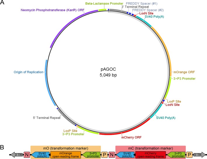
Figure 1—figure supplement 1. The pAGOC vector.

Figure 1—figure supplement 1.

(A) Vector map of pAGOC, which is based on the pAVOIAF{#1–#2–#3–#4} vector (Figure 1—figure supplement 3). In this vector, #1 and #2 remain empty, while mO and mC together with their flanking upstream LoxP and downstream LoxN sites were inserted into #3 and #4, respectively. The unlabeled dark blue boxes represent the same restriction enzyme sites as shown for the pAVOIAF{#1–#2–#3–#4} vector. The light gray band on the inside indicates the transgene. (B) Scheme of mO and mC that are embedded into interweaved but incompatible LoxN and LoxP site pairs. Restriction enzyme sites are not shown. Extents of genetic elements are not to scale. ORF, open-reading frame; TR, piggyBac terminal repeat.
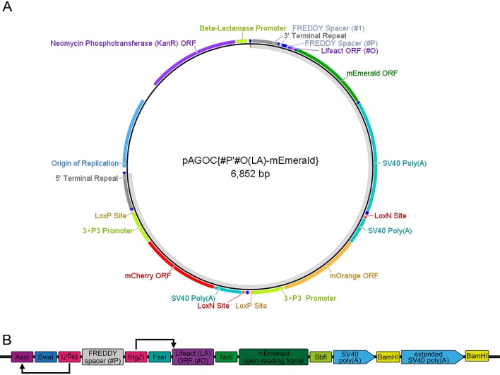
Figure 1—figure supplement 2. The pAGOC{#P’#O(LA)-mEmerald} vector.

Figure 1—figure supplement 2.

(A) Vector map of pAGOC{#P’#O(LA)-mEmerald}, which is based on the pAGOC vector (Figure 1—figure supplement 1). In this vector, #1 remains empty, while the #P’#O(LA)-mEmerald two-slot cloning site was inserted into #2. The unlabeled dark blue boxes represent the same restriction enzyme sites as shown for the pAVOIAF{#1–#2–#3–#4} vector (Figure 1—figure supplement 3) as well as several new restriction enzyme sites shown in (B). The light gray band on the inside indicates the transgene. (B) Scheme of the #P’#O(LA)-mEmerald two-slot cloning site. To insert a promoter, the #P slot can be accessed by the AscI/FseI site pair, but alternatively by the double BtgZI site pair, which flanks a FREDDY spacer. BtgZI is a type I restriction enzyme with a non-palindromic recognition sequence. It digests the sequence several bp (10/14) downstream, resulting in a 4 bp sticky end. In this vector, the upstream BtgZI site (in reverse orientation) allows the opening of the AscI restriction enzyme site, while the downstream BtgZI site (in forward orientation) allows the opening of the Lifeact open-reading frame start codon and the first bp of the subsequent codon, which allows scarless insertion of respectively digested promoter sequences (indicated by arrows). The Lifeact (LA) open-reading frame, which is in #O per default, can be substituted with another open-reading frame to change the intracellular localization by the FseI/NotI site pair, while the mEmerald open-reading frame can be substituted with another fluorescence protein open-reading frame by the NotI/SbfI site pair. Extends of the genetic elements are not to scale. ORF, open-reading frame.
Figure 1—figure supplement 3. The pAVOIAF{#1–#2–#3–#4} vector.

Figure 1—figure supplement 3.

(A) Vector map of pAVOIAF{#1–#2–#3–#4}. The vector is based on the pUC57-Kan vector, from which only the kanamycin resistance cassette and the origin of replication remain. The four-slot cloning site together with the 3’ and 5’ piggyBac terminal repeats is located between the AatII and PciI sites. The light gray band on the inside indicates the transgene. (B) Scheme of the four-slot cloning site. Each slot consists of an 18 bp spacer that translates into the amino acids Phe-Arg-Glu-Asp-Asp-Tyr and thus was termed FREDDY spacer. The slots can be accessed individually by unique restriction enzyme site pairs (XmaI/SpeI for #1, HindIII/XbaI for #2, XhoI/NheI for #3 and AflII/AvrII for #4). They are embedded into five PmeI restriction enzyme sites that allow a simple one-enzyme control digestion to determine the size of the sequences that were inserted into the slots. For convenience, the downstream restriction enzyme sites for each slot (SpeI for #1, XbaI for #2, NheI for #3 and AvrII #4) result in identical sticky ends, facilitating cloning procedures that cannot utilize the suggested restriction enzyme site pairs. Extends of the genetic elements are not to scale. ORF, open-reading frame.
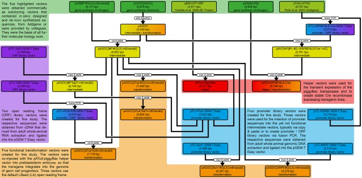
Figure 1—figure supplement 4. Development of the 24 vectors used in this study.

Figure 1—figure supplement 4.

Each vector belongs to either one or two of five types, as indicated by the differently colored backgrounds. Green depicts gene synthesis and previously published vectors, blue depicts promoter library vectors, purple depicts open reading frame (ORF) library vectors, red depicts helper vectors and orange depicts transformation vectors. The ‘copy and paste’ boxes indicate a molecular biology-based procedure with PCR-based amplification of the insert, while the ‘cut and paste’ boxes indicate that the respective insert was extracted from another vector and inserted into the respective backbone without any amplification. See also the Materials and methods section.