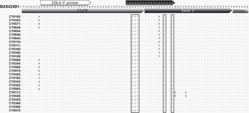
Fig. 1. KIR2DL4 genomic alignment.

Genomic reads mapped to BX842591 KIR2DL4 exon 1. Each sequence represents a consensus of all reads mapped. Degenerate bases are used to represent sites of heterogeneity. Detected genotypes are represented by two to five animals each. Previous forward primer sequence is shown in the black arrow. Mismatches within the previous forward primer targeted region are boxed. The revised primer binding region is shown by the white arrow.
