
Fig. 4. Overall view of the Fab fragment (chains L and H) in PDB 5K8A.
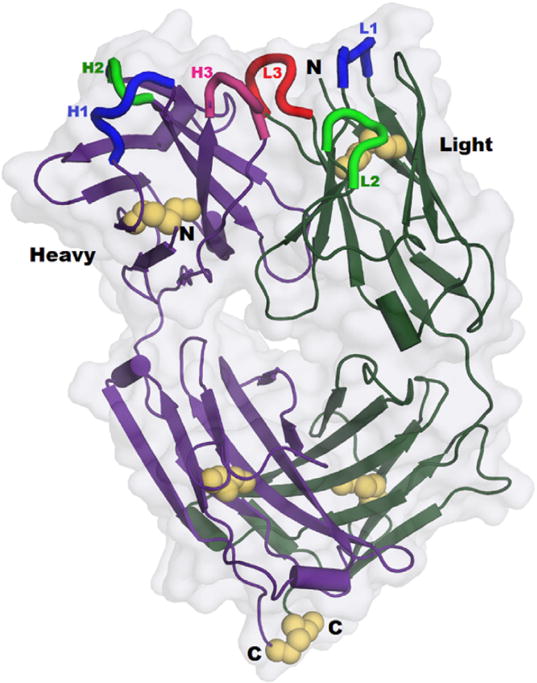
The 5 disulfides are emphasized using yellow spheres, including the sole interchain link, between the observed C-termini at bottom. Cores of the CDR loops are shown in bright colors at top. Molecular surface shows the ‘hole’ through the center of the Fab fragment, between the elbows. (For interpretation of the references to colour in this figure legend, the reader is referred to the web version of this article.)