



Abstract
The early stages of ovarian folliculogenesis generally progress independent of gonadotropins, whereas later stages require signaling initiated by FSH. In Siberian hamsters, cycles of folliculogenesis are mediated by changes in photoperiod which depress the hypothalamic pituitary gonadal axis. Reduced gonadotropins lead to decreases in mature follicle development and ovulation; however, early stages of folliculogenesis have not been explored in regressed ovaries. We hypothesized that intraovarian factors that contribute predominantly to later stages of folliculogenesis would react to changes in photoperiod, whereas factors contributing to earlier stages would not change. To probe if the early stages of folliculogenesis continue in the photoinhibited ovary while late stages decline, we measured the mRNA abundance of factors that interact with FSH signaling (Fshr, Igf1, Cox2) and factors that can function independently of FSH (c-Kit, Kitl, Foxo3, Figla, Nobox, Sohlh1, Lhx8). While plasma FSH, antral follicles, and corpora lutea numbers declined with exposure to inhibitory photoperiod, the numbers of primordial, primary, and secondary follicles did not change. Expression of factors that interact with FSH signaling changed with changes in photoperiod; however, expression of factors that do not interact with FSH were not significantly altered. These results suggest that the photoinhibited ovary is not completely quiescent, as factors important for follicle selection and early follicle growth are still expressed in regressed ovaries. Instead, the lack of gonadotropin support that characterizes the non-breeding season appears to inhibit only final stages of folliculogenesis in Siberian hamsters.
Keywords: Seasonal reproduction, folliculogenesis, follicle stimulating hormone, ovary
1. Introduction
The pituitary gonadotropins, follicle stimulating hormone (FSH)1 and luteinizing hormone (LH), are critical signals for ovarian function via their stimulation of follicle growth and subsequent ovulation. While gonadotropins are essential for these later stages of folliculogenesis, their necessity is reduced in earlier pre-antral stages, which include the transition of the resting primordial follicles into the actively-developing primary follicle pool, and the subsequent granulosa cell proliferation and follicle differentiation to the secondary follicle stage. Early folliculogenesis in these pre-antral stages is generally considered gonadotropin-independent, or requiring only basal levels of gonadotropins (McGee et al., 1997; Hartshorne et al., 1994; Kol and Adashi, 1995; Richards and Midgley, 1976; Oktay et al., 1997).
FSH, working through its receptor (FSHR) interacts with a number of genes within the ovary responsible for late stages of follicle development, such as insulin-like growth factor-1 (Igf1), and cyclooxygenase-2 (Cox2). Within the ovary, IGF1 is produced by granulosa cells and mediates FSHR activity to influence granulosa cell proliferation, as FSHR itself is necessary for the transition of the secondary follicles with multiple granulosa cell layers to the mature tertiary or antral follicles which contain the fluid filled antrum and specialized granulosa and theca cell layers (Hammond et al., 1985; Oktay et al., 1998; Khamsi et al., 2001). Follicles from IGF1 null mice fail to develop past the pre-antral stage as granulosa cell proliferation is disrupted, whereas treatment of cultured granulosa cells with IGF1 and FSH increases Fshr mRNA transcription and stability (Minegishi et al., 2000, Baker et al., 1996; Kadakia et al., 2001). COX2 is the rate limiting enzyme in prostaglandin production; mice lacking COX2 are infertile due to depressed rates of ovulation, although follicular development appears normal (Lim et al., 1997). In addition to cytokines, Cox2 mRNA is also stimulated by FSH in vitro, and increases in vivo with the LH surge (Sirois et al., 1992). The interaction of many of these factors with FSH is critical for the development and maturation of antral follicles during late folliculogenesis.
In contrast to needing large amounts of FSH for survival, earlier stages in folliculogenesis are considered FSH-independent or requiring only basal levels of FSH. The entry of primordial follicles to the growing primary stage, and the passing of primary follicles to the secondary stage involves a number of transcription and growth factors. The transcription factor spermatogenesis and oogenesis helix-loop-helix 1 (SOHLH1) mediates aspects of the FSH-independent transition of primordial follicles into primary follicles through the transcription factors newborn ovary homeobox gene (NOBOX), folliculogenesis specific BHLH transcription factor (FIGLA), and lim-homeobox protein 8 (LHX8) (Jagarlamundi and Rajkovic, 2011; Pangas et al., 2006; Belli et al., 2013). Sohlh genes largely affect the development, stimulation, and survival of primordial follicles; with Sohlh mRNA expression significantly reduced in later stages of folliculogenesis (Jagarlamundi and Rajkovic, 2011; Pangas et al., 2006; Choi et al., 2008). The transition from primordial follicle to consecutive stages is disrupted in NOBOX-null mice, which exhibit a reduction in the number of follicles in subsequent stages (Belli et al., 2013). The helix-loop-helix transcription factor Figla is expressed in germ cells of primordial follicles and regulates aspects of follicle maintenance, including the zona pellucida 1, 2 and 3 proteins (Pangas et al., 2006; Joshi et al., 2007). Finally, while the LHX8 transcription factor is present in follicles throughout folliculogenesis, it is essential for the transition between primordial and primary follicles (Choi et al., 2008). In addition to transcription factors, growth factors also play a role in early follicular growth. The interaction of c-KIT (Mast/stem cell growth factor receptor SCFR or CD117), expressed by oocytes, and kit ligand (stem cell factor, KITL) from somatic cells, is critical for early stages of folliculogenesis (Thomas and Vanderhyden, 2006). In vitro supplementation of KITL promotes primordial follicle development of cultured rat and mouse ovaries (Parrott et al., 1999; Hutt et al., 2006) and induces the FSH-independent primordial to primary follicle transition (Nilsson and Skinner, 2004); a process that doesn’t occur when c-KIT is blocked (Yoshida et al., 1997). Similarly, eliminating expression of growth factor forkhead box O3 (FOXO3) leads to exhaustion of the ovarian follicle reserve, and FOXO3 hyperphosphorylation results in follicle activation (Castrillon et al., 2003; John et al., 2008; Liu et al., 2007). Together, these and other intra-ovarian factors mediate the development of follicles during the FSH-independent phase of folliculogenesis.
While the cycle of hormones and folliculogenic factors is constantly moving follicle development forward in cycling ovaries, most vertebrates do not maintain constant ovarian activity. In many species, reproductive ability is limited to particular seasons of the year to ensure optimal survival of offspring. These seasonal changes in reproductive function are often regulated by photoperiod, or the number of hours of light per day (Ortavant et al., 1988; Nelson et al., 1983). Changes in photoperiod affect endocrine secretions from the hypothalamic pituitary gonadal (HPG) axis via pineal secretion of melatonin (Reiter, 1980; Turek and Campbell, 1979). Melatonin is secreted in the dark, with longer secretion duration occurring during the winter and shorter duration of melatonin occurring during the summer. In seasonally-breeding individuals, exposure to inhibitory photoperiod produces a duration of melatonin secretion that dampens the HPG axis. The subsequent reduction in ovarian function in the non-breeding season has been well established across a number of species, with the change in function typically referring to the decline in gonadotropin-dependent antral follicles and corpora lutea, as is observed in Siberian hamsters (Schlatt et al., 1993; Moffatt-Blue et al., 2006; Salverson et al., 2008), Egyptian buffaloes (Bubalus bubalis) (Abdoon et al., 2015; Ali, 2015), and Western white-faced ewes (Bartlewski et al., 1998). Interestingly, this follicular decline is not typically observed in pre-antral gonadotropin-independent populations of follicles (Schlatt et al., 1993; Moffatt-Blue et al., 2006; Salverson et al., 2008), suggesting that early stages of folliculogenesis may not be affected by changes in photoperiod. To further explore this idea, we hypothesized that 1) mRNA expression of growth factors that interact with FSH (Fshr, Igf1, Cox2) will change in a photoperiod dependent manner that parallels plasma FSH along with tertiary follicle and corpora lutea numbers, and 2) pre-antral follicle numbers and mRNA expression of growth factors (c-Kit, Kitl, Foxo3) and transcription factors (Sohlh1, Nobox, Figla, and Lhx8) that are critical for stages not directly mediated by FSH will not change across photoperiods.
2. Materials and Methods
2.1 Animals
Adult, female Siberian hamsters obtained from our breeding colony were treated in compliance with California State University Long Beach and NRC guidelines for the use of laboratory animals, and under the requirements of approved CSULB IACUC protocol #316. All animals were housed in individual polypropylene cages prepared with bedding and tap water, and were given ad libitum access to food (Laboratory Rodent Diet 5001 from LabDiet, St. Louis, MO) and water. After 2 weeks of acclimation, hamsters (n=55) were exposed to long day photoperiods (16 hours of light per day: 8 hours of dark per day) for 2 weeks. One randomly assigned group of hamsters was kept in long photoperiod for 14 weeks to serve as controls (LD). The remaining hamsters were transferred to short day photoperiods (8L: 16D) for 14 weeks. At 14 weeks of short photoperiod exposure, ovarian and uterine tissues were collected from the regressed group exposed to short days (SD), and the remaining hamsters were transferred to long photoperiod in one of three post transfer (PT) groups representing ovarian recrudescence. In these PT groups, tissues were collected after a total of 14 weeks of SD + 2- (PT2), 4-(PT4), or 8-weeks (PT8) of long photoperiod exposure. At the time of collection, ovaries were dissected and weighed. One ovary was fixed in 10% neutral buffered formalin for seven days; the contralateral ovary was flash frozen for mRNA extraction. Blood samples were collected from the retro-orbital sinus at the time of tissue collection and plasma was stored at −80°C until use.
2.2 Follicle counts
Formalin fixed tissues were processed through a series of PBS washes, dehydrated through a graded series of ethanol, and embedded in paraffin wax. Six-micron sections were collected serially every 60 mm and mounted onto Superfrost-plus microscope slides (ThermoFisher, Waltham, MA). Tissues were stained with hematoxylin and eosin to differentiate ovarian structures and 5-6 cross sections per animal were quantified. Structures counted included primordial follicles (oocyte segregated from surrounding environment by squamous granulosa cells), primary follicles (one layer of cuboidal granulosa cells), secondary follicles (two or more layers of cuboidal granulosa cells), tertiary/antral follicles (multiple layers of granulosa cells, clear antrum present), hypertrophied granulosa cells (large potentially atretic follicles lacking oocytes and characteristic of ovaries from SD exposed females; e.g., van den Hurk et al., 2002; Park et al., 2014) and corpora lutea.
2.3 Follicle Stimulating Hormone and 17β-Estradiol Immunoassays
FSH and 17β-estradiol levels were determined by enzyme immunoassay using FSH ELISA and 17β-Estradiol ELISA kits (Caymen Chemical, MI) following included instructions. All plasma samples and standard curves were run in duplicate per kit instructions, and R2 values from the standard curves were 0.97 and 0.98 for FSH and 17β–estradiol, respectively. Reported intra-assay coefficient of variation (%CV) values are between 8.5-9.4 for FSH and 7.8-18.8 for 17β-estradiol. Hormone concentrations were calculated using previously reported methods (Shahed and Young, 2011; Shahed et al., 2015a, Shahed et al., 2015b).
2.4 cDNA synthesis and RT-PCR
Trizol LS reagent (Invitrogen Life Technologies, Carlsbad, CA) was used to extract total RNA from the ovaries as per manufacturer’s protocol. RNA quality was assured using RNA electrophoresis, and samples were treated with DNase to remove potential DNA contamination. One microgram of total RNA was used for cDNA synthesis using the ImProm Reverse Transcription System according to manufacture’s instructions (Promega, Madison, WI). For PCR analysis, cDNA was diluted 1: 2.5 with DNAase/RNAase free water. Relative real-time PCR was conducted on the StepOnePlus Real-Time PCR System (Applied Biosystems ThermoFisher, Waltham, MA) using Absolute qPCR SYBR green mix (ThermoFisher, Waltham, MA), with no changes to dNTP or MgCl2 concentrations. The PCR reaction mix contained 1 μl cDNA + 1 μl each of forward and reverse primers (80 nM concentration) + 6 μl SYBRgreen mix + 3 μl water to a total volume of 12 μl. PCR cycles consisted of a 15 min hold at 95 °C (1 cycle), then 40 amplification cycles at appropriate melting temperature (Table 1), extension (1 min at 72°C) followed by dissociation. Melt curves for all products were produced to identify the presence of non-specific products, and efficiency ranged from 61-89% for all reactions. Non-template negative controls were included in each PCR analysis. In addition, PCR products were analyzed on agarose gels to confirm that the correct size products were obtained (all primers designed for this study were designed to generate products that were approximately 200 bp) and to visualize potential secondary and nonspecific amplification. For the standard curve, cDNA from all samples was pooled and a 4-point curve was included with each run. The standard curve was used to calculate the relative amounts of mRNA expression. Ratios of the values for the genes of interest compared to the mRNA expression of reference gene glyceraldehyde 3-phosphate dehydrogenase (Gapdh) were analyzed for statistical differences. The expression of Gapdh mRNA did not change significantly between different photoperiod exposure groups when quantified by real-time RT PCR, as we have reported previously (Shahed and Young, 2013).
Table 1.
Primer description for real time PCR
| Gene | Primer Sequence | Tm (°C) | Accession number/Reference |
|---|---|---|---|
| c - Kit | F-TAAGTCAGATGCTGCCATGACGGT R-TCATGTGATTGCCCAGGTAGCTCA |
64 | XM_013121056.2 |
| Cox 2 | F- CAACTCCCTTGGGTGTGA R- TCCTCGTTTCTGATCTGTCT |
56 | XM_005073469.3 |
| Foxo 3 | F-ACAAACGGCTCACTTTGTCC R-TTCTGAACTCGCATGAATCG |
55 | XM_021729722.1 |
| Figla | F- AATCTCAACCGTGGCTTTGC R- GCTACTGTGGGTCTGTTCCT |
54 | XM_006998309.1 |
| Fshr | F-TGTCATCACTGGCTGTGTCA R-TTGGTGAGCACAAACCTCAG |
56 | XM_021231294.1 |
| Gapdh | F-GGAGAAAGCTGCCAAGTA2 R-TGTCATTGAGAGCGATGC |
55 | Shahed and Young, 2013 |
| Igf1 | F-CTGGTGGATGCTCTTCAGTTC R-CCAGTCTCCTCAGATCACAGC |
65 | NM_010512.5 |
| Kitl | F-GAATCTCCGAAGAGGCCAGA R-GCTGCAAGACCCCCTAACAT |
58 | NM_013598.3 |
| Lhx8 | F- CAGTTCGCTCAGGACAACAA R- CCTGCAGTTCTGAAACCACA |
58 | Zhang et al., 2012 |
| Sohlh1 | F- CTGTGGGGCTTGGATTTTGG R- TGCCAGCCTCCCTCAAATTA |
54 | XM_005083759.3 |
2.5 Statistical Analysis
All data were analyzed by a one-way ANOVA using the PRISM statistical software package (GraphPad Prism 6, San Diego, CA). Because our goal was to study the differences between cycling, fully regressed, and recrudescing ovaries, females that did not respond to 14 weeks of SD exposure, based on ovarian mass and lack of pelage change, were removed from the study (n=11, after Schlatt et al., 1993). In addition, outliers falling outside two standard deviations of the mean for each statistical test were removed, resulting in a final n of 5-11 per group. In the case of 17β-Estradiol, data were not normal; therefore, a Kruskal Wallis/Fishers LSD test was used. All other data were log transformed prior to ANOVA analysis. No significant differences across standard deviations via the Brown-Forsythe and Bartlett’s test were noted. Results are indicated with ±SEM, and the Newman Keuls post-hoc test was used to determine if significant (p<0.05) differences existed across the groups.
3 Results
3.1 Short Photoperiod Exposure Inhibits Plasma FSH, 17β-Estradiol, and Reproductive Organ Masses
To establish the effect of inhibitory and stimulatory photoperiod in the present study, we assessed key reproductive parameters known to change with different photoperiod exposures. Plasma FSH was reduced in females exposed to 14 weeks of short photoperiod as compared to FSH concentrations in long day exposed females (p<0.05), while transfer of reproductively regressed females to long photoperiod restored FSH to levels no different than observed in the LD group (p>0.05; Figure 1A). Similarly, plasma 17β-estradiol was reduced in both SD females and post-transfer week 2 females as compared to all other groups (p<0.05; Figure 1B). Both ovarian and uterine tissues also responded to inhibitory photoperiod exposure with reductions in mass, with ovaries from SD females showing reduced mass as compared to all groups, and ovaries in post-transfer groups increasing in mass as compared to SD groups (p<0.05; Figure 1C). Uterine mass was also reduced in both SD and post-transfer week 2 females as compared to all other groups (p<0.05; Figure 1D).
Figure 1.

A) Mean plasma FSH concentrations (mIU/ml) B) mean plasma 17-β estradiol concentrations (pg/ml) C) mean paired ovarian mass (mg) D) mean paired uterine mass (mg) from females exposed to long photoperiods (LD), short photoperiods (SD), and 2 weeks, 4 weeks, and 8 weeks post transfer from SD to LD photoperiod (PT2, PT4, PT8). Data are presented as mean ± SEM, columns with different letters indicate significant differences between groups (p < 0.05).
3.2 Antral, but not preantral follicles are reduced by exposure to inhibitory photoperiod
Follicle types and ovarian structures were identified and quantified across photoperiod groups (Figure 2). The mean number of primordial, primary, and secondary follicles per ovarian cross section did not change significantly with photoperiod exposure (p>0.05; Figure 3A-C). In contrast, short photoperiod exposure reduced the number of tertiary follicles and the number of corpora lutea as compared to both LD controls and post-transfer week 8 ovaries (p<0.05; Figure 3D-E). Hypertrophied granulosa cells, the eosinic potentially-atretic follicles typical of photoregressed Siberian hamsters (Moffatt-Blue et al., 2004; Kabithe and Place, 2008), peaked in ovaries from females exposed to SD, and were present in early recrudescence groups, PT2 and PT4, at significantly lower numbers (p<0.05; Figure 3F).
Figure 2.
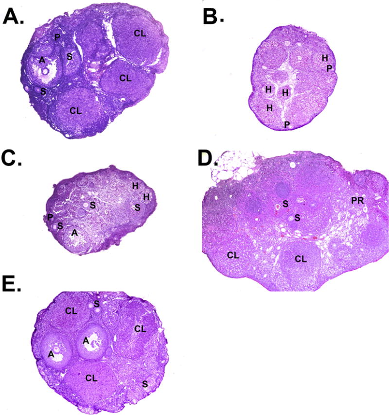
A) Representative ovarian cross sections stained with hematoxylin and eosin. A) long day control ovaries, B) short day regressed ovaries, C) post transfer from SD to LD for two, D) four, or E) eight weeks, representing recrudescing ovaries. Primordial follicles (PR); primary follicles (P), secondary follicles (S), antral follicles (A); corpus luteum (CL); hypertrophied granulosa cells (H).
Figure 3.

Follicle/structure counts presented as average number per section for A) primordial B) primary C) secondary D) tertiary E) corpora lutea and F) hypertrophied granulosa cells. Counts were made across serially sectioned ovaries from females exposed to long photoperiods (LD), short photoperiods (SD), and 2 weeks, 4 weeks, and 8 weeks post transfer from SD to LD photoperiod (PT2, PT4, PT8). Data are presented as mean ± SEM, columns with different letters indicate significant differences between groups (p < 0.05).
3.3 mRNA expression of folliculogenic factors which interact with FSH
FSH-receptor mRNA was expressed in ovaries from females housed in long days; however, this was significantly decreased with exposure to 14 weeks of short day photoperiod exposure (p<0.05; Figure 4A). Transfer to long photoperiods resulted in a significant return of Fshr mRNA at 2, 4, and 8 weeks above levels in SD animals (Figure 4A). A similar pattern was observed for Igf1 mRNA, which was reduced in ovaries from females exposed to short as compared to long photoperiods, with levels remaining low in post-transfer week 2 (Figure 4B). By eight weeks post transfer to photostimulation, Igf1 mRNA expression returned to levels no different from LD (p>0.5; Figure 4B). Cox2 mRNA was present in the LD group, declined significantly in SD, and slowly increased with photostimulation by post-transfer week 8; however, levels were still reduced as compared to LD controls (Figure 4C).
Figure 4.
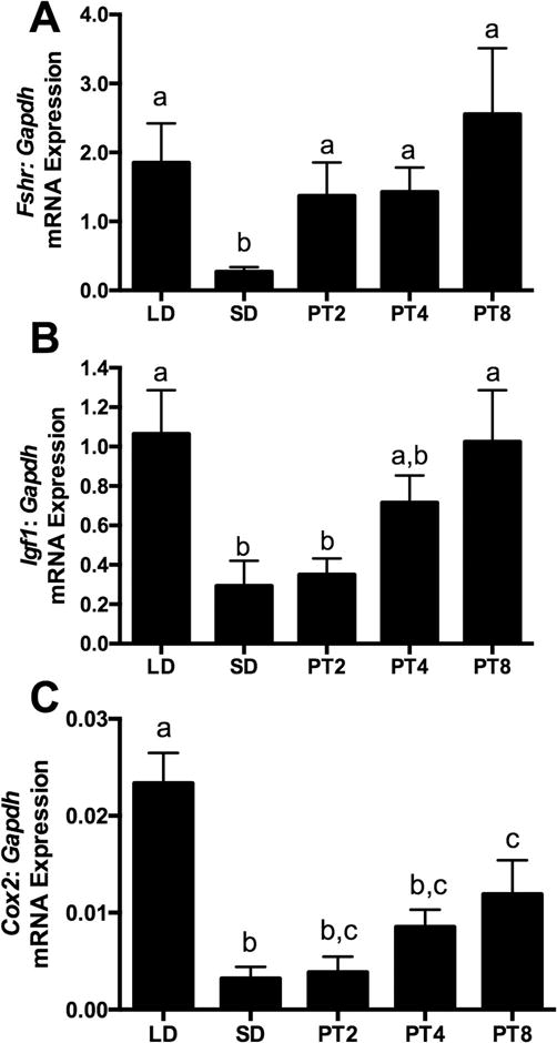
Ovarian mRNA expression of genes involved with FSH signaling A) follicle stimulating hormone receptor (Fshr), B) Insulin like growth factor 1 (Igf1) and C) Cyclooxygenase-2 (Cox2) from Siberian hamsters exposed to long day (LD) stimulatory photoperiod, short day (SD) inhibitory photoperiod, and two, four, or eight weeks of long photoperiod following short day exposure (PT2, PT4, PT8). Graphical results are presented as mean ± SEM, relative to Gapdh, and columns with different letters are significantly different (p < 0.05).
3.4 mRNA expression of FSH-independent transcription factors
Ovarian mRNA expression of folliculogenic factors critical for early follicle development, Nobox1, Sohlh, Lhx8, and Figla were present in ovaries of Siberian hamsters; however, did not significantly change across photoperiod exposures (p > 0.05 for all four analyses; Figure 5A-D).
Figure 5.

Ovarian mRNA expression of transcription factors not dependent on FSH-signaling: A) Newborn ovary homeobox gene (Nobox), B) Spermatogenesis and oogenesis basic helix–loop–helix 1(Sohlh1), C) LIM-homeobox transcription factor 8 (Lhx8) and D) factor in the germ-line alpha (Figla) from Siberian hamsters exposed to long day (LD) stimulatory photoperiod, short day (SD) inhibitory photoperiod, and two, four, or eight weeks of long photoperiod following short day exposure (PT2, PT4, PT8). Graphical results are presented as mean ± SEM, relative to Gapdh, and columns with different letters are significantly different (p < 0.05).
3.5 mRNA expression of FSH-independent growth factors
Growth factors important for early aspects of folliculogenesis were present in cycling, regressed, and recrudescing hamster ovaries; however, mRNA expression for c-Kit, Kitl, and Foxo3 did not change across photoperiod groups (p >0.05 for all three analyses; Figure 6A-C).
Figure 6.

Ovarian mRNA expression of growth factors not dependent on FSH-signaling: A) c-Kit (Kit), B) Kit ligand (Kitl), and C) Forkhead box O3 (Foxo3) from Siberian hamsters exposed to long day (LD) stimulatory photoperiod, short day (SD) inhibitory photoperiod, and two, four, or eight weeks of long photoperiod following short day exposure (PT2, PT4, PT8). Graphical results are presented as mean ± SEM, relative to Gapdh, and columns with different letters are significantly different (p < 0.05).
4 Discussion
Data from the present study show for the first time that growth factors necessary for early follicle growth are expressed regardless of photoperiod exposure in Siberian hamster ovaries, in parallel with the lack of change observed in the numbers of these gonadotropin-independent follicles across photoperiod groups. In contrast, exposure to inhibitory short photoperiods reduced ovarian mass, plasma FSH concentrations, antral follicles, corpora lutea, and mRNA for folliculogenic factors known to interact with FSH. Ovaries in photoinhibited females are often termed ‘quiescent’ or ‘inactive’; however, our results suggest that ovarian activity continues, at least among the follicle populations not dependent on gonadotropin support.
Ovarian function is systemically maintained by the HPG axis, with adenohypophyseal secretion of FSH and LH critical to aspects of folliculogenesis and steroidogenesis. In long day breeding rodents, this gonadotropin release is seasonally-regulated through the interaction of the hypothalamus and pineal gland, with long winter durations of melatonin secretion effectively shutting down release of GnRH, and thus reducing FSH and LH (Reiter, 1980; Turek and Campbell, 1979). Data from the current experiment correlate with previous studies demonstrating that exposure to inhibitory photoperiods reduces FSH in hamsters (Shahed et al., 2015b; Schlatt et al., 1993; Benson and McAsey, 1998; Berndtson and Desjardins, 1974), ewes (Legan and Karsch, 1980) and mares (Freedman et al., 1979). Among antral follicles, FSH is a potent stimulator of follicle growth and survival (Richards, 1994; Vanderhyden, 2002), and administration of FSH preferentially stimulates development of large follicles (Hartshorne et al., 1994). Because antral follicles require tonic FSH for survival (Markström et al., 2002; Hillier, 1994), reductions in FSH increase follicular atresia (Hsueh et al., 1994; Lin and Rui, 2010). In Siberian hamsters, the loss of gonadotropin support is concomitant with reductions in the number of healthy antral follicles and corpora lutea in SD as compared to LD females (e.g., Moffat-Blue et al., 2006; Salverson et al., 2008; van den Hurk et al., 2002, Figure 3). Because regressed ovaries lack antral follicle development and ovulation, they are often characterized as inactive organs.
To better understand how FSH functions within the photoregressed and photostimulated ovary, we quantified the mRNA expression of factors known to interact directly with FSH. Intraovarian factors that respond directly to FSH stimulation that were examined in this study (Fshr, Igf1, Cox2) and previous studies (anti-Müllerian hormone, inhibin- [inh-α], aromatase, estrogen receptor β) mirror the changes in plasma FSH concentrations observed when female hamsters are exposed to different photoperiods (Figure 4, Shahed and Young, 2013; Shahed et al., 2015a; Shahed et al., 2015b). In cycling ovaries from mice and rats, FSH acts through the FSH receptor to initiate granulosa cell proliferation and folliculogenesis (Knecht et al., 1981; Kumar et al., 1997), whereas mRNA expression of the FSH receptor declines in ovaries from cats in the non-breeding season (Hobbs et al., 2012). In Siberian hamsters, declines in plasma FSH parallel reductions in ovarian Fshr and proliferation factor Pcna mRNA expression (Figure 1, Figure 4, Shahed et al., 2015), suggesting that FSH interaction with its receptor may mediate aspects of photoperiod driven changes observed in follicle development. Granulosa cell proliferation is also mediated by Igf1, likely through the FSH receptor by enhancing cAMP action downstream of FSHR while maintaining the stability of Fshr mRNA transcripts (Khamsi et al., 2001; Kadakia et al., 2001). FSH itself induces granulosa cell synthesis of Igf1 (Adashi et al., 1991a and b), further amplifying Igf1 action. Because the number of granulosa cells increases exponentially as follicles grow, thereby increasing the number of FSH receptors present and production of Inh-α and Igf1, it may be that the drop in mRNA expression from genes like Fshr, Igf1, and Inh-α in SD females simply reflects a loss of larger follicles. This lack of large follicles also correlates with the decline in mRNA for the prostaglandin synthesis enzyme important for oocyte maturation and ovulation, Cox2 (Lim et al., 1997; Dinchuk et al., 1995). Experiments blocking FSHR action during photostimulated recrudescence could tease apart the direct effects of the loss of FSH as compared solely to the loss of follicles. Short photoperiod induced declines in plasma FSH, Fshr, Igf1, Cox2, and Inh-α mRNA values which are restored upon photostimulation as antral follicle growth is again stimulated, suggesting that downstream action of FSH is critical during early recrudescence (Figure 4, Shahed and Young, 2013). Further studies are needed to understand if FSH is directly responsible for these photoperiodic fluctuations, if changes simply reflect loss of granulosa cells, or if the changes in genes such as Igf1 drive the sensitivity of the ovarian response to FSH. Because some genes critical for full follicle development do not show this typical pattern of SD induced decline, for example TGFβ superfamily members growth differentiation factor 9 (GDF9), and bone morphogenic protein-15 (BMP15) both increase with SD exposure (Shahed and Young, 2013), it is evident that factors other than gonadotropins are involved in seasonal changes in ovarian function.
While FSH is a critical mediator of follicle development, successful folliculogenesis depends on multiple reproductive hormones, including LH and estrogen. Stimulation from the LH surge triggers ovulation and is responsible for luteinizing follicle cells during formation of the corpus luteum. LH is also critical for initiating ovarian steroidogenesis, and therefore is important in aspects of follicle development. However, LH can inhibit granulosa cell proliferation (Yong et al., 1992), and dominates only in the periovulatory stages of follicular maturation. LH has been reported not to change across photoperiod exposure in Siberian hamsters, and transfer of male hamsters from SD to LD fails to produce a rapid increase in LH (Greives, et al., 2011; Anand et al., 2002). It may be that major changes in LH do not mediate much of the photoperiod-driven change in ovarian function. In contrast, plasma 17β-estradiol does decline in adult females exposed to short photoperiods for 14 weeks (Moffatt-Blue et al., 2006; Salverson et al., 2008; Shahed et al., 2015a), and ovarian mRNA expression for key enzymes of the steroidogenic pathway, such as steroid acute regulatory protein, 3β hydroxysteroid dehydrogenase, and aromatase/Cyp19 also decline with short photoperiod exposure (Shahed et al., 2015a). Because estrogen itself can serve as a granulosa cell mitogen (Goldenberg et al., 1972) and estrogen action, along with FSH, is needed for development of antral follicles and granulosa cell differentiation (Vanderhyden, 2002), the observed short photoperiod-induced reduction in estrogen may also impede late folliculogenic development. In women with low serum FSH, LH, and 17β-estradiol, the number of antral follicles, but not smaller follicles, was decreased (Deb et al., 2012), illustrating the importance of these hormones to late folliculogenesis.
In contrast to antral follicle development, early stages of folliculogenesis are not fully-dependent on FSH, and are often considered FSH-independent (Elvin and Matsuk, 1998; Hillier, 1994), although the idea of full gonadotropin-independence is controversial. Primordial and preantral follicles, but not antral follicles or corpora lutea are present in in FSHβ and FSHR knockout mice (Kumar, 2005; Kumar et al., 1997, Durlinger et al., 2001). Other studies have shown a moderate benefit of FSH to early follicle growth (reviewed by Dewailly et al., 2016). Photoperiod does not affect the number of preantral follicles in adult female hamsters (Figure 3; Shahed et al., 2015a; Shahed et al., 2015b; Salverson et al., 2008), despite the changes observed in plasma FSH (Figure 1). In this natural model of changing ovarian function, FSH is not completely ablated with SD exposure; plasma concentrations decline but are still present even in photoinhibited females (Figure 1). Therefore, short photoperiod-exposed hamsters have the basal FSH levels that may be necessary for early follicle growth; however, these hamsters lack the higher concentrations of FSH present in cycling females that are needed to stimulate full antral follicle development.
Concomitant with this lack of change in primordial, primary, and secondary follicles (Figure 3), mRNA expression from growth (c-Kit, Kitl, Foxo3) and transcription (Nobox, Sohlh1, Lhx8, Figla) factors that do not directly interact with FSH did not significantly change with differential photoperiod exposure (Figures 4, 5). These factors are critical for the early stages of follicle development, a time when follicles have not yet developed FSH receptors (Yoshida et al., 1997), and therefore the reduced concentrations of plasma FSH observed in photoinhibited hamsters would not be predicted to affect their function. Some of these factors are also important in later stages of follicle development, for example c-kit and Kitl are involved throughout folliculogenesis (Thomas and Vanderhyden, 2006). However, reductions in mRNA expression may not have been observed because the number of primordial, primary, and secondary follicles exceeds the number of tertiary follicles, the only follicle type observed to decline significantly with photoinhibition in the present study (Figure 3). Previous studies in Siberian hamsters (Moffatt-Blue et al., 2006; Kabithe and Place, 2008), ewes (McNatty et al., 1984), and mares (Sharp and Ginther, 1975) also show photoperiod induced declines in tertiary follicles. Pathways that are important for primordial follicle activation, such as the PI3K/FOXO3 signaling pathway may likely be constitutively active in the photoinhibited ovary, and folliculogenesis may only be interrupted at stages requiring FSH stimulation. Indeed, the eosinic hypertrophied granulosa cells (aka luteinized atretic follicles) observed in photoregressed hamster ovaries are postulated to be atretic secondary follicles that had been previously exposed to gonadotropins, based on their size and lack of antrum (van den Hurk et al., 2002). These hypertrophied pockets of cells are not observed in long day exposed hamsters, and their increase with photoinhibition may be the alternative fate for pre-antral follicles deprived of FSH during short photoperiod exposure. If ovarian function was indeed ‘frozen’ during the non-breeding season, the transcripts important for follicle selection and early follicular growth should decline as does the expression of mRNA from genes involved in later stages of folliculogenesis. Instead, the identification that this pool of transcripts important for follicle selection is maintained in ovaries regardless of photoperiod exposure suggests that folliculogenesis in photoinhibited ovaries is not completely arrested. Additional studies are required to determine if this pool of constitutively transcribed mRNAs is indeed important for uninterrupted early follicular development in regressed ovaries.
In conclusion, because both ovulation and 17β-estradiol production decline in female Siberian hamsters exposed to chronic short photoperiod regimes, ovaries from photoinhibited females are considered “quiescent” or “inactive”. However, data from this study suggest that early folliculogenesis continues during gonadal regression whereas only late folliculogenesis is halted due to a lack of FSH support. These findings highlight the gonadotropin dependence of factors contributing to photostimulated return of ovulation, and suggest a potential new role of early folliculogenesis in restoring ovarian function. Because photostimulation rapidly stimulates antral follicle development and ovulation (Shahed et al., 2015b), maintaining early stages of folliculogenesis may allow the photoinhibited ovary to respond immediately to the return of systemic FSH, thus enabling a rapid return to reproductive function.
Highlights.
Late, but not early, stages of folliculogenesis decline with photoinhibition
Expression of early folliculogenic factors do not change with photoperiod
Intraovarian factors related to FSH signaling change with photoperiod
FSH-independent stages may continue in the photoinhibited ovary
The photoinhibited ovary may not be completely inactive
Acknowledgments
We thank Chelsea Berens for tissue collection and isolation of mRNA, Tim Tusso for the Nobox PCR assay, Angela Hoang for her work with Kit. We are grateful to Dr. Asha Shahed for her guidance with PCR, designing the Cox2 primers, and the hormone assays. We are also appreciative of the thoughtful and valuable comments provided by two anonymous reviewers. This project was supported by: NIH MARC fellowship from T34GM008074 (AKS), NIH RISE fellowship R25GM071638 (KL), NIH SCORE grants 1SC3GM089611-01 and 1SC3GM116696-01 (KAY).
Footnotes
Publisher's Disclaimer: This is a PDF file of an unedited manuscript that has been accepted for publication. As a service to our customers we are providing this early version of the manuscript. The manuscript will undergo copyediting, typesetting, and review of the resulting proof before it is published in its final citable form. Please note that during the production process errors may be discovered which could affect the content, and all legal disclaimers that apply to the journal pertain.
Abbreviations: Follicle stimulating hormone (FSH), Follicle stimulating hormone receptor (FSHR), Insulin-like growth factor 1 (IGF1), Cyclooxygenase (COX2), Spermatogenesis and oogenesis specific basic helix-loop-helix 1 (SOHLH1), Newborn ovary homeobox (NOBOX), Factor in the germline alpha (FIGLA), Lim homeobox 8 (LHX8), c-kit receptor tyrosine kinase (c-KIT), Kit ligand (KITL), Forkhead box O3 (FOXO3), hypothalamic pituitary gonadal (HPG) axis, inhibin-α (INHα).
Declaration of interest: Conflicts of interest: none
References
- Abdoon AS, Gabler C, Holder C, Kandil OM, Einspanier R. Seasonal variations in developmental competence and relative abundance of gene transcripts in buffalo (Bubalus bubalis) oocytes. Theriogenology. 2014;82:1055–1067. doi: 10.1016/j.theriogenology.2014.07.008. [DOI] [PubMed] [Google Scholar]
- Adashi EY, Resnick CE, Hernandez ER, Hurwitz A, Roberts CT, Leroith D, Rosenfeld R. Insulin-like growth factor I as an intraovarian regulator: basic and clinical implications. Ann N Y Acad Sci. 1991a;626:161–168. doi: 10.1111/j.1749-6632.1991.tb37910.x. [DOI] [PubMed] [Google Scholar]
- Adashi EY, Resnick CE, Hurwitz A, Ricciarelli E, Hernandez ER, Roberts CT, Leroith D, Rosenfeld R. Insulin-like growth factors: the ovarian connection. Hum Reprod. 1991b;6:1213–1219. doi: 10.1093/oxfordjournals.humrep.a137514. [DOI] [PubMed] [Google Scholar]
- Ali A. Seasonal variations of the ovarian activity and pregnancy rate in the Egyptian buffalo cows (Bubalus bubalis) Trop Anim Health Prod. 2015;47:815–818. doi: 10.1007/s11250-015-0793-8. [DOI] [PubMed] [Google Scholar]
- Anand S, Losee-Olson S, Turek FW, Horton TH. Differential regulation of luteinizing hormone and follicle-stimulating hormone in male Siberian hamsters by exposure to females and photoperiod. Endocrinology. 2002;143:2178–2188. doi: 10.1210/endo.143.6.8839. [DOI] [PubMed] [Google Scholar]
- Baker J, Hardy MP, Zhou J, Bondy C, Lupu F, Bellv AR, Efstratiadis A. Effects of an Igf1 gene null mutation on mouse reproduction. Molecular Endocrinology. 1996;10:903–918. doi: 10.1210/mend.10.7.8813730. [DOI] [PubMed] [Google Scholar]
- Bartlewski PM, Beard AP, Cook SJ, Rawlings NC. Ovarian follicular dynamics during anoestrus in ewes. J Reprod Fertil. 1998;113:275–85. doi: 10.1530/jrf.0.1130275. [DOI] [PubMed] [Google Scholar]
- Belli M, Cimadomo D, Merico V, Redi CA, Garagna S, Zuccotti M. The NOBOX protein becomes undetectable in developmentally competent antral and ovulated oocytes. Int J Dev Biol. 2013;57:35–39. doi: 10.1387/ijdb.120125mz. [DOI] [PubMed] [Google Scholar]
- Benson B, McAsey ME. An essential role for ovarian inhibin in pineal gland-mediated anestrus in Syrian hamsters. J Pineal Res. 1998;25:5–11. doi: 10.1111/j.1600-079x.1998.tb00379.x. [DOI] [PubMed] [Google Scholar]
- Berndtson WE, Desjardins C. Circulating LH and FSH levels and testicular function in hamsters during light deprivation and subsequent photoperiodic stimulation. Endocrinology. 1974;95:195–205. doi: 10.1210/endo-95-1-195. [DOI] [PubMed] [Google Scholar]
- Castrillon DH, Miao L, Kollipara R, Horner JW, DePinho RA. Suppression of ovarian follicle activation in mice by the transcription factor Foxo3a. Science. 2003;301:215–218. doi: 10.1126/science.1086336. [DOI] [PubMed] [Google Scholar]
- Choi Y, Ballow DJ, Xin Y, Rajkovic A. Lim homeobox gene, lhx8, is essential for mouse oocyte differentiation and survival. Biol Reprod. 2008;79:442–449. doi: 10.1095/biolreprod.108.069393. [DOI] [PMC free article] [PubMed] [Google Scholar]
- Deb S, Campbell BK, Pincott-Allen C, Clewes JS, Cumberpatch G, Raine-Fenning NJ. Quantifying effect of combined oral contraceptive pill on functional ovarian reserve as measured by serum anti-Mullerian hormone and small antral follicle count using three-dimensional ultrasound. Ultrasound Obstet Gynecol. 2012;39:574–580. doi: 10.1002/uog.10114. [DOI] [PubMed] [Google Scholar]
- Dewailly D, Robin G, Peigne M, Decanter C, Pigny P, Catteau-Jonard S. Interactions between androgens, FSH, anti-Mullerian hormone and estradiol during folliculogenesis in the human normal and polycystic ovary. Hum Reprod Update. 2016;22:709–724. doi: 10.1093/humupd/dmw027. [DOI] [PubMed] [Google Scholar]
- Dinchuk JE, Car BD, Focht RJ, Johnston JJ, Jaffee BD, Covington MB, Contel NR, Eng VM, Collins RJ, Czerniak PM. Renal abnormalities and an altered inflammatory response in mice lacking cyclooxygenase II. Nature. 1995;378:406–409. doi: 10.1038/378406a0. [DOI] [PubMed] [Google Scholar]
- Durlinger AL, Gruijters MJ, Kramer P, Karels B, Kumar TR, Matzuk MM, Rose UM, de Jong FH, Uilenbroek JT, Grootegoed JA, Themmen AP. Anti-Mullerian hormone attenuates the effects of FSH on follicle development in the mouse ovary. Endocrinology. 2001;142:4891–4899. doi: 10.1210/endo.142.11.8486. [DOI] [PubMed] [Google Scholar]
- Elvin JA, Matzuk MM. Mouse models of ovarian failure. Rev Reprod. 1998;3:183–195. doi: 10.1530/ror.0.0030183. [DOI] [PubMed] [Google Scholar]
- Freedman LJ, Garcia MC, Ginther OJ. Influence of Photoperiod and Ovaries on Seasonal Reproductive Activity in Mares. Biology of Reproduction. 1979;20:567–574. doi: 10.1095/biolreprod20.3.567. [DOI] [PubMed] [Google Scholar]
- Galas J, Słomczyńska M, Pierściński A. Effect of photoperiod on the distribution patterns of androgen receptors and steroid hormone concentrations in ovaries of bank voles. Acta Histochemica. 2003;105:175–181. doi: 10.1078/0065-1281-00691. [DOI] [PubMed] [Google Scholar]
- Goldenberg RL, Bridson WE, Kohler PO. Estrogen stimulation of progesterone synthesis by porcine granulosa cells in culture. Biochem Biophys Res Commun. 1972;48:101–107. doi: 10.1016/0006-291x(72)90349-x. [DOI] [PubMed] [Google Scholar]
- Greives TJ, Long KL, Burns CM, Demas GE. Response to exogenous kisspeptin varies according to sex and reproductive condition in Siberian hamsters (Phodopus sungorus) Gen Comp Endocrinol. 2011;170:172–179. doi: 10.1016/j.ygcen.2010.10.001. [DOI] [PMC free article] [PubMed] [Google Scholar]
- Hammond JM, Baranao JLS, Skaleris D, Knight AB, Romanus JA, Rechler MM. Production of insulin-like growth factors by ovarian granulosa cells. Endocrinology. 1985;117:2553–2555. doi: 10.1210/endo-117-6-2553. [DOI] [PubMed] [Google Scholar]
- Hartshorne GM, Sargent IL, Barlow DH. Meiotic progression of mouse oocytes throughout follicle growth and ovulation in vitro. Human Reproduction. 1994;9:352–359. doi: 10.1093/oxfordjournals.humrep.a138507. [DOI] [PubMed] [Google Scholar]
- Hillier SG. Current concepts of the roles of follicle stimulating hormone and luteinizing hormone in folliculogenesis. Human Reproduction. 1994;9:188–191. doi: 10.1093/oxfordjournals.humrep.a138480. [DOI] [PubMed] [Google Scholar]
- Hsueh AJ, Billig H, Tsafriri A. Ovarian follicle atresia: a hormonally controlled apoptotic process. Endocr Rev. 1994;15:707–724. doi: 10.1210/edrv-15-6-707. [DOI] [PubMed] [Google Scholar]
- Hutt KJ, McLaughlin EA, Holland MK. Kit ligand and c-Kit have diverse roles during mammalian oogenesis and folliculogenesis. Mol Hum Reprod. 2006;12:61–69. doi: 10.1093/molehr/gal010. [DOI] [PubMed] [Google Scholar]
- Jagarlamudi KR, Rajkovic A. Lhx8 in Oocytes Is Required for Survival of Primordial Follicle Pool and Follicular Development. Biology of Reproduction. 2011;85:31. [Google Scholar]
- John GB, Gallardo TD, Shirley LJ, Castrillon DH. Foxo3 is a PI3K-dependent molecular switch controlling the initiation of oocyte growth. Dev Biol. 2008;321:197–204. doi: 10.1016/j.ydbio.2008.06.017. [DOI] [PMC free article] [PubMed] [Google Scholar]
- Joshi S, Davies H, Sims LP, Levy SE, Dean J. Ovarian gene expression in the absence of FIGLA, an oocyte-specific transcription factor. BMC Dev Biol. 2007;7:67. doi: 10.1186/1471-213X-7-67. [DOI] [PMC free article] [PubMed] [Google Scholar]
- Kabithe EW, Place NJ. Photoperiod-dependent modulation of anti-Mullerian hormone in female Siberian hamsters, Phodopus sungorus. Reproduction. 2008;135:335–342. doi: 10.1530/REP-07-0423. [DOI] [PMC free article] [PubMed] [Google Scholar]
- Kadakia R, Arraztoa JA, Bondy C, Zhou J. Granulosa cell proliferation is impaired in the Igf1 null ovary. Growth Hormone & IGF Research. 2001;11:220–224. doi: 10.1054/ghir.2001.0201. [DOI] [PubMed] [Google Scholar]
- Khamsi F, Roberge S, Yavas Y, Lacanna IC, Zhu X, Wong J. Recent discoveries in physiology of insulin-like growth factor-1 and its interaction with gonadotropins in folliculogenesis. Endocrine. 2001;16:151–165. doi: 10.1385/ENDO:16:3:151. [DOI] [PubMed] [Google Scholar]
- Knecht M, Katz MS, Catt KJ. Gonadotropin-releasing hormone inhibits cyclic nucleotide accumulation in cultured rat granulosa cells. J Biol Chem. 1981;256:34–36. [PubMed] [Google Scholar]
- Kol S, Adashi EY. Intraovarian factors regulating ovarian function. Current Opinion in Obstetrics and Gynecology. 1995;7:209–213. doi: 10.1097/00001703-199506000-00010. [DOI] [PubMed] [Google Scholar]
- Kumar TR. What have we learned about gonadotropin function from gonadotropin subunit and receptor knockout mice? Reproduction. 2005;130:293–302. doi: 10.1530/rep.1.00660. [DOI] [PubMed] [Google Scholar]
- Kumar TR, Wang Y, Lu N, Matzuk MM. Follicle stimulating hormone is required for ovarian follicle maturation but not male fertility. Nat Genet. 1997;15:201–204. doi: 10.1038/ng0297-201. [DOI] [PubMed] [Google Scholar]
- Legan SJ, Karsch FJ. Photoperiodic Control of Seasonal Breeding in Ewes: Modulation of the Negative Feedback Action of Estradiol12. Biology of Reproduction. 1980;23:1061–1068. doi: 10.1095/biolreprod23.5.1061. [DOI] [PubMed] [Google Scholar]
- Lim H, Paria BC, Das SK, Dinchuk JE, Langenbach R, Trzaskos JM, Dey SK. Multiple female reproductive failures in cyclooxygenase 2–deficient mice. Cell. 1997;91:197–208. doi: 10.1016/s0092-8674(00)80402-x. [DOI] [PubMed] [Google Scholar]
- Lin P, Rui R. Effects of follicular size and FSH on granulosa cell apoptosis and atresia in porcine antral follicles. Mol Reprod Dev. 2010;77:670–678. doi: 10.1002/mrd.21202. [DOI] [PubMed] [Google Scholar]
- Liu L, Rajareddy S, Reddy P, Du C, Jagarlamudi K, Shen Y, Gunnarsson D, Selstam G, Boman K, Liu K. Infertility caused by retardation of follicular development in mice with oocyte-specific expression of Foxo3a. Development. 2007;134:199–209. doi: 10.1242/dev.02667. [DOI] [PubMed] [Google Scholar]
- Markstrom E, Svensson EC, Shao R, Svanberg B, Billig H. Survival factors regulating ovarian apoptosis – dependence on follicle differentiation. Reproduction. 2002;123:23–30. doi: 10.1530/rep.0.1230023. [DOI] [PubMed] [Google Scholar]
- McGee EA, Perlas E, LaPolt PS, Tsafriri A, Hsueh AJ. Follicle-stimulating hormone enhances the development of preantral follicles in juvenile rats. Biol Reprod. 1997;57:990–998. doi: 10.1095/biolreprod57.5.990. [DOI] [PubMed] [Google Scholar]
- Minegishi T, Hirakawa T, Kishi H, Abe K, Abe Y, Mizutani T, Miyamoto K. A role of insulin-like growth factor I for follicle-stimulating hormone receptor expression in rat granulosa cells. Biol Reprod. 2000;62:325–333. doi: 10.1095/biolreprod62.2.325. [DOI] [PubMed] [Google Scholar]
- Moffatt-Blue C, Sury JJ, Young KA. Short photoperiod-induced ovarian regression is mediated by apoptosis in Siberian hamsters (Phodopus sungorus) Reproduction. 2006;131:771–782. doi: 10.1530/rep.1.00870. [DOI] [PMC free article] [PubMed] [Google Scholar]
- Nelson RJ, Dark J, Zucker I. Influence of photoperiod, nutrition and water availability on reproduction of male California voles (Microtus californicus) Journal of Reproduction and Fertility. 1983;69:473–477. doi: 10.1530/jrf.0.0690473. [DOI] [PubMed] [Google Scholar]
- Nilsson EE, Skinner MK. Kit ligand and basic fibroblast growth factor interactions in the induction of ovarian primordial to primary follicle transition. Mol Cell Endocrinol. 2004;214:19–25. doi: 10.1016/j.mce.2003.12.001. [DOI] [PubMed] [Google Scholar]
- Oktay K, Newton H, Aubard Y, Salha O, Gosden RG. Cryopreservation of immature human oocytes and ovarian tissue: an emerging technology? Fertil Steril. 1998;69:1–7. doi: 10.1016/s0015-0282(97)00207-0. [DOI] [PubMed] [Google Scholar]
- Oktay K, Briggs D, Gosden RG. Ontogeny of follicle-stimulating hormone receptor gene expression in isolated human ovarian follicles. The Journal of Clinical Endocrinology & Metabolism. 1997;82:3748–3751. doi: 10.1210/jcem.82.11.4346. [DOI] [PubMed] [Google Scholar]
- Ortavant R, Bocquier F, Pelletier J, Ravault JP, Thimonier J, Volland-Nail P. Seasonality of reproduction in sheep and its control by photoperiod. Aust J Biol Sci. 1988;41:69–86. [PubMed] [Google Scholar]
- Pangas SA, Choi Y, Ballow DJ, Zhao Y, Westphal H, Matzuk MM, Rajkovic A. Oogenesis requires germ cell-specific transcriptional regulators Sohlh1 and Lhx8. Proc Natl Acad Sci U S A. 2006;103:8090–8095. doi: 10.1073/pnas.0601083103. [DOI] [PMC free article] [PubMed] [Google Scholar]
- Park SU, Bernstein AN, Place NJ. Complementary histological and genomic analyses reveal marked differences in the developmental trajectories of ovaries in Siberian hamsters raised in long- and short-day lengths. Mol Reprod Dev. 2014;81:248–256. doi: 10.1002/mrd.22292. [DOI] [PubMed] [Google Scholar]
- Parrott JA, Skinner MK. Kit-ligand/stem cell factor induces primordial follicle development and initiates folliculogenesis. Endocrinology. 1999;140:4262–4271. doi: 10.1210/endo.140.9.6994. [DOI] [PubMed] [Google Scholar]
- Reiter RJ. The Pineal and Its Hormones in the Control of Reproduction in Mammals. Endocrine Reviews. 1980;1:109–131. doi: 10.1210/edrv-1-2-109. [DOI] [PubMed] [Google Scholar]
- Richards JS. Hormonal control of gene expression in the ovary. Endocr Rev. 1994;15:725–751. doi: 10.1210/edrv-15-6-725. [DOI] [PubMed] [Google Scholar]
- Richards JS, Midgley AR. Protein hormone action: a key to understanding ovarian follicular and luteal cell development. Biol Reprod. 1976;14:82–94. doi: 10.1095/biolreprod14.1.82. [DOI] [PubMed] [Google Scholar]
- Salverson TJ, McMichael GE, Sury JJ, Shahed A, Young KA. Differential expression of matrix metalloproteinases during stimulated ovarian recrudescence in Siberian hamsters (Phodopus sungorus) Gen Comp Endocrinol. 2008;155:749–761. doi: 10.1016/j.ygcen.2007.09.003. [DOI] [PMC free article] [PubMed] [Google Scholar]
- Schlatt S, Niklowitz P, Hoffmann K, Nieschlag E. Influence of short photoperiods on reproductive organs and estrous cycles of normal and pinealectomized female Djungarian hamsters, Phodopus sungorus. Biol Reprod. 1993;49:243–250. doi: 10.1095/biolreprod49.2.243. [DOI] [PubMed] [Google Scholar]
- Shahed A, Simmons JJ, Featherstone SL, Young KA. Matrix metalloproteinase inhibition influences aspects of photoperiod stimulated ovarian recrudescence in Siberian hamsters. Gen Comp Endocrinol. 2015a;216:46–53. doi: 10.1016/j.ygcen.2015.04.010. [DOI] [PMC free article] [PubMed] [Google Scholar]
- Shahed A, Young KA. Intraovarian expression of GnRH-1 and gonadotropin mRNA and protein levels in Siberian hamsters during the estrus cycle and photoperiod induced regression/recrudescence. Gen Comp Endocrinol. 2011;170:356–364. doi: 10.1016/j.ygcen.2010.10.008. [DOI] [PMC free article] [PubMed] [Google Scholar]
- Shahed A, Young KA. Anti-Mullerian hormone (AMH), inhibin-alpha, growth differentiation factor 9 (GDF9), and bone morphogenic protein-15 (BMP15) mRNA and protein are influenced by photoperiod-induced ovarian regression and recrudescence in Siberian hamster ovaries. Mol Reprod Dev. 2013;80:895–907. doi: 10.1002/mrd.22215. [DOI] [PMC free article] [PubMed] [Google Scholar]
- Shahed A, McMichael CF, Young KA. Rapid changes in ovarian mRNA induced by brief photostimulation in Siberian hamsters (Phodopus sungorus) Journal of Experimental Zoology Part A: Ecological Genetics and Physiology. 2015b;323:627–636. doi: 10.1002/jez.1953. [DOI] [PMC free article] [PubMed] [Google Scholar]
- Sirois J, Simmons DL, Richards JS. Hormonal regulation of messenger ribonucleic acid encoding a novel isoform of prostaglandin endoperoxide H synthase in rat preovulatory follicles. Induction in vivo and in vitro. J Biol Chem. 1992;267:11586–11592. [PubMed] [Google Scholar]
- Thomas FH, Vanderhyden BC. Oocyte-granulosa cell interactions during mouse follicular development: regulation of kit ligand expression and its role in oocyte growth. Reprod Biol Endocrinol. 2006;4:19. doi: 10.1186/1477-7827-4-19. [DOI] [PMC free article] [PubMed] [Google Scholar]
- Turek FW, Campbell CS. Photoperiodic regulation of neuroendocrine-gonadal activity. Biol Reprod. 1979;20:32–50. doi: 10.1093/biolreprod/20.1.32. [DOI] [PubMed] [Google Scholar]
- van den Hurk R, Dijkstra G, De Jong FH. Enhanced serum oestrogen levels and highly steroidogenic, luteinized atretic follicles in the ovaries of the Djungarian hamster (Phodopus sungorus) kept under a short photoperiod from birth. Eur J Endocrinol. 2002;147:701–710. doi: 10.1530/eje.0.1470701. [DOI] [PubMed] [Google Scholar]
- Vanderhyden B. Molecular basis of ovarian development and function. Front Biosci. 2002;7:2006. doi: 10.2741/A895. [DOI] [PubMed] [Google Scholar]
- Yong EL, Baird DT, Hillier SG. Mediation of gonadotrophin-stimulated growth and differentiation of human granulosa cells by adenosine-3′,5′-monophosphate: one molecule, two messages. Clin Endocrinol (Oxf) 1992;37:51–58. doi: 10.1111/j.1365-2265.1992.tb02283.x. [DOI] [PubMed] [Google Scholar]
- Yoshida H, Takakura N, Kataoka H, Kunisada T, Okamura H, Nishikawa SI. Stepwise requirement of c-kit tyrosine kinase in mouse ovarian follicle development. Dev Biol. 1997;184:122–137. doi: 10.1006/dbio.1997.8503. [DOI] [PubMed] [Google Scholar]
- Zhang LJ, Pan B, Chen B, Zhang XF, Liang GJ, Feng YN, Wang LQ, Ma JM, Li L, Shen W. Expression and epigenetic dynamics of transcription regulator Lhx8 during mouse oogenesis. Gene. 2012;506:1–9. doi: 10.1016/j.gene.2012.06.093. [DOI] [PubMed] [Google Scholar]