



Abstract
Background
Hip osteoarthritis typically manifests with groin or thigh pain. Other atypical pain patterns, including knee pain, have been described. Except for 2 case reports, there is no literature on this subject.
Methods
From our institutional database, between 2011 and 2016, we identified 21 patients who were referred for treatment of knee pain but ultimately diagnosed with hip pathology as the cause of their pain. This group was evaluated for duration of symptoms prior to diagnosis, previous interventions, presence of walking aids, and symptom resolution after treatment of the hip pathology.
Results
Fifteen of the 21 patients were referred from musculoskeletal providers (12 from orthopaedic surgeons). Prior to diagnosis of the hip etiology, 16 patients were reduced to major assistive devices including wheelchairs. Twelve of 21 patients had undergone surgical knee interventions, including total knee arthroplasty, with minimal to no relief of their pain. Seventeen of 21 referred patients underwent total hip arthroplasty at our institution. Fourteen patients had complete resolution of knee pain after total hip arthroplasty.
Conclusions
Although knee pain referred from hip disease may be considered a basic and common knowledge, it continues to be an overlooked phenomenon. Most of the cases were misdiagnosed by musculoskeletal providers including orthopaedic surgeons and this highlights the need for continued education and awareness of this clinical scenario.
Keywords: Hip pain, Knee pain, Referred pain, Delay in diagnosis, Unnecessary surgery
Introduction
Osteoarthritis (OA) is a common diagnosis in ambulatory primary care visits, with an incidence of 21.7 million, and accounts for 47.5% of all arthritis-related hospitalizations annually [1], [2]. In individuals >45-years-old, prevalence of hip and knee OA is 28% and 37%, respectively [3], [4]. Patients usually present with pain, stiffness, and progressive loss of function [5]. With hip OA, groin pain is the classic and most common presenting symptom [6], [7], [8], [9]. However, less commonly, hip pathology can also refer to the buttock, thigh, knee, and distal leg [6], [7], [8], [9], [10].
Atypical presentation of hip arthritis can be a diagnostic challenge. Referred pain from the hip to the buttock or low back can be misdiagnosed as spine pathology. Lee et al [11] described a prevalence of 32.5% of hip pathology on preoperative abdominal radiographs of patients who underwent spine surgery. In addition, the prevalence of knee pain caused by hip pathology has been reported between 2% and 29% [6], [7], [9], [10], [12]. When a patient presents with persistent knee pain, healthcare providers may continue to consider it as the sole source of the pain even if it is not the pain generator. This continued focus can be driven by the limited soft tissue information provided by plain radiographs and the broad differential for knee pain due to its intricate anatomy.
In the setting of a benign knee examination and inconclusive imaging, clinicians must consider extrinsic sources for knee pain such as the thigh, hip, or low back. Hip pathology presenting as knee pain is a well-described phenomenon in the pediatric literature, typically related to a slipped capital femoral epiphysis or Legg-Calves-Perthes disease [13], [14], [15]. To our knowledge, the adult literature regarding this topic is sparse and limited to a few small case series [16], [17]. The primary purpose of this study is to describe a cohort of patients evaluated, treated, and then referred for recalcitrant knee pain, which was eventually determined to be due to underlying hip arthritis.
Material and methods
This retrospective study was approved by the Institutional Review Board. Between 2011 and 2016, we identified patients who were referred primarily for the evaluation and treatment of persistent knee pain but were found to have hip arthritis as the cause of the knee pain. In our practice, all patients who present with knee pain also get a physical examination of the hips; when a difference in range of motion is noted between sides, an anterior-posterior pelvis and lateral hip radiograph is obtained, sometimes leading to a diagnosis of hip OA. We reviewed the medical records, as well as radiographic imaging, consisting of standardized hip and knee radiographs. Tönnis classification and Kellgren and Lawrence system were used by the authors for grading radiographic hip and knee arthritis, respectively [18], [19], [20]. Demographic information, previous treatments (surgical and nonsurgical), specialty of referring provider, use of walking aids, severity of knee and hip arthritis, delay in diagnosis, treatment received after diagnosis, and response to treatment were obtained from the medical records.
Results
Twenty-one patients were identified and included in our review. Fifteen of the 21 patients were referred from musculoskeletal providers, including orthopaedic surgeons, primary sports medicine practitioners, and physiatrists. Of that subgroup, 12 patients were sent by orthopaedic surgeons for a second opinion. Delay in diagnosis was greater than a year for 18 of 21 patients.
Prior to presentation in our clinic, all the patients had undergone surgical or nonsurgical knee intervention for their pain. Eleven of 21 patients had undergone intra-articular knee injection (viscosupplementation or corticosteroid) and 5 had advanced imaging performed, such as knee or spine magnetic resonance imaging. Eleven of 21 patients had undergone knee surgery including 3 arthroscopies and 8 total knee arthroplasties (TKAs). Two of the arthroplasty patients underwent revision TKA at outside institutions due to unrelenting knee pain.
Interestingly, significant disability was noted in our patients, with 12 of 21 patients requiring use of a walker or wheelchair for ambulation. That number was even greater (16 patients) when including a cane as a walking aid.
Seventeen of 21 patients eventually underwent ipsilateral total hip arthroplasty (THA) for their pain, with 2 of these 17 patients having undergone bilateral THA for knee pain on both sides. Fourteen of the 17 patients experienced complete resolution of their knee pain. Postoperatively, 12 patients experienced improvements in their disability as noted by use of walking aids. The patients requiring preoperative use of a wheelchair typically downgraded to a walker or cane, postoperatively, while those using a walker preoperatively were able to be downgraded to use of a cane or no walking aids.
Three patients reported only partial improvement of their knee pain after THA. One patient was referred to chronic pain management for geniculate nerve block injections with otherwise unremarkable workup of her previously done TKA. Another was referred to sports medicine for meniscal pathology found on magnetic resonance imaging of the knee. Interestingly, the patient with meniscal pathology had a previous history of contralateral knee pain due to hip arthritis and he had complete resolution of knee pain with THA. The third patient is being treated conservatively with anti-inflammatories.
Four patients in our series did not undergo definitive treatment of their hip disease at our institution. One patient expired prior to surgery; 1 was referred for treatment closer to home; and another patient was lost to follow-up. Finally, 1 patient underwent an intra-articular steroid injection of his hip with temporary complete relief of his knee pain; he is awaiting THA pending medical optimization. Complete patient details and outcomes are included in Table 1.
Table 1.
Patient information highlighting diagnosis, prior knee treatments, and outcomes after treatment of the hip pathology.
| Patient (age/gender) | Prior treatment/diagnostic studies |
Assistive device prior to diagnosis | Delay in diagnosis | Knee osteoarthritis classificationa | Hip osteoarthritis classificationb | Treatment | Assistive device after intervention | Knee pain resolution | |
|---|---|---|---|---|---|---|---|---|---|
| Nonoperative | Operative | ||||||||
| 85/Male (Figs. 1 and 2) | CSI, PT | Right revision TKA | Walker | >1 y | Prosthesis | Grade 3 | Right THA | None | Complete relief |
| 70/Female | PT | Right revision TKA | Walker | >1 y | Prosthesis | Grade 3 | Right THA | None | Complete relief |
| 93/Male | CSI, VS | Right TKA | Wheelchair | <1 y | Prosthesis | Grade 3 | Right THA | Cane | Complete relief |
| 61/Male | PT | Left TKA | None | >1 y | Prosthesis | Grade 3 | Left THA | None | Complete relief |
| 69/Male | PT | Bilateral TKA | Walker | >1 y | Prosthesis | Grade 3 | Bilateral THA | Cane | Complete relief |
| 86/Female | CSI | Left TKA | Walker | <1 y | Grade 3 | Grade 3 | Left THA | None | Complete relief |
| 91/Male | None | None | Wheelchair | >1 y | Prosthesis | Grade 3 | Right THA | Cane | Complete relief |
| 52/Female | CSI, PT, knee MRI × 2 | None | Wheelchair | >1 y | Grade 2 | Grade 3 | Left THA | None | Complete relief |
| 66/Male (Figs. 3 and 4) | PT | None | None | >1 y | Grade 0 | Grade 3 | Left THA | None | Complete relief |
| 83/Male | CSI | None | Walker | >1 y | Grade 1 | Grade 3 | Left THA | Cane | Complete relief |
| 78/Female | PT | None | Walker | <1 y | Grade 0 | Grade 3 | Right THA | Cane | Complete relief |
| 71/Male | None | None | Cane | >1 y | Grade 2 | Grade 3 | Right THA | None | Complete relief |
| 66/Male (Figure 5, Figure 6, Figure 7) | PT | None | Cane | >1 y | Grade 1: left Grade 2: right |
Grade 3: bilateral | Bilateral THA | None | Complete relief |
| 85/Female | CSI, VS, right common peroneal block, MRI lumbar spine, lumbar spine ESI | None | Cane | >1 y | Grade 2 | Grade 3 | Right THA | Cane | Complete relief |
| 64/Male | CSI, PT, brace | None | Wheelchair | >1 y | Grade 1 | Grade 3 | Right THA | Cane | Partial relief |
| 77/Male | CSI, PT, knee MRI | Right knee arthroscopy | Not documented | >1 y | Grade 1 | Grade 3 | Right THA | None | Partial reliefc |
| 76/Female | PT | Right TKA | None | >1 y | Prosthesis | Grade 3 | Right THA | None | Partial reliefd |
| 51/Male | CSI, VS knee MRI | None | Cane | >1 y | Grade 2 | Grade 3 | THA pending | Pending | Knee pain resolved with hip CSI |
| 83/Female | PT | Left TKA | Wheelchair | >1 y | Prosthesis | Grade 3 | Lost to follow-up | Lost to follow-up | Lost to follow-up |
| 64/Female | CSI | Left knee arthroscopy | Not documented | <1 y | Grade 1 | Grade 3 | Referred for THA | Lost to follow-up | Lost to follow-up |
| 74/Male | CSI, PT, knee MRI | Left knee arthroscopy | Walker | >1 y | Grade 2 | Grade 3 | Died before treatment | Died before treatment | Died before treatment |
CSI, corticosteroid injection; ESI, epidural steroid injection; MRI, magnetic resonance imaging; PT, physical therapy; VS, viscosupplementation.
Kellgren and Lawrence system for classification of osteoarthritis of the knee.
Tönnis classification of osteoarthritis of the hip.
Patient was referred to sports medicine for meniscal pathology.
Patient was referred to pain management for geniculate nerve blockade.
We highlight 3 particularly illustrative cases in Figure 1, Figure 2, Figure 3, Figure 4, Figure 5, Figure 6, Figure 7.
Figure 1.

Eighty-five-year-old male who underwent revision knee surgery for several years of right knee pain but without relief. Knee radiographs revealed revision knee prosthesis without any obvious etiology of the knee pain.
Figure 2.

Eighty-five-year-old male who underwent revision knee surgery for several years of right knee pain but without relief. Right hip radiographs (a) revealed Grade 3 OA. Knee pain completely resolved after right THA (b).
Figure 3.

Sixty-six-year-old male with 18 months of left knee pain. Knee radiographs revealed Grade 0 OA.
Figure 4.

Sixty-six-year-old male with 18 months of left knee pain. Hip radiographs (a) revealed Grade 3 OA. Knee pain completely resolved after left THA (b).
Figure 5.

Sixty-six-year-old male with several years of bilateral knee pain. Knee radiographs revealed Grade 2 right knee OA and Grade 1 left knee OA.
Figure 6.
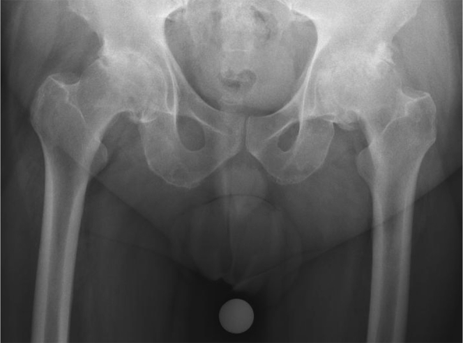
Sixty-six-year-old male with several years of bilateral knee pain. Hip radiographs revealed Grade 3 bilateral hip OA.
Figure 7.

Sixty-six-year-old male with several years of bilateral knee pain. Knee pain completely resolved after bilateral THA.
Discussion
To our knowledge, there have been only isolated case reports of hip pathology masquerading as knee pain in the adult population, and this is the largest series to date on this subject [16], [17]. We highlight the importance of maintaining a high index of suspicion when patients present with knee pain, and disability disproportionate to their radiographic disease, especially if their symptoms are refractory to interventions. If patients have predominantly knee pain but are dependent on a walker for ambulation or in a wheelchair, the suspicion for a more proximal source of the pain or disability should be high.
The hip and knee joints receive multiple sensory innervations. In the hip, the obturator and femoral nerves supply the hip capsule anteriorly, while sciatic and superior gluteal nerves supply it posteriorly [21]. In the knee, anterior fibers originate from the femoral, saphenous, and common peroneal nerves, while posterior fibers originate from the tibial and obturator nerves [22]. Perhaps the crossover in innervation could explain the referral patterns of hip disease; however, the neural mechanisms have not been fully elucidated.
Early theories explaining the pathophysiology of referred pain include Ruch's [23]convergence-projection theory, which describes the convergence of somatic and visceral fibers directly onto one dorsal horn neuron in the spinal cord. Sinclair postulated that the somatic and visceral fibers converged onto an afferent neuron before reaching the spinal cord [24]. Since their original descriptions, some studies seem to support Ruch's theory; however, the topic remains poorly understood [25], [26]. Miura et al [27], using a rat model, demonstrated that a small percentage of all dorsal root ganglion neurons innervating the hip joints had other axons that extended to the medial portion of knee skin. These dichotomizing fibers may also explain referred knee pain originating from hip joint pathology.
In humans, the precise sensory innervation pattern and correlation of the sensory nerves in the hip and knee remains unknown. This is especially true in patients with persistent knee pain after THA [28]. One potential explanation for persistent knee pain could be that the muscle to which pain is referred for a prolonged period of time may itself become a generator of pain [10]. This highlights the importance of a swift diagnosis in patients that could be prone to such reorganization of their neural pain pathways.
In the current healthcare economic climate, the burden of arthritis is projected to increase in the coming years. By the year 2040, an estimated 78 million adults, older than 18 years of age, will have a diagnosis of arthritis [29]. Currently, total direct and indirect annual costs of managing OA per patient are estimated at $5700 [30]. With the anticipated projection of future patients and the standard high cost associated with OA, patients cannot afford unnecessary testing and interventions, especially when they result in a delay in accurate diagnosis.
Moreover, patients suffering from OA have a higher morbidity count and are 2-3 times more likely to suffer from anxiety and depression, which leads to limitations in activity and more pain and overall disability [31], [32]. Quickly and adequately addressing their osteoarthritic needs is essential to expedite their return to activities and ultimately restore overall health and well-being.
There are certain limitations to this study. The retrospective nature of this case series imparts observational and selection biases. Cases presented in this series were recorded over 5 years by the treating surgeons who diagnosed their hip disease. It is likely that, during the period studied, patients with knee pain from hip pathology could have been missed if the index of suspicion was low or the pain severity and related disability were low. Additionally, it would be very difficult to determine if patients with hip pathology presenting as knee pain sought treatment elsewhere before this was recognized. Considering this, the authors do not feel that estimating a prevalence of knee pain related to hip arthritis would be accurate and would in fact likely grossly underestimate the prevalence. The prevalence of this has been highly variable in the literature and is between 2% and 29% [6], [7], [9], [10], [12]. This further confirms that an estimation would not be accurate. In select patients, there was incomplete information regarding previous treatments received outside our institution, as well as prior radiographic studies, which would have provided additional valuable information to our study. Finally, there were no patient-reported outcome data before and after their hip replacement, only subjective reports of relief of their knee pain.
Conclusions
Referred pain from the hip to the knee can be misleading to healthcare providers including musculoskeletal providers who evaluated 15 of the 21 patients in our series. Sometimes the referred pain can be difficult to believe by the patient themselves. We highlight the following warning signs that should prompt investigation of the hip as a potential underlying source of knee pain: knee pain out of proportion to clinical and radiographic findings related to the knee; significant disability and use of walking aids (especially a wheelchair or walker); abnormal hip motion during physical examination and knee pain that fails to improve with knee interventions. This constellation of findings should raise the index of suspicion to evaluate extrinsic sources for knee pain, especially the hip. Improved awareness of this issue may more quickly lead to an accurate diagnosis, timely treatment of the source of pain, and prevent unnecessary interventions for affected patients. Although it is considered a basic knowledge in orthopaedic surgery, this case series highlights the importance of continued education and awareness of this clinical scenario which can still lead to misdiagnosis and mistreatment by musculoskeletal providers.
Acknowledgements
Publication of this article was funded in part by the University of Florida Open Access Publishing Fund.
Footnotes
One or more of the authors of this paper have disclosed potential or pertinent conflicts of interest, which may include receipt of payment, either direct or indirect, institutional support, or association with an entity in the biomedical field which may be perceived to have potential conflict of interest with this work. For full disclosure statements refer to http://dx.doi.org/10.1016/j.artd.2017.06.008.
Appendix A. Supplementary data
References
- 1.Sacks J.J., Luo Y.H., Helmick C.G. Prevalence of specific types of arthritis and other rheumatic conditions in the ambulatory health care system in the United States, 2001–2005. Arthritis Care Res (Hoboken) 2010;62(4):460. doi: 10.1002/acr.20041. [DOI] [PubMed] [Google Scholar]
- 2.Arthritis and related conditions, chapter 4. http://www.boneandjointburden.org/2013-report/iv-arthritis/iv [accessed 27.01.17]
- 3.Jordan J.M., Helmick C.G., Renner J.B. Prevalence of knee symptoms and radiographic and symptomatic knee osteoarthritis in African Americans and Caucasians: the Johnston County Osteoarthritis Project. J Rheumatol. 2007;34(1):172. [PubMed] [Google Scholar]
- 4.Jordan J.M., Helmick C.G., Renner J.B. Prevalence of hip symptoms and radiographic and symptomatic hip osteoarthritis in African Americans and Caucasians: the Johnston County Osteoarthritis Project. J Rheumatol. 2009;36(4):809. doi: 10.3899/jrheum.080677. [DOI] [PMC free article] [PubMed] [Google Scholar]
- 5.Hasan M., Shuckett R. Clinical features and pathogenetic mechanisms of osteoarthritis of the hip and knee. BCMJ. 2010;52(8):393. [Google Scholar]
- 6.Nakamura J., Oinuma K., Ohtori S. Distribution of hip pain in osteoarthritis patients secondary to developmental dysplasia of the hip. Mod Rheumatol. 2013;23(1):119. doi: 10.1007/s10165-012-0638-5. [DOI] [PubMed] [Google Scholar]
- 7.Lesher J.M., Dreyfuss P., Hager N., Kaplan M., Furman M. Hip joint pain referral patterns: a descriptive study. Pain Med. 2008;9(1):22. doi: 10.1111/j.1526-4637.2006.00153.x. [DOI] [PubMed] [Google Scholar]
- 8.Khan A.M., McLoughlin E., Giannakas K., Hutchinson C., Andrew J.G. Hip osteoarthritis: where is the pain? Ann R Coll Surg Engl. 2004;86(2):119. doi: 10.1308/003588404322827518. [DOI] [PMC free article] [PubMed] [Google Scholar]
- 9.Khan N.Q., Woolson S.T. Referral patterns of hip pain in patients undergoing total hip replacement. Orthopedics. 1998;21(2):123. doi: 10.3928/0147-7447-19980201-05. [DOI] [PubMed] [Google Scholar]
- 10.Kawada T., Ushida T., Ikeuchi M. Clinical study of hip joint referred pain. Pain Res. 2006;21(3):127. [Google Scholar]
- 11.Lee B.H., Moon S.H., Lee H.M., Kim T.H., Lee S.J. Prevalence of hip pathology in patients over age 50 with spinal conditions requiring surgery. Indian J Orthop. 2012;46(3):291. doi: 10.4103/0019-5413.96386. [DOI] [PMC free article] [PubMed] [Google Scholar]
- 12.Sakamoto J., Morimoto Y., Ishii S. Investigation and macroscopic anatomical study of referred pain in patients with hip disease. J Phys Ther Sci. 2014;26(2):203. doi: 10.1589/jpts.26.203. [DOI] [PMC free article] [PubMed] [Google Scholar]
- 13.Ledwith C.A., Fleisher G.R. Slipped capital femoral epiphysis without hip pain leads to missed diagnosis. Pediatrics. 1992;89(4):660. [PubMed] [Google Scholar]
- 14.Matava M.J., Patton C.M., Luhmann S., Gordon J.E., Schoenecker P.L. Knee pain as the initial symptom of slipped capital femoral epiphysis: an analysis of initial presentation and treatment. J Pediatr Orthop. 1999;19(4):455. doi: 10.1097/00004694-199907000-00007. [DOI] [PubMed] [Google Scholar]
- 15.Atanda A., Shah S.A., O'Brien K. Osteochondrosis: common causes of pain in growing bones. Am Fam Physician. 2011;83(3):285. [PubMed] [Google Scholar]
- 16.Gannon J.M., Gustilo R.B. Treat the hip and not the knee: a report of four cases. Orthopedics. 1992;15(4):474. doi: 10.3928/0147-7447-19920401-13. [DOI] [PubMed] [Google Scholar]
- 17.Emms N.W., O'Connor M., Montgomery S.C. Hip pathology can masquerade as knee pain in adults. Age Ageing. 2002;31(1):67. doi: 10.1093/ageing/31.1.67. [DOI] [PubMed] [Google Scholar]
- 18.Kellgren J.H., Lawrence J.S. Radiological assessment of osteo-arthrosis. Ann Rheum Dis. 1957;16(4):494. doi: 10.1136/ard.16.4.494. [DOI] [PMC free article] [PubMed] [Google Scholar]
- 19.Fernando D., Thakar A.B., Shukla V.D. Clinical efficacy of Eranda Muladi Yapana Basti in the management of Kati Graha (Lumbar spondylosis) Clin Res. 2013;34(10):36. doi: 10.4103/0974-8520.115444. [DOI] [PMC free article] [PubMed] [Google Scholar]
- 20.Society of Preventive Hip Surgery Tönnis classification of osteoarthritis by radiographic changes. http://www.preventivehip.org/hip-scores/tonnis-classification [accessed 27.01.17]
- 21.Birnbaum K., Prescher A., Hessler S., Keller K.D. The sensory innervation of the hip joint—an anatomical study. Surg Radiol Anat. 1997;19(6):371. doi: 10.1007/BF01628504. [DOI] [PubMed] [Google Scholar]
- 22.Hirasawa Y., Okajima S., Ohta M., Tokioka T. Nerve distribution to the human knee joint: anatomical and immunohistochemical study. Int Orthop. 2000;24(1):1. doi: 10.1007/s002640050001. [DOI] [PMC free article] [PubMed] [Google Scholar]
- 23.Ruch T.C. Visceral sensation and referred pain. In: Fulton J.F., editor. Howell's textbook of physiology. 15th ed. WB Saunders; Philadelphia (PA): 1946. p. 385. [Google Scholar]
- 24.Sinclair D.C., Weddell G., Feindel W.H. Referred pain and associated phenomena. Brain. 1948;71:184. doi: 10.1093/brain/71.2.184. [DOI] [PubMed] [Google Scholar]
- 25.Rucker H.K., Holloway J.A. Viscerosomatic convergence onto spinothalamic tract neurons in the cat. Brain Res. 1982;243(1):155. doi: 10.1016/0006-8993(82)91130-1. [DOI] [PubMed] [Google Scholar]
- 26.Selzer M., Spencer W.A. Convergence of visceral and cutaneous afferent pathways in the lumber spinal cord. Brain Res. 1969;14:331. doi: 10.1016/0006-8993(69)90114-0. [DOI] [PubMed] [Google Scholar]
- 27.Miura Y., Ohtori S., Nakajima T. Dorsal root ganglion neurons with dichotomizing axons projecting to the hip joint and the knee skin in rats: possible mechanism of referred knee pain in hip joint disease. J Orthop Sci. 2011;16(6):799. doi: 10.1007/s00776-011-0144-1. [DOI] [PubMed] [Google Scholar]
- 28.Morimoto Y., Kondo Y., Shimosako J. Investigation of pain in hip disease patients before and after arthroplasty. J Phys Ther Sci. 2001;23:535. [Google Scholar]
- 29.Hootman J.M., Helmick C.G., Barbour K.E., Theis K.A., Boring M.A. Updated projected prevalence of self-reported doctor-diagnosed arthritis and arthritis-attributable activity limitation among US adults, 2015-2040. Arthritis Rheumatol. 2016;68(7):1582. doi: 10.1002/art.39692. [DOI] [PMC free article] [PubMed] [Google Scholar]
- 30.Maetzel A., Li L.C., Pencharz J., Tomlinson G., Bombardier C. The economic burden associated with osteoarthritis, rheumatoid arthritis, and hypertension: a comparative study. Ann Rheum Dis. 2004;63(4):395. doi: 10.1136/ard.2003.006031. [DOI] [PMC free article] [PubMed] [Google Scholar]
- 31.Katon W. Epidemiology and treatment of depression in patients with chronic medical illness. Dialogues Clin Neurosci. 2011;13(1):7. doi: 10.31887/DCNS.2011.13.1/wkaton. [DOI] [PMC free article] [PubMed] [Google Scholar]
- 32.van Dijk G.M., Veenhof C., Schellevis F. Comorbidity, limitations in activities and pain in patients with osteoarthritis of the hip or knee. BMC Musculoskelet Disord. 2008;9:95. doi: 10.1186/1471-2474-9-95. [DOI] [PMC free article] [PubMed] [Google Scholar]
Associated Data
This section collects any data citations, data availability statements, or supplementary materials included in this article.